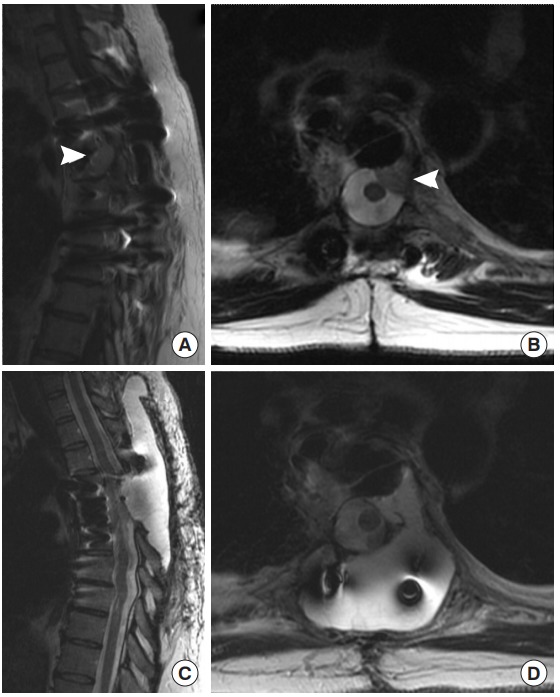
Fig. 5.

Preoperative magnetic resonance imaging (MRI) images from case 3, a 39-year-old female with a recurrent, extradural paraganglioma. The patient had a previous resection with thoracic spine stabilization years prior. (A) Sagittal T1 gadolinium-enhanced and (B) axial T2 MRI prior to her second surgery show recurrence of the extradural paraganglioma (arrowheads), abutting the dura. During her surgery for repeat resection, three small durotomies were encountered and repaired primarily and reinforced with muscle. Despite these precautions, she developed a pseudomeningocele postoperatively. T2 sagittal (C) and axial (D) images showed the T2 bright cerebrospinal fluid filling a pseudomeningocele above the resection site and spreading through the paraspinal musculature. She was treated conservatively with percutaneous drainage of the collection.
