Abstract
The purpose of an experimental design is to improve the productivity of experimentation. It is an efficient procedure for planning experiments, so the data obtained can be analyzed to yield a valid and objective conclusion. This approach has been used as an important tool in the optimization of different analytical approaches. A D-optimal experimental design was used here, for the first time, to optimize the experimental conditions for the detection of reactive oxygen species (ROS) produced by human blood from healthy donors, a biological matrix that better resembles the physiologic environment, following stimulation by a potent inflammatory mediator, phorbol-12-myristate-13-acetate (PMA). For that purpose, different fluorescent probes were used, as 2′,7′-dichlorodihydrofluorescein diacetate (DCFH-DA), 2-[6-(4′-amino)-phenoxy-3H-xanthen-3-on-9-yl] benzoic acid (APF), and 10-acetyl-3,7-dihydroxyphenoxazine (amplex red). The variables tested were the human blood dilution, and the fluorescent probe and PMA concentrations. The experiments were evaluated using the Response Surface Methodology and the method was validated using specific compounds. This model allowed the search for optimal conditions for a set of responses simultaneously, enabling, from a small number of experiments, the evaluation of the interaction between the variables under study. Moreover, a cellular model was implemented and optimized to detect the production of ROS using a yet nonexplored matrix, which is human blood.
1. Introduction
The scientific research on reactive oxygen species (ROS), for a deeper insight into their biological functions and/or deleterious effects, still is a matter of intense research. Fluorescent probes have been mainly used to detect ROS in isolated cells, namely neutrophils [1, 2]. However, the isolation process itself often leads to artifactual cell activation, which represents an experimental confounder, being also expensive and time-consuming [3]. Moreover, in the detection of ROS, it is important to account the interaction of all blood components to resemble as closely as possible the in vivo physiologic state. In that sense, human blood is the most complex biological matrix that better resembles the physiological environment. There are just a few reports in literature about the detection of reactive species in human blood [3–5], but none of them described the experimental optimization of the method.
In this work, we use a D-optimal experimental design. This type of design is particularly useful when full factorial design cannot be applied due to experimental constrains, for example, when biological samples are used, as human blood. In a D-optimal design, the best subset of experiments is selected in order to maximize the determinant of the matrix X'X for a predetermined regression model. This means that the experimental runs chosen span the largest volume possible in the experimental region [6, 7]. Despite the usefulness of the D-optimal experimental design, this method is not usually applied to biologic matrices, being used here, for the first time, to optimize the experimental conditions for the in vitro detection of ROS produced by human blood cells, from healthy donors, following stimulation by a potent inflammatory mediator, phorbol-12-myristate-13-acetate (PMA), using different fluorescent probes, 2′,7′-dichlorodihydrofluorescein diacetate (DCFH-DA), 2-[6-(4′ -amino)-phenoxy-3H-xanthen-3-on-9-yl] benzoic acid (APF), and 10-acetyl-3,7-dihydroxyphenoxazine (amplex red). The variables tested were the human blood dilution, and the fluorescent probe and PMA concentrations. The experiments were evaluated using the Response Surface Methodology (RSM), and the method was validated using specific inhibitors of ROS production, for example, aminobenzoyl hydrazide (ABAH), diphenyleneiodonium chloride (DPI), N,N-dimethylurea (DMTU), and also a known antioxidant, the flavonoid luteolin.
2. Material and Methods
2.1. Chemicals
Dulbecco's phosphate buffer saline, without calcium chloride and magnesium (PBS), DCFH-DA, diphenyleneiodonium chloride (DPI), horseradish peroxidase (HRP), amplex red, catalase (from bovine liver), luteolin, and N,N-dimethylurea (DMTU), and phorbol-12-myristate-13-acetate (PMA) were obtained from Sigma-Aldrich Co. LLC (St. Louis, USA). 4-Aminobenzoyl hydrazide (ABAH) was obtained from Calbiochem (San Diego, CA, USA). APF was obtained from Invitrogen, Life Technologies Ltd. (Paisley, UK). The erythrocyte-lysing buffer (BD Pharm Lyse) was obtained from BD Biosciences (San Jose, CA, USA).
2.2. Blood Samples
All patient-related procedures and protocols were performed in accordance with Helsinki Declaration. Following informed consent, venous blood was collected, in the morning, from healthy human male and nonpregnant female volunteers aged 18–65 years. Experiments were performed within 30 min following blood collection.
2.3. Experimental Design
The optimization of the experimental conditions for the in vitro detection of ROS by DCFH-DA, amplex red, and APF was undertaken by using the RSM and an interaction D-optimal experimental design with 3 levels, two quantitative factors: probe and PMA concentrations, and a qualitative factor: blood dilution. The RSM methodology allows a deeper understanding of a product or process by optimizing and stablishing a robust experimental process.
All analyses were carried out at least six times, and the mean data obtained in the experiments was analyzed using the RSM so as to fit the model equation that related the response to the factors varied by the Modde software version 10.1.1 (Umetrics AB, Umeå, Sweden). In order to correlate the response variable to the independent variables, multiple linear regression was used to fit the coefficient of the model. The model goodness-of-fit was evaluated using analysis of variance (ANOVA) at the level of 95% of significance. For all models, the R2 and the Q2 values were calculated. R2 is an indication of the model fit, and Q2 shows an estimate of the future prediction precision. Q2 should be greater than 0.1 for a significant model and greater than 0.5 for a good model.
To validate the optimization, at least six experiments were conducted under the chosen optimal conditions to compare to the results obtained by the experimental design and, in this way, allowing to verify the predictability ability of the model.
2.4. Detection of ROS
Experimental optimization was conducted for each of the three probes under study. After this optimization, the method effectiveness was attested by using the following compounds: specific inhibitors of the enzymes responsible for the generation of reactive species as DPI (NADPH oxidase inhibitor) [8] and ABAH [myeloperoxidase (MPO) inhibitor] [9], the scavenger of hydrogen peroxide (H2O2), DMTU [10], and catalase, that catalyzes H2O2 decomposition into H2O [11].
The known antioxidant flavonoid luteolin [12] was also tested to validate the method. The antioxidant activity of luteolin has been associated with their ability to scavenge ROS, as anion radical superoxide (O2·-) and hypochlorous acid (HOCl) [13], to inhibit the prooxidant enzymes NADPH oxidase [14–16] and myeloperoxidase (MPO) [17], and to chelate transition metals involved in Fenton reaction [18]. All the concentrations cited in the following sections are final concentrations.
2.4.1. DCFH-DA Assay
(1) Experimental Optimization. DCFH-DA is a nonpolar and nonfluorescent molecule that has the ability to diffuse through cell membranes into the cytoplasm where it is enzymatically cleaved by intracellular esterases to the polar nonfluorescent 2′,7′-dichlorodihydrofluorescein (DCFH). This molecule then becomes trapped inside the cell and is oxidized by ROS, producing the highly fluorescent 2′,7′ dichlorofluorescein (DCF) [19].
Whole blood (630 μL), diluted in PBS (ratio of 1 : 5, 1 : 10, and 1 : 20), was placed in 24-well plates and incubated in a humidified atmosphere with 5% CO2 at 37°C, with 100 μL of DCFH-DA (50, 100, and 200 μM) during 30 minutes. Then, 20 μL of PBS (same volume used for inhibitors) was added and incubated with the mixture for 15 minutes. In sequence, 50 μL of PMA (100, 200, and 400 nM) was added. The fluorescence was measured at λexcitation = 485 ± 20 nm and λemission = 528 ± 20 nm in a microplate reader (Cytation 3, Biotek, Vermont, USA). The D-optimal design consisted on a total of 16 experiments with 2 central points. Effects are expressed as the percentage of DCFH-DA oxidation, comparing with the blank (without PMA).
(2) Effect of ROS Production Inhibitors. Whole blood (diluted 1 : 20 in PBS) was placed in 24-well plates and incubated in a humidified atmosphere with 5% CO2 at 37°C, with DCFH-DA (120 μM) during 30 minutes. Then, DPI (0-5 μM), ABAH (0-2.5 mM), DMTU (0-60 mM), catalase (0-1300 U/mL), or luteolin (0-100 μM) were added and incubated with the reaction mixture for 15 minutes. In sequence, PMA (120 nM) was added. The fluorescence was measured as previously described in the Experimental Optimization section (2.4.1. Effects are expressed as the percentage of inhibition of DCFH-DA oxidation, comparing with the control (with PMA).
2.4.2. Amplex Red Assay
(1) Experimental Optimization. Amplex red is a highly specific and sensitive fluorogenic probe for the detection of extracellular H2O2. Amplex red is a colourless and nonfluorescent compound that, when oxidized by H2O2, in the presence of HRP, originates resofurin, which is a highly fluorescent product [20].
Whole blood (630 μL), diluted in PBS (ratio of 1 : 5, 1 : 10, and 1 : 20), was placed in 24-well plates and incubated in a humidified atmosphere with 5% CO2 at 37°C, with 25 μL of HRP (1 U/mL) and 25 μL of amplex red (6.3, 12.5, and 25 μM) during 10 minutes. Then, 20 μL of PBS (same volume used for inhibitors) was added and incubated with the mixture for 15 minutes. In sequence, 50 μL of PMA (25, 100, and 200 nM) was added. The fluorescence was measured at λexcitation = 560 ± 20 nm and λemission = 585 ± 20 nm in a microplate reader (Cytation 3, Biotek, Vermont, USA). The D-optimal design consisted on a total of 17 experiments with 3 central points. Effects are expressed as the percentage of amplex red oxidation, comparing with the blank (without PMA).
(2) Effect of ROS Production Inhibitors. Whole blood (diluted 1 : 20 in PBS) was placed in 24-well plates and incubated in a humidified atmosphere with 5% CO2 at 37°C, with HRP (1 U/mL) and amplex red (10 μM) during 10 minutes. Then, DPI (0-2.5 μM), ABAH (0-500 μM), catalase (0-1000 U/mL), DMTU (0-60 mM), or luteolin (0-100 μM) were added and incubated with the reaction mixture for 15 minutes. In sequence, 150 nM of PMA was added. The fluorescence was measured as previously described in the Experimental Optimization section (2.4.2). Effects are expressed as the percentage of inhibition of amplex red oxidation, comparing with the control (with PMA).
2.4.3. APF Assay
(1) Experimental Optimization. APF is a nonfluorescent derivative of fluorescein that originates fluorescein intracellularly, by O-dearylation, upon reaction with HOCl, hydroxyl radical (HO·) and peroxynitrite anion (ONOO−) leading to its characteristic fluorescence [21].
Whole blood (630 μL), diluted in PBS (ratio of 1 : 5, 1 : 10, and 1 : 20), was placed in 24-well plates and incubated in a humidified atmosphere with 5% CO2 at 37°C, with 100 μL of APF (1, 5, and 10 μM) during 10 minutes. Then, 20 μL of PBS was added and incubated with the mixture for 15 minutes. In sequence, 50 μL of PMA (50, 200, and 400 nM) was added and incubated for 30 minutes.
The samples were subsequently transferred to conic tubes with 8 mL of an erythrocyte lysing solution (BD Pharm Lyse), according to the manufacturer specifications and incubated at room temperature, protected from light, for 15 minutes. Then, samples were centrifuged at 200 g for 5 minutes, followed by the removal of the supernatant. The pellet was resuspended in 300 μL of PBS. The fluorescence was measured at λexcitation = 485 ± 20 nm and λemission = 528 ± 20 nm in a microplate reader (Cytation 3, Biotek, Vermont, USA). The D-optimal design consisted on a total of 16 experiments with 2 central points. Effects are expressed as the percentage of APF oxidation, comparing with the blank (without PMA).
(2) Effect of ROS Production Inhibitors. Whole blood (diluted 1 : 20 in PBS) was placed in 24-well plates and incubated in a humidified atmosphere with 5% CO2 at 37°C, with APF (5.5 μM) during 10 minutes. Then, DPI (0-5 μM), ABAH (0-2.5 mM), DMTU (0-60 mM), or luteolin (0-100 μM) were added and incubated with the reaction mixture for 15 minutes. In sequence, PMA (150 nM) was added and incubated for 30 minutes.
Then the samples were treated as mentioned in the Experimental Optimization section (2.4.3). Effects are expressed as the percentage of inhibition APF oxidation, comparing with the control (with PMA).
2.5. Statistical Analysis
The IC50 value (concentration that reduces the studied effect by 50%) was calculated using GraphPad Prism™ (version 7.0; GraphPad Software). Results are expressed as mean ± standard error of the mean (SEM) (from at least three individual experiments, performed in triplicate in each experiment).
3. Results
3.1. Optimization of Experimental Settings in the DCFH-DA Assay
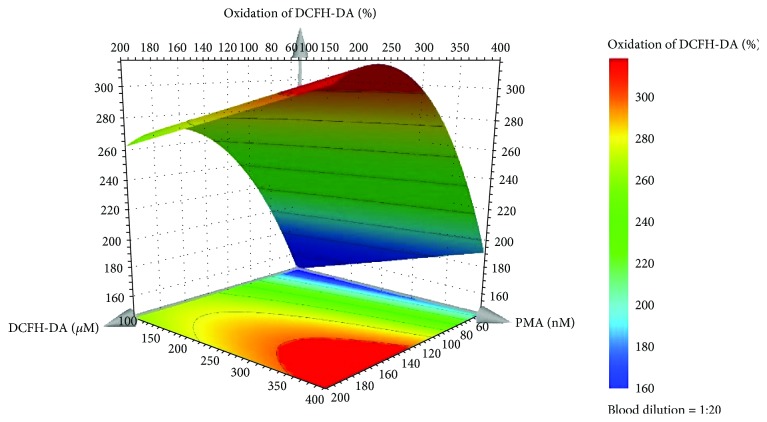
The examination of the model regression coefficients (p < 0.05) showed that the qualitative factor (blood dilution) was not significant for the model and that the oxidation increases with the increase of DCFH-DA and PMA concentrations. The model for the percentage of DCFH-DA oxidation [equation (1)] fitted the experimental data with a R2 of 0.95 and a Q2 value of 0.88, demonstrating that a model with a good fit and good predictability ability was obtained.
| (1) |
In equation (1), X1 is the DCFH-DA concentration (μM) and X2 is the PMA concentration (nM). Taking into account that all tested dilutions of human blood originated a good range of oxidation percentage of the probe, we choose the 1 : 20 dilution in order to use less quantity of human blood in each assay, making the biologic sample more profitable. Equation (1) and the response surface plot (Figure 1) allow us to choose the percentage of oxidation of the probe that better fits our aims. In this case, to obtain a probe oxidation around 250% (indicative value to validate the Equation), the concentrations of DCFH-DA and PMA should be 120 μM and 120 nM, respectively. The validation of the model was executed (n = 6), and the results showed that the percentage of oxidation of the probe was around the expected value (232 ± 45%). No significant difference (p < 0.05) was found between the validation experiments and those predicted by the model, confirming the good model prediction ability.
Figure 1.
Response surface plot obtained for the percentage of oxidation of the DCFH-DA by the predictive model of the D-optimal design for the 1 : 20 blood dilution.
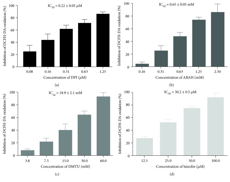
To better understand which reactive species are involved in the oxidation of DCFH-DA in this model, we used DPI (inhibit NADPH oxidase), catalase (catalyzes H2O2 decomposition into H2O), DMTU (scavenges H2O2), ABAH (inhibitor of MPO), and luteolin (a known antioxidant). As it can be seen in Figure 2, among the inhibitors used, only DPI, ABAH, and DMTU avoided the oxidation of DFCH-DA in a concentration-dependent manner. The IC50 values obtained for DPI was IC50 = 0.22 ± 0.05 μM, for ABAH was 0.65 ± 0.05 mM, and for DMTU was IC50 = 18.9 ± 2.1 mM. Catalase increased the fluorescence value per se, suggesting a possible interference with the methodology. These results indicate that, using human blood as cellular model, DCFH-DA preferentially detects NADPH oxidase-derived ROS as H2O2 and HOCl. Luteolin originated an IC50 = 30.2 ± 0.5 μM.
Figure 2.
Inhibitory effects of DPI (0.08-1.25 μM) (a), ABAH (0.16-2.50 mM) (b), DMTU (3.8-60 mM) (c), and luteolin (12.5-100 μM) (d) on the oxidation of DCFH-DA, by whole blood-generated ROS, when stimulated by PMA. Values are given as mean ± SEM (n ≥ 3).
3.2. Optimization of Experimental Setting in the Amplex Red Assay
The examination of the model regression coefficients (p < 0.05) showed that the qualitative factor (blood dilution) was not significant for the model and that the oxidation increases with the increase of amplex red and PMA concentration. The model for the percentage of amplex red oxidation [equation (2)] fitted the experimental data with a R2 of 0.85 and a Q2 value of 0.67 demonstrating that a model with a good fit and good predictability ability was obtained.
| (2) |
In equation (2), X1 is the amplex red concentration (μM) and X2 is the PMA concentration (nM). Taking into account that all tested dilutions of human blood originated a good range of oxidation percentage of the probe, we choose the 1 : 20 dilution in order to use less quantity of human blood in each assay, making the biologic sample more profitable.
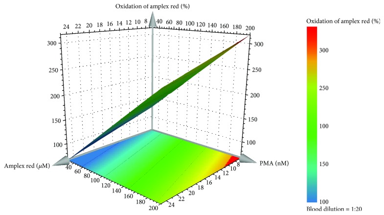
In this case, we also choose a probe oxidation around 250% as an indicative value to validate the Equation. According to equation (2) and as it can be seen in the response surface plot (Figure 3), to obtain a percentage of probe oxidation around 250%, a concentration of amplex red and PMA of 10 μM and 150 nM, respectively, was chosen. The validation of the model was executed (n = 6), and the results showed that the percentage of oxidation of the probe was around the expected value (268 ± 18%).
Figure 3.
Response surface plot obtained for the percentage of oxidation of amplex red by the predictive model of the D-optimal design for the 1 : 20 blood dilution.
No significant difference (P < 0.05) was found between the validation experiments and those predicted by the model, confirming the good model prediction ability.
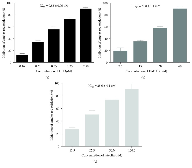
To guarantee that, in this model, amplex red continues to be a specific probe to detect H2O2, we tested the enzymatic inhibitors DPI (inhibitor of NADPH oxidase) and ABAH (inhibitor of MPO). Figure 4 shows that 2.5 μM of DPI totally hindered the oxidation of amplex red, demonstrating that the detection of H2O2 by amplex red is dependent on the NADPH oxidase activation. The lack of inhibition of amplex red oxidation by ABAH (data not shown) corroborates that HOCl production was not detected by this probe, in this cellular model. Catalase interferes with this method by inducing an HRP-independent oxidation of amplex red in the presence or absence of blood. This interference with the probe led us to use a scavenger of H2O2, DMTU, which inhibited the oxidation of amplex red in a concentration-dependent manner, proving that amplex red mainly detects H2O2. In addition, we also studied the flavonoid luteolin that was very effective in inhibiting the H2O2 production, presenting an IC50 of 25.6 ± 4.4 μM.
Figure 4.
Inhibitory effects of DPI (0.16-2.5 μM) (a), DMTU (7.5-60.0 mM) (b), and luteolin (12.5-100 μM) (c) on the oxidation of amplex red, by whole blood-generated ROS, when stimulated by PMA. Values are given as mean ± SEM (n ≥ 3).
3.3. Optimization of Experimental Settings in the APF Assay
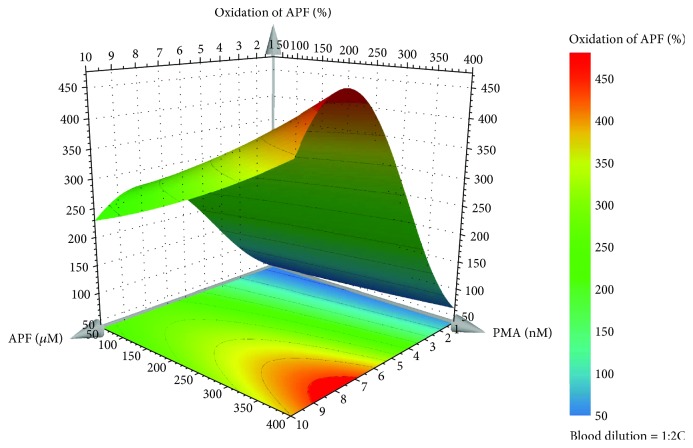
The examination of the model regression coefficients (P < 0.05) showed that the qualitative factor (blood dilution) was not significant for the model and that the oxidation increases with the increase of APF and PMA concentration. The model for the percentage of APF oxidation [equation (3)] fitted the experimental data with a R2 of 0.98 and a Q2 value of 0.95, demonstrating that a model with a good fit and good predictability ability was obtained. In this case, a logarithmic transformation was made to the data to improve the model robustness.
| (3) |
In equation (3), X1 is the APF concentration (μM) and X2 is the PMA concentration (nM). Taking into account that all the tested dilutions of human blood originated a good range of oxidation percentage of the probe, we choose the 1 : 20 dilution in order to use less quantity of human blood in each assay, making the biologic sample more profitable. Once again, we choose 250% of the probe oxidation as an indicative value to validate the Equation. According to equation (3) and as it can be seen in the response surface plot (Figure 5), to obtain a percentage of oxidation of the probe around 250%, a concentration of APF and PMA of 5.5 μM and 150 nM, respectively, was chosen. There were no significant differences (P < 0.05) between the validation experiments and those predicted by the model, confirming its good prediction ability. The validation of the model was executed (n = 6), and the results showed that the percentage of oxidation of the probe was around the expected value (257 ± 45%).
Figure 5.
Response surface plot obtained for the percentage of oxidation of APF by the predictive model of the D-optimal design for the 1 : 20 blood dilution.
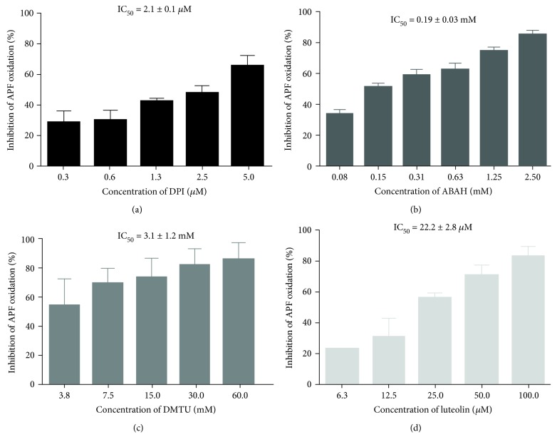
As it can be seen in Figure 6, DPI, ABAH and DMTU almost avoided the oxidation of APF at the concentrations of 5.0 μM, 2.5 mM, and 60 mM, respectively. Since DMTU scavenges H2O2, this could indicate that APF can detect H2O2 or other ROS that derived from H2O2, as HOCl. To test this cellular model, we also studied the flavonoid luteolin that was very effective in inhibiting the ROS production, presenting an IC50 = 22.2 ± 2.8 μM.
Figure 6.
Inhibitory effects of DPI (0.31-5.00 μM) (a), ABAH (0.08-2.50 mM) (b), DMTU (3.75-60.0 mM) (c), and luteolin (6.25-100 μM) (d) on the oxidation of APF by whole blood-generated reactive species, when stimulated by PMA. Values are given as mean ± SEM (n ≥ 3).
4. Discussion
The detection of reactive species has been a matter of intense debate. Most of the studies described the detection of ROS in different cells types, primary or cell line, as neutrophils [14, 22], monocytes [23, 24], and macrophages [25]. However, in these studies lack the interaction among all the cells present in the blood, which could interfere and dictate a different behavior than that obtained using a single cell type. Therefore, here, we used human blood, from healthy donors, as cellular model, since it is a more complex and physiologic in vitro system, preserving all cell-cell and cell-matrix interactions. Besides that, the manipulation of this cellular model is easier, faster, and cheaper than the isolation process and the maintenance of a cell culture of a single cell type.
A D-optimal experimental design was used, for the first time, to optimize the experimental conditions for the in vitro detection of ROS produced by PMA-stimulated human blood cells, using fluorescent probes. The use of the D-optimal experimental design allows the optimization of the experimental conditions in a single step using few experiments. In this way, the region of interest is covered optimally by the chosen experimental settings. Moreover, this optimization and the provided equations increase the time and cost effectiveness of the experiments. For that purpose, three different but complementary fluorescent probes were used for ROS detection, namely, DCFH-DA, amplex red, and APF. The detection of reactive species was done using a common equipment of microanalysis, a microplate reader, which is cheaper and easy to use than other equipments used in this type of assays, as flow cytometer [26, 27] or HPLC [28]. In the DCFH-DA system, a quadratic equation was obtained with PMA concentration interacting with itself. This means that there is a quadratic relationship between the percentage of oxidation and the PMA concentration. The APF system also has a quadratic relationship between the response and the concentration of PMA. Moreover, in this system, the response was logarithmized to normalize the distribution of the response in order to improve estimates and statistics.
DCFH-DA has been used for many years for the detection of ROS in isolated cells such as leukocytes [3, 29, 30]. However, to the best of our knowledge, there are only two reports using DCFH-DA to detect ROS in human blood [3, 31]. Here, we innovate and optimized the experimental conditions using a D-optimal experimental design in order to achieve the conditions that best fit the objectives. Analyzing the response surface plot, and to obtain a percentage of oxidation of the probe of approximately 250%, it was possible to fix the concentrations of DCFH-DA and PMA into 120 μM and 120 nM, respectively, and the blood dilution in 1 : 20.
DCFH-DA is a small, nonpolar, and nonfluorescent molecule that can diffuse into the cell, where intracellular esterases hydrolyze the acetate groups resulting in dichlorofluorescin that then reacts with a variety of ROS such as H2O2, HO·, and ROO· and also with reactive nitrogen species such as ·NO, ·NO2, and ONOO− [20], resulting in an increase in the fluorescent signal. Nevertheless, there is no information about which ROS are detected by this probe using a complex matrix as human blood. To clarify this point, we used the inhibitors of the most important enzymes responsible for the ROS production, such as DPI (inhibitor of several flavoenzymes, including NADPH oxidase [32]) and ABAH (inhibitor of MPO [33]). DPI and ABAH avoided the oxidation of DFCH-DA in a concentration-dependent manner. These results indicate that DCFH-DA can detect several ROS as O2·- or H2O2 and also ROS derived from MPO, as HOCl. To understand which ROS, O2·- or H2O2, were detected by DCFH-DA, we also tested catalase (catalyzes H2O2 decomposition into H2O) and DMTU (a cell-permeable scavenger of H2O2 [34]). Surprisingly, catalase increased the fluorescence of the probe by itself. As reported before, catalase may act as an intracellular factor able to oxidize fluorescent probes and also act as a cofactor for the reaction with H2O2, due to peroxidase activity [35]. Therefore, we used DMTU, which decreased the fluorescent signal, indicating that DCFH-DA essentially detects H2O2.
One of the main advantages of DCFH-DA assay is its simplicity of use, as it can be seen in the materials and methods section, but it also has other advantages as high sensitivity, an affordable price, and a nonselective detection of intracellular ROS, providing an overview of the overall prooxidant status. Nevertheless, the use of this probe has also some limitations that should be taken into account. One of them is the autooxidation and photoreduction, whether in the presence of visible light or by action of UVA radiation, that can be easily overcome by properly protecting the probe [20, 36]. The presence of antioxidants, naturally present in the cells or in the culture media, could compete with the probe for reaction with ROS resulting in the underestimation of ROS production. As such, it is important to use a high probe concentration to establish appropriate intracellular concentration or stimulate the cells properly to overcome this interference [36].
We also tested amplex red, which selectively reacts with H2O2 at the stoichiometry of 1 : 1 in a reaction catalyzed by HRP to generate the highly fluorescent product resorufin [37]. H2O2 is one of the most stable forms of ROS; thus, its detection allows the observation of oxidation processes in real time. Similar to DCFH-DA, amplex red has been used in isolated leukocytes [29, 38]. There is only one paper describing the use of amplex red in rat blood applied for the measurement of acetylcholinesterase activity [39]. The use of amplex red as probe for the detection of H2O2 in human blood is described here for the first time.
Interestingly, our results show that the percentage of amplex red oxidation increases with the decrease of amplex red concentration, indicating a higher sensitivity for lower concentrations of amplex red. This could be related with the higher baseline background fluorescence caused by higher concentrations of amplex red. This effect was already reported by Mohanty et al. [40], which stated that reducing the concentration of amplex red from 50 μM to 10 μM, increased the detection limiting, enabling the detection of 2 pmol instead of 0.1 to 2 nmol of H2O2. Accordingly, by analyzing the response surface plot derived from a D-optimal experimental design, the concentration of amplex red was fixed at 10 μM together with 150 nM of PMA and the blood dilution 1 : 20 to obtain a percentage of oxidation of the probe around 250%. DPI totally inhibited the oxidation of amplex red, showing that it detects H2O2 that can derive from NADPH oxidase and/or mitochondria activities. Catalase and DMTU were also tested. Once again, catalase interfered with the assay, increasing by itself, the fluorescent signal, independently of the presence of blood and PMA in the reaction mixture. The depletion of fluorescent signal induced by DMTU, together with the absence of an effect of ABAH, reinforces the idea that amplex red selectively detects H2O2. As such, using blood as cellular model, amplex red continues to be highly sensitive and selective for H2O2 detection. In addition, resofurin, the highly fluorescent product of amplex red oxidation, is stable, and its longwave spectrum avoids inference from autofluorescence originated in biological samples [37]. It is important to note that this methodology, as other HRP-dependent methodologies, is susceptible to the interference from substances that oxidize this enzyme [1]. Nevertheless, given its high sensitivity, specificity, and chemical stability, amplex red is suitable for the detection of H2O2 in human blood.
APF was successfully used to quantify MPO activity in isolated cells by few researchers [14, 15]. To the best of our knowledge, there is only one report from our group, using APF in human blood [31]. Once again, we felt that the literature lacks information about this issue, and most importantly, the optimization process should be clarified. As it was mentioned above, there is a current need to use human blood as a cellular model to truthfully understand the influence of all blood components in the activity of anti- or prooxidant compounds. In the case of APF, we have to perform a hypotonic lysis of erythrocytes before the reading of the fluorescent signal in the microplate reader. This step was essential to obtain a difference of values between the blank (without PMA) and the control (with PMA) assays. Our procedure is in agreement with Flemmig et al. [41], who used a flow cytometer to detect the APF fluorescence in human blood and also reported the elimination of erythrocytes. However, in contrast with what was reported in the work of Flemmig et al. [41], in the present study, the blood was incubated with PMA and/or the compounds under study, before the erythrocytes lysis, precisely to guarantee their interaction with the different type of the cells, including erythrocytes. In addition, here, we proposed an analysis in a microplate reader that is of easy use and cheaper than a flow cytometer.
APF is a nonfluorescent derivative of fluorescein that detects HOCl, HO·, and ONOO− intracellularly [42]. Our previous reports have shown that APF is more sensitive to HOCl than to the other ROS [14, 15, 43]. As it was done with the other probes, we also tested several inhibitors of the ROS production. ABAH also avoided the production of reactive species, suggesting that HOCl strongly contributes to APF oxidation. Interestingly, DMTU also inhibited the APF fluorescent signal. This indicates that H2O2 influences, directly or indirectly, the oxidation of APF. As the majority of the produced H2O2 is used by MPO to originate HOCl, it seems that DMTU removes the produced H2O2 that will not be consumed by MPO and consequently originates a decrease in the fluorescent signal.
Compared to the other probes, APF is not light sensitive; however, it is the more expensive and time-consuming probe [42]. Nevertheless, APF allowed the selective detection of one of the main ROS produced during the inflammatory process. The overproduction of HOCl is related to the development of several human diseases such as arthritis, cancer, and neurodegeneration [44]. As such, due to its biological importance, the use of sensitive and selective probes for its detection is of utmost importance.
To validate the methods discussed here, luteolin, a known antioxidant, was used as a positive control. Luteolin decreased the production of ROS, using all of the three probes, proving its ability to modulate the production of reactive species, also in a complex cellular model, as human blood, as it was already described in other type of in vitro assays, using isolated human neutrophils [14–16].
5. Conclusions
In this work, a D-optimal experimental design was used for the first time to optimize the experimental conditions for the in vitro detection of ROS produced by human blood, from healthy donors, using three different and complementary fluorescent probes, DCFH-DA, amplex red, and APF. Our results will help researchers to accurately choose the conditions that better fit their scientific objectives, saving time and money, and most importantly, using a physiological in vitro system that mimics the in vivo settings and which is yet unexplored.
Acknowledgments
The authors acknowledge the financial support from the European Union (FEDER funds POCI/01/0145/FEDER/007265) and the national funds (FCT/MEC, Fundação para a Ciência e Tecnologia and Ministério da Educação e Ciência) under the Partnership Agreement PT2020 UID/QUI/50006/2013. Tânia Soares acknowledges FCT, the financial support for the PhD grant (SFRH/BD/128647/2017), in the ambit of “QREN - POPH - Tipologia 4.1 - Formação Avançada”, co-sponsored by the Fundo Social Europeu (FSE) and by the national funds of the Ministério da Ciência, Tecnologia e Ensino Superior (MCTES). Daniela Ribeiro acknowledges FEDER, through COMPETE and FCT, the financial support for the post-doc grant in the ambit of the project PTDC/QEQ-QAN/1742/2014 - POCI-01-0145-FEDER-016530.
Contributor Information
Eduarda Fernandes, Email: egracas@ff.up.pt.
Marisa Freitas, Email: marisafreitas@ff.up.pt.
Data Availability
The data used to support the findings of this study are included within the article.
Conflicts of Interest
The authors declare no conflict of interest.
References
- 1.Freitas M., Lima J. L. F. C., Fernandes E. Optical probes for detection and quantification of neutrophils' oxidative burst. A review. Analytica Chimica Acta. 2009;649(1):8–23. doi: 10.1016/j.aca.2009.06.063. [DOI] [PubMed] [Google Scholar]
- 2.Walrand S., Valeix S., Rodriguez C., Ligot P., Chassagne J., Vasson M.-P. Flow cytometry study of polymorphonuclear neutrophil oxidative burst: a comparison of three fluorescent probes. Clinica Chimica Acta. 2003;331(1-2):103–110. doi: 10.1016/S0009-8981(03)00086-X. [DOI] [PubMed] [Google Scholar]
- 3.Himmelfarb J., Hakim R. M., Holbrook D. G., Leeber D. A., Ault K. A. Detection of granulocyte reactive oxygen species formation in whole blood using flow cytometry. Cytometry. 1992;13(1):83–89. doi: 10.1002/cyto.990130113. [DOI] [PubMed] [Google Scholar]
- 4.Kampen A. H., Tollersrud T., Lund A. Flow cytometric measurement of neutrophil respiratory burst in whole bovine blood using live Staphylococcus aureus. Journal of Immunological Methods. 2004;289(1-2):47–55. doi: 10.1016/j.jim.2004.03.008. [DOI] [PubMed] [Google Scholar]
- 5.Richardson M. P., Ayliffe M. J., Helbert M., Davies E. G. A simple flow cytometry assay using dihydrorhodamine for the measurement of the neutrophil respiratory burst in whole blood: comparison with the quantitative nitrobluetetrazolium test. Journal of Immunological Methods. 1998;219(1-2):187–193. doi: 10.1016/S0022-1759(98)00136-7. [DOI] [PubMed] [Google Scholar]
- 6.Eriksson L., Johansson E., Kettaneh-Wold N., WikstrÄom C., Wold S. Design of Experiments: Principles and Applications. Umetrics AB; 2008. [Google Scholar]
- 7.Armstrong N. A. Pharmaceutical Experimental Design and Interpretation. CRC/Taylor & Francis; 2006. [Google Scholar]
- 8.Ellis J. A., Mayer S. J., Jones O. T. The effect of the NADPH oxidase inhibitor diphenyleneiodonium on aerobic and anaerobic microbicidal activities of human neutrophils. The Biochemical Journal. 1988;251(3):887–891. doi: 10.1042/bj2510887. [DOI] [PMC free article] [PubMed] [Google Scholar]
- 9.Dypbukt J. M., Bishop C., Brooks W. M., Thong B., Eriksson H., Kettle A. J. A sensitive and selective assay for chloramine production by myeloperoxidase. Free Radical Biology & Medicine. 2005;39(11):1468–1477. doi: 10.1016/j.freeradbiomed.2005.07.008. [DOI] [PubMed] [Google Scholar]
- 10.Brown J. M., Terada L. S., Grosso M. A., et al. Xanthine oxidase produces hydrogen peroxide which contributes to reperfusion injury of ischemic, isolated, perfused rat hearts. The Journal of Clinical Investigation. 1988;81(4):1297–1301. doi: 10.1172/JCI113448. [DOI] [PMC free article] [PubMed] [Google Scholar]
- 11.Keilin D., Hartree E. F. Properties of catalase. Catalysis of coupled oxidation of alcohols. The Biochemical Journal. 1945;39(4):293–301. doi: 10.1042/bj0390293. [DOI] [PMC free article] [PubMed] [Google Scholar]
- 12.Lopez-Lazaro M. Distribution and biological activities of the flavonoid luteolin. Mini Reviews in Medicinal Chemistry. 2009;9(1):31–59. doi: 10.2174/138955709787001712. [DOI] [PubMed] [Google Scholar]
- 13.Gomes A., Fernandes E., Silva A. M. S., et al. 2-Styrylchromones: novel strong scavengers of reactive oxygen and nitrogen species. Bioorganic & Medicinal Chemistry. 2007;15(18):6027–6036. doi: 10.1016/j.bmc.2007.06.046. [DOI] [PubMed] [Google Scholar]
- 14.Freitas M., Ribeiro D., Tomé S. M., Silva A. M. S., Fernandes E. Synthesis of chlorinated flavonoids with anti-inflammatory and pro-apoptotic activities in human neutrophils. European Journal of Medicinal Chemistry. 2014;86:153–164. doi: 10.1016/j.ejmech.2014.08.035. [DOI] [PubMed] [Google Scholar]
- 15.Ribeiro D., Freitas M., Tomé S. M., Silva A. M. S., Porto G., Fernandes E. Modulation of human neutrophils' oxidative burst by flavonoids. European Journal of Medicinal Chemistry. 2013;67:280–292. doi: 10.1016/j.ejmech.2013.06.019. [DOI] [PubMed] [Google Scholar]
- 16.Yang S. C., Chen P. J., Chang S. H., et al. Luteolin attenuates neutrophilic oxidative stress and inflammatory arthritis by inhibiting Raf1 activity. Biochemical Pharmacology. 2018;154:384–396. doi: 10.1016/j.bcp.2018.06.003. [DOI] [PubMed] [Google Scholar]
- 17.Shiba Y., Kinoshita T., Chuman H., et al. Flavonoids as substrates and inhibitors of myeloperoxidase: molecular actions of aglycone and metabolites. Chemical Research in Toxicology. 2008;21(8):1600–1609. doi: 10.1021/tx8000835. [DOI] [PubMed] [Google Scholar]
- 18.Mira L., Tereza Fernandez M., Santos M., Rocha R., Helena Florêncio M., Jennings K. R. Interactions of flavonoids with iron and copper ions: a mechanism for their antioxidant activity. Free Radical Research. 2002;36(11):1199–1208. doi: 10.1080/1071576021000016463. [DOI] [PubMed] [Google Scholar]
- 19.Chen Y. F., Jobanputra P., Barton P., et al. A systematic review of the effectiveness of adalimumab, etanercept and infliximab for the treatment of rheumatoid arthritis in adults and an economic evaluation of their cost-effectiveness. Health Technology Assessment. 2006;10(42):1–229. doi: 10.3310/hta10420. [DOI] [PubMed] [Google Scholar]
- 20.Kalyanaraman B., Darley-Usmar V., Davies K. J. A., et al. Measuring reactive oxygen and nitrogen species with fluorescent probes: challenges and limitations. Free Radical Biology & Medicine. 2012;52(1):1–6. doi: 10.1016/j.freeradbiomed.2011.09.030. [DOI] [PMC free article] [PubMed] [Google Scholar]
- 21.Setsukinai K., Urano Y., Kakinuma K., Majima H. J., Nagano T. Development of novel fluorescence probes that can reliably detect reactive oxygen species and distinguish specific species. The Journal of Biological Chemistry. 2003;278(5):3170–3175. doi: 10.1074/jbc.M209264200. [DOI] [PubMed] [Google Scholar]
- 22.Houston N., Stewart N., Smith D. S., Bell S. C., Champion A. C., Reid D. W. Sputum neutrophils in cystic fibrosis patients display a reduced respiratory burst. Journal of Cystic Fibrosis. 2013;12(4):352–362. doi: 10.1016/j.jcf.2012.11.004. [DOI] [PubMed] [Google Scholar]
- 23.Wu W., Muller R., Berhane K., et al. Inflammatory response of monocytes to ambient particles varies by highway proximity. American Journal of Respiratory Cell and Molecular Biology. 2014;51(6):802–809. doi: 10.1165/rcmb.2013-0265OC. [DOI] [PMC free article] [PubMed] [Google Scholar]
- 24.Arora M., Arora R., Kumar A., Das N., Srivastava L. M. Monocytes from patients with rheumatoid arthritis release increased amount of reactive oxygen intermediates. Current Science. 2000;78:989–992. [Google Scholar]
- 25.Koo S.-j., Chowdhury I. H., Szczesny B., Wan X., Garg N. J. Macrophages promote oxidative metabolism to drive nitric oxide generation in response to Trypanosoma cruzi. Infection and Immunity. 2016;84(12):3527–3541. doi: 10.1128/IAI.00809-16. [DOI] [PMC free article] [PubMed] [Google Scholar]
- 26.Chen Y., Junger W. G. Measurement of oxidative burst in neutrophils. Methods in Molecular Biology. 2012;844:115–124. doi: 10.1007/978-1-61779-527-5_8. [DOI] [PMC free article] [PubMed] [Google Scholar]
- 27.Okano K., Kimura K., Tanaka Y., Tsuchiya K., Akiba T., Nitta K. Direct measurement of reactive oxygen species in leukocytes during hemodialysis therapy. International Journal of Clinical and Experimental Medicine. 2015;8:20959–20964. [PMC free article] [PubMed] [Google Scholar]
- 28.Daiber A., Oelze M., Steven S., Kröller-Schön S., Münzel T. Taking up the cudgels for the traditional reactive oxygen and nitrogen species detection assays and their use in the cardiovascular system. Redox Biology. 2017;12:35–49. doi: 10.1016/j.redox.2017.02.001. [DOI] [PMC free article] [PubMed] [Google Scholar]
- 29.Rinaldi M., Moroni P., Paape M. J., Bannerman D. D. Evaluation of assays for the measurement of bovine neutrophil reactive oxygen species. Veterinary Immunology and Immunopathology. 2007;115(1-2):107–125. doi: 10.1016/j.vetimm.2006.09.009. [DOI] [PubMed] [Google Scholar]
- 30.Caldefie-Chézet F., Walrand S., Moinard C., Tridon A., Chassagne J., Vasson M.-P. Is the neutrophil reactive oxygen species production measured by luminol and lucigenin chemiluminescence intra or extracellular? Comparison with DCFH-DA flow cytometry and cytochrome c reduction. Clinica Chimica Acta. 2002;319(1):9–17. doi: 10.1016/S0009-8981(02)00015-3. [DOI] [PubMed] [Google Scholar]
- 31.Proença C., Ribeiro D., Soares T., et al. Chlorinated flavonoids modulate the inflammatory process in human blood. Inflammation. 2017;40(4):1155–1165. doi: 10.1007/s10753-017-0559-8. [DOI] [PubMed] [Google Scholar]
- 32.Massart C., Giusti N., Beauwens R., Dumont J. E., Miot F., Sande J. V. Diphenyleneiodonium, an inhibitor of NOXes and DUOXes, is also an iodide-specific transporter. FEBS Open Bio. 2014;4(1):55–59. doi: 10.1016/j.fob.2013.11.007. [DOI] [PMC free article] [PubMed] [Google Scholar]
- 33.Han L., Shen X., Pan L., et al. Aminobenzoic acid hydrazide, a myeloperoxidase inhibitor, alters the adhesive properties of neutrophils isolated from acute myocardial infarction patients. Heart and Vessels. 2012;27(5):468–474. doi: 10.1007/s00380-011-0178-5. [DOI] [PubMed] [Google Scholar]
- 34.Parker N. B., Berger E. M., Curtis W. E., Muldrow M. E., Linas S. L., Repine J. E. Hydrogen peroxide causes dimethylthiourea consumption while hydroxyl radical causes dimethyl sulfoxide consumption in vitro. Journal of Free Radicals in Biology & Medicine. 1985;1(5-6):415–419. doi: 10.1016/0748-5514(85)90155-2. [DOI] [PubMed] [Google Scholar]
- 35.Hempel S. L., Buettner G. R., O’Malley Y. Q., Wessels D. A., Flaherty D. M. Dihydrofluorescein diacetate is superior for detecting intracellular oxidants: comparison with 2′,7′-dichlorodihydrofluorescein diacetate, 5(and 6)-carboxy-2′,7′-dichlorodihydrofluorescein diacetate, and dihydrorhodamine 123. Free Radical Biology & Medicine. 1999;27(1-2):146–159. doi: 10.1016/S0891-5849(99)00061-1. [DOI] [PubMed] [Google Scholar]
- 36.Chen X., Zhong Z., Xu Z., Chen L., Wang Y. 2′,7′-Dichlorodihydrofluorescein as a fluorescent probe for reactive oxygen species measurement: forty years of application and controversy. Free Radical Research Communications. 2010;44(6):587–604. doi: 10.3109/10715761003709802. [DOI] [PubMed] [Google Scholar]
- 37.Rhee S. G., Chang T. S., Jeong W., Kang D. Methods for detection and measurement of hydrogen peroxide inside and outside of cells. Molecules and Cells. 2010;29(6):539–549. doi: 10.1007/s10059-010-0082-3. [DOI] [PubMed] [Google Scholar]
- 38.Rochael N. C., Guimarães-Costa A. B., Nascimento M. T. C., et al. Classical ROS-dependent and early/rapid ROS-independent release of neutrophil extracellular traps triggered by Leishmania parasites. Scientific Reports. 2015;5(1, article 18302) doi: 10.1038/srep18302. [DOI] [PMC free article] [PubMed] [Google Scholar]
- 39.Santillo M. F., Liu Y. A fluorescence assay for measuring acetylcholinesterase activity in rat blood and a human neuroblastoma cell line (SH-SY5Y) Journal of Pharmacological and Toxicological Methods. 2015;76:15–22. doi: 10.1016/j.vascn.2015.07.002. [DOI] [PubMed] [Google Scholar]
- 40.Mohanty J. G., Jaffe J. S., Schulman E. S., Raible D. G. A highly sensitive fluorescent micro-assay of H2O2 release from activated human leukocytes using a dihydroxyphenoxazine derivative. Journal of Immunological Methods. 1997;202(2):133–141. doi: 10.1016/S0022-1759(96)00244-X. [DOI] [PubMed] [Google Scholar]
- 41.Flemmig J., Schwarz P., Bäcker I., Leichsenring A., Lange F., Arnhold J. Fast and specific assessment of the halogenating peroxidase activity in leukocyte-enriched blood samples. Journal of Visualized Experiments. 2016;(113, article e54484) doi: 10.3791/54484. [DOI] [PMC free article] [PubMed] [Google Scholar]
- 42.Shepherd J., Hilderbrand S. A., Waterman P., Heinecke J. W., Weissleder R., Libby P. A fluorescent probe for the detection of myeloperoxidase activity in atherosclerosis-associated macrophages. Chemistry & Biology. 2007;14(11):1221–1231. doi: 10.1016/j.chembiol.2007.10.005. [DOI] [PMC free article] [PubMed] [Google Scholar]
- 43.Freitas M., Costa V. M., Ribeiro D., et al. Acetaminophen prevents oxidative burst and delays apoptosis in human neutrophils. Toxicology Letters. 2013;219(2):170–177. doi: 10.1016/j.toxlet.2013.03.007. [DOI] [PubMed] [Google Scholar]
- 44.Wang E., Qiao H., Zhou Y., et al. A novel “turn-on” fluorogenic probe for sensing hypochlorous acid based on BODIPY. RSC Advances. 2015;5(89):73040–73045. doi: 10.1039/C5RA14118D. [DOI] [Google Scholar]
Associated Data
This section collects any data citations, data availability statements, or supplementary materials included in this article.
Data Availability Statement
The data used to support the findings of this study are included within the article.


