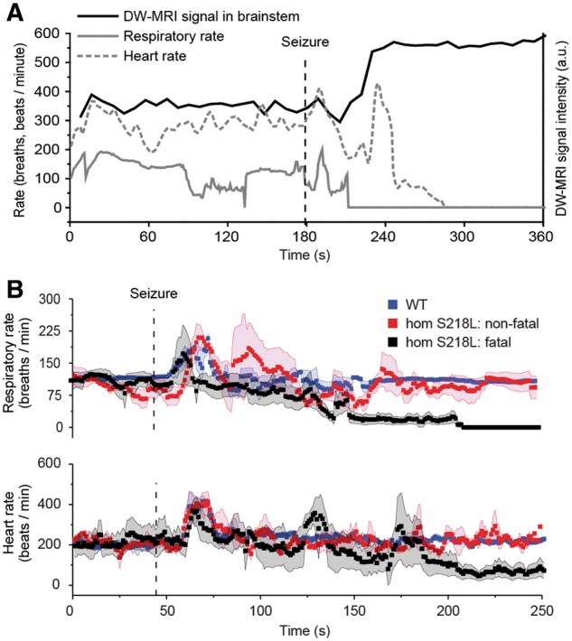
Figure 7.

Physiological function following induced seizures during DW-MRI in anaesthetized homozygous Cacna1aS218L mice. (A) Representative example from a single animal of respiratory rate, heart rate and spreading depolarization aligned temporally in relation to seizure onset (dashed line). (B) Physiological parameters (aligned with spreading depolarization dynamics shown in Fig. 6E) from homozygous Cacna1aS218L (hom S218L) mice, following non-fatal (n = 10) and fatal seizures (n = 4), and wild-type (WT) mice, following non-fatal seizures (n = 15). Simultaneous respiratory (upper trace) and heart rate (lower trace) data acquired during seizures from the same animals showing that, for fatal seizures, respiratory rate decreased until complete arrest and preceded cardiac arrest. A.u. = arbitrary units. Data shown as mean ± SD.
