Background
Surgical exploration of historic wounds for removal of foreign bodies can be challenging, both in correctly identifying their location and in the careful surgical dissection required when neurovascular structures are nearby. Ultrasonography represents an inexpensive, non-invasive method of dynamically imaging soft tissues1 and is available in the majority operating departments, where it is commonly used for securing vascular access or delivering regional anaesthesia. We report our experience of intraoperative portable ultrasonography for the removal of deep foreign bodies.
Technique
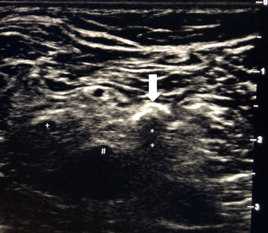
After appropriate patient positioning, an ultrasonography probe can be used to determine penetration depth and to mark the precise location of the foreign body in order to guide the skin incision and plan subsequent surgical dissection with due consideration to neurovascular structures (Fig 1).
Figure 1.

Ultrasonography showing tip of pellet (arrow) and its long acoustic shadow (*). The tibial (#) and fibular (+) nerve components are in close proximity
Discussion
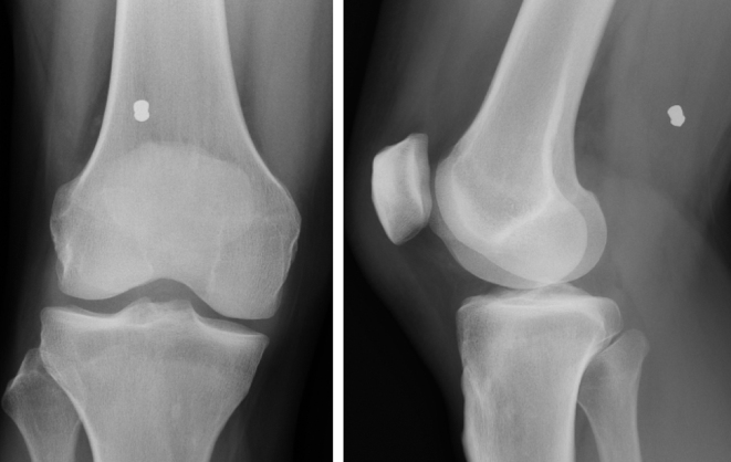
This method can help minimise unnecessary surgical exposure by correctly locating the foreign body. Furthermore, it can be more effective than plain radiography (Fig 2), which may be less accurate due to its timing (owing to further migration of the foreign body after the time of the radiography) or because of challenges in interpretation (owing to limb rotation and radiographic projection). Expertise in interpretation can be readily gained in most operating departments from anaesthetists, who are trained in the use of ultrasonography as part of their curriculum.
Figure 2.
Radiography showing location of pellet
Finally, this technique can be employed to facilitate the localisation and excision of symptomatic, deep, benign soft tissue tumours (most commonly nerve sheath tumours), which are often very mobile and can occur deep within the peripheral skeleton.
Reference
- 1.Kent MJ, Melton JT. Use of portable ultrasound for exploration and removal of superficial foreign bodies. Ann R Coll Surg Engl 2009; : 344–345. [DOI] [PMC free article] [PubMed] [Google Scholar]


