Abstract
Gold nanoparticles (AuNPs) have found a wide range of biomedical and environmental monitoring applications (viz. drug delivery, diagnostics, biosensing, bio-imaging, theranostics, and hazardous chemical sensing) due to their excellent optoelectronic and enhanced physico-chemical properties. The modulation of these properties is done by functionalizing them with the synthesized AuNPs with polymers, surfactants, ligands, drugs, proteins, peptides, or oligonucleotides for attaining the target specificity, selectivity and sensitivity for their various applications in diagnostics, prognostics, and therapeutics. This review intends to highlight the contribution of such AuNPs in state-of-the-art ventures of diverse biomedical applications. Therefore, a brief discussion on the synthesis of AuNPs has been summarized prior to comprehensive detailing of their surface modification strategies and the applications. Here in, we have discussed various ways of AuNPs functionalization including thiol, phosphene, amine, polymer and silica mediated passivation strategies. Thereafter, the implications of these passivated AuNPs in sensing, surface-enhanced Raman spectroscopy (SERS), bioimaging, drug delivery, and theranostics have been extensively discussed with the a number of illustrations.
Keywords: Gold nanoparticles, Synthesis approaches, Surface functionalization strategies, Biomedical applications
Introduction
Advancement in nanomaterial researches have shown a great impact in clinical diagnostics, therapeutics, and energy generations (Chandra et al. 2010; Kumar et al. 2018; Mahato et al. 2018a; Prasad et al. 2016). The common properties shown by nanoparticles (NPs) are (1) high surface-to-volume ratio, (2) ease of functionalization enabling specific target-binding properties, (3) tuneable optoelectronic properties, and (4) high robustness of the NPs (Baranwal et al. 2016), which enables them in various biomedical applications. In recent years, NPs of various materials viz. metals, non-oxide ceramics, metal oxides, silicates, polymers, biopolymers, and carbon have been used for such applications (He et al. 2000; Baranwal et al. 2018a). Noble metal NPs especially gold (Au) and silver (Ag) have fascinated researchers for decades and are being extensively used due to their excellent compatibility towards the biological systems (Baranwal et al. 2018b; Elahi et al. 2018). Among all, AuNPs are considered as a most important candidate due to its chemical inertness, environmentally benign nature, and biocompatibility when functionalized with appropriate ligand/group of ligands (Blanco et al. 2015; Sahoo et al. 2017). The first scientific note on AuNPs synthesis was reported by Michael Faraday in the early nineteenth century where the synthesis was done using chloroauric acid and phosphorous as the reducing agent (Faraday 1857). In this context, Turkevich and co-workers delivered the major breakthrough by demonstrating the synthetic mechanism of AuNPs formation in colloidal systems (Turkevich et al. 1951; Daniel and Astruc 2004). In recent years, numerous methods of AuNPs synthesis along with its numerous applications have been reported (Chen et al. 2018; Strozyk et al. 2018), however, the whole essence of these advancements was driven for obtaining the facile synthetic process, characterization, functionalization, and applications of these uniquely fabricated AuNPs (Mandal et al. 2018; Chandra et al. 2013b). Chemically, elemental gold ([Xe]4f145d106s1) contains electrons that can move freely throughout the metal and exhibits three oxidation states including Au [0], aurous (+ 1 Au [I]), and auric (+ 3 Au [III]) form. The availability of free electrons at the atomic surface and multiple oxidation states of metal facilitates the formation of stable nanostructures. Depending on the synthetic procedure, nature of solvent, solution pH, and surface passivating agents, these nanostructures are obtained of varied sizes and shapes. Due to its immense capabilities of exhibiting varied tunable optical, fluorescence, SPR, and magnetic properties, it has found a wide range of clinical and biomedical applications (Biju 2014). The other factors including greater stability, biocompatibility, selectivity, and lesser toxicity in the biological environment have led AuNPs for reliable commercial usage (Kumar et al. 2018). Surface modifications of AuNPs play a crucial role in achieving the enhanced properties for various biomedical applications. So far, various molecules of chemical and biochemical origin have been used to obtain such functionalized AuNPs by tuning the physicochemical behavior of AuNPs, i.e., the surface charges, ligand-binding ability, etc. These modifications of AuNPs commonly done using thiols, amines, phosphines, silica, carboxy-terminated groups, etc., eventually help to conjugate a number of biomolecules (Alex and Tiwari 2015).
Methodologically, these passivating processes follow either covalent-based modifications or non-covalent interactions. A strong Au–S covalent interaction has been reported using organothiols, disulfides, and cysteine groups, whereas the non-covalent interaction has been achieved by physiosorption and electrostatic interactions of surface-ionized ligands (Alex and Tiwari 2015). Based on their reaction involved in the covalent process, these modifications have been categorized under the direct and indirect covalent coupling. The direct coupling rely on the attachment of the ligand on the AuNPs surface, however, when the direct binding is not favorable, the linking process is done with the shell of stabilizing molecules encapsulating the AuNPs using various bio-conjugation techniques viz. carbodiimide, biotin-streptavidin, and silane coupling reactions (Craig et al. 2010). So far, the functionalized AuNPs have been exploited for a wide range of biomedical applications, not only in research and development sector but also in various commercially viable point of care systems viz. cyto-sensors (Koh et al. 2011), immuno-sensors (Noh et al. 2012), drug delivery (Baranwal et al. 2018b), cancer imaging (Wu et al. 2015), apta-sensing (Chandra et al. 2013a), and most advanced theranostics devices (Song et al. 2016). In this context, theranostic devices are one of the modern advents in biomedical devices, which delivers the precise sensing and accurate therapeutic effect synergistically.
The intention of this review is to summarize the extensively used various strategies for AuNPs synthesis and functionalization followed by its biomedical applications. For that, we have provided a brief introduction to the AuNPs synthesis before discussing its functionalization strategies to put wider insight to the readers. Thereafter, we have discussed diverse kind of passivating strategies used for AuNPs, where we have covered majorly employed techniques viz. thiol, amine, polymer, and silica-based modification. In the next section, we have described the application of such passivated AuNPs in various domains viz. biosensing, bioimaging, therapeutics, drug delivery, and most advent theranostics.
Synthesis of AuNPs
Methodologically, the synthesis of AuNPs follows two types of approaches, including “top-down” (physical manipulations) and “bottom-up” (chemical transformations) approaches (Mandal et al. 2018; Teimouri et al. 2018; Abalde-Cela et al. 2018; Elahi et al. 2018; Catherine and Olivier 2017). In the top-down strategy, bulk gold is gradually eroded by physicochemical mechanisms until the desired size and shape is achieved. For example, gold clusters have been made from the bulk using attrition and pyrolysis. In attrition-based techniques, the bulk gold have been grounded into macro- or micro-scale particles by reducing the size, however, these size reducing mechanisms rarely produce a homogenous range of NPs. In pyrolysis, the bulk gold is heated to atoms and those atoms reform to gold clusters viz. physical vapor deposition and chemical vapor deposition. In these processes, the bulk gold is thermally heated to atoms under an inert atmosphere and the cooled metal atoms are deposited on a cold finger to form metal clusters. When the process is finished, the metal clusters can be collected from the cold finger (Schmid 2005). The limitations associated with top-down strategies are the requirement of stronger interactions between metal and capping ligands and the techniques used in this strategy involve expensive cumbersome instruments.
For the biomedical applications, NPs synthesized by the bottom-up approaches are considered to be more suitable, due to their relatively uniform shapes and sizes (Baranwal et al. 2016). It involves the reduction of Au+3 salts in the presence of various reducing and stabilizing agents, where Au atoms form clusters and subsequently to the particles by undergoing the nucleation process (Turkevich et al. 1951; Yeh et al. 2012). In this process, the stabilizing agent passivates the nanoparticle’s surface and thereby prevents further aggregation. Commonly, there are two types of passivating agents used for stabilizing the AuNPs formed in bottom-up approaches, which are either from chemicals or extracted biochemical (Baranwal et al. 2016). There are a number of methods been reported for AuNPs synthesis using such chemicals or extracted bio-chemicals. These reducing bio-chemicals are commonly obtained from various vegetation sources including plant, algae, bacteria, and fungi (Shankar et al. 2003a, b; Dhas et al. 2012; Nair and Pradeep 2002). Since, the chemical-based AuNPs syntheses were achieved with well-defined compositions of pure reducing agents found a great advantage of scaling up of the synthetic process, while the extracted reducing soup from the biological sources facilitates the complex synthetic process that might lead to complex downstream purification and lower scaling capabilities (Sau and Rogach 2012). Due to easy fabrication and facile nano-manipulations, the chemically synthesized AuNPs are widely used in various application including biosensing, bio-imaging and nano-medicine (Chandra et al. 2010, 2012). Figure 1 shows a schematic representation of the bottom-up and top-down synthesis strategies.
Fig. 1.
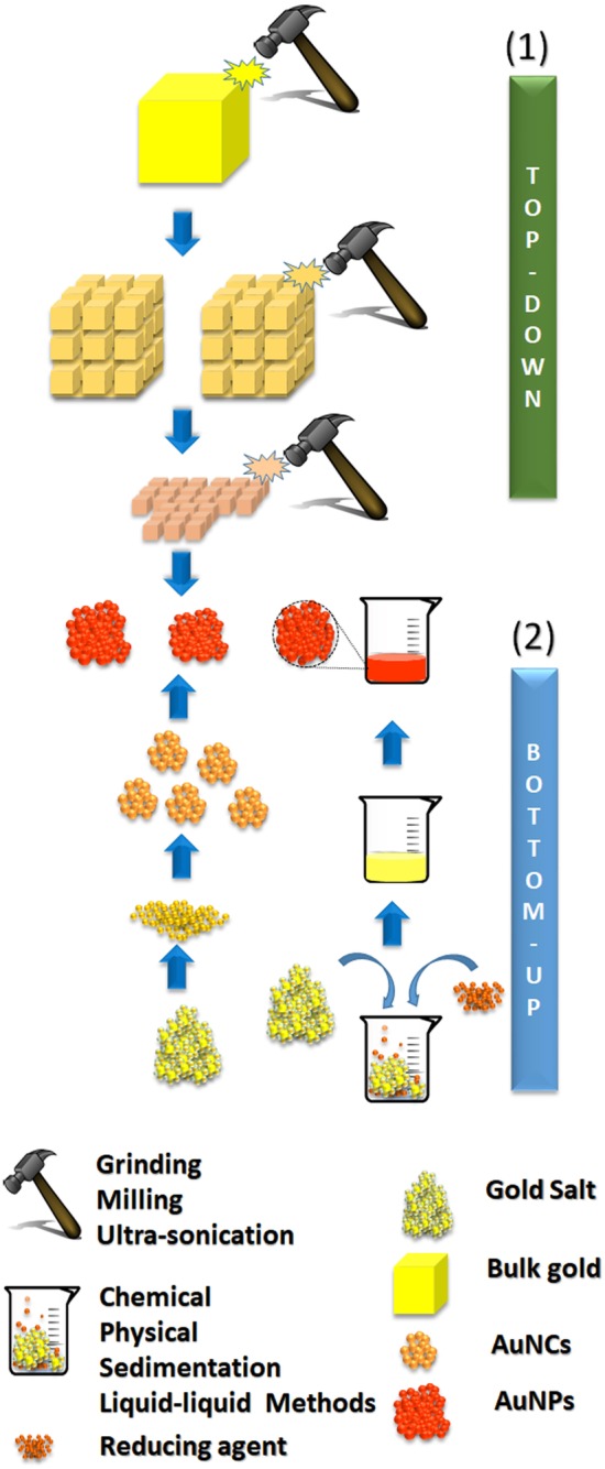
Schematic representation of NP syntheses using (1) top-down and (2) bottom up approaches
Surface modification strategies
Surface modification using sulfur-containing ligands
Direct conjugation to form thiol-protected AuNPs
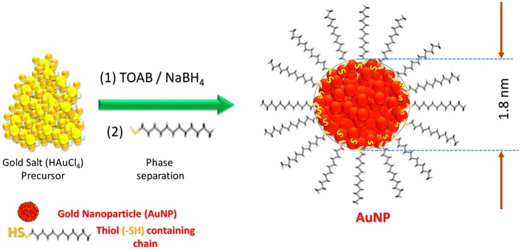
Surface modifications of AuNPs involve the binding of linker molecule onto the surface, where thiol-based coupling have extensively been employed. These alterations provide the control over reactivity and induction of hydrophobicity/hydrophilicity to the NPs. These NPs are obtained by following the direct conjugation of thiol containing molecules during the synthesis of AuNPs (shown in Fig. 2). In an example, synthetic process follows the dissolution of chloroauric salts (HAuCl4) to obtain AuCl4− ions in water followed by phase extraction of AuCl4− ions at tetraoctyl ammonium bromide (TOAB) dissolved in toluene. Thereafter, the process was followed by the treatment of reducing agent (sodium borohydride; NaBH4) and 1-dodecanethiol in the solution. The rapid color change (orange to deep brown) of the solution in presence of 1-dodecanethiol authenticates the formation of thiol-capped AuNPs. In final steps, the thiolated AuNPs were purified following the phase extraction and thorough washing of AuNPs. The overall reaction has been summarized in Eqs. (1) and (2):
Fig. 2.
The pictorial representation of thiol-stabilized AuNPs synthesis based on the phase extraction process using (1) tetraoctylammonium bromide [TOAB; (C8H17)4NBr] followed by the reduction with sodium borohydrite (NaBH4) (2) Particles are capped using dodecane thiol (C12H25SH)
| 1 |
| 2 |
In the above case, mainly AuNPs of cuboctahedral and icosahedral structures shape were synthesized in size range of 2–8 nm with a dispersion of about 4–6%, however, the particle’s shapes and sizes can be tuned by altering the reaction conditions; such as, gold/thiol molar ratio, temperature, and reduction rates. For instance, the fast reductant addition and low solution temperature have been reported with the smaller and monodisperse particles. In addition, the tuning of size has also been achieved by introducing a sterically bulky group for immediate quenching of reaction (Templeton et al. 2000). In addition to these, the optimization of reducing temperature and rate of injection of reductant have also been reported to alter the size and dispersity index of AuNPs (Schaaff and Whetten 2000). To achieve thiol-coated AuNPs, a number of methods have been employed to synthesize, where Brust–Schiffrin procedure is well known for monolayer-protected clusters (MPCs) formation, wherein further steps, the purification was performed using TOAB to obtain high yield (Waters et al. 2003). Apart from these, catalyst-less synthesis of MPCs has also been reported without trace ionic impurities by coupling the two different single-phase steps. For instance, thiol-derivatized AuNPs in a two-phase liquid–liquid system was synthesized by simultaneous reduction of the gold salt in the presence of passivating ligand using a water/methanol solution. This method was originally demonstrated with 4-mercaptophenol as capping agent (Brust et al. 1995) and is widely used for the incorporation of water-soluble ligands (Chen and Kimura 1999; Templeton et al. 1999). In another single-phase synthetic approach (Rowe method), tetrahydrofuran (THF) is used as the solvent which provides several advantages including compatibility for a wide range of ligands. The usage of strong reducing agents [lithium triethyl borohydride (super-hydride)] in this method has increased the efficiency by multiple folds than Brust method due to its capability of reducing various functional groups including esters and amides (Brown et al. 1980), where the two-phase Brust method limits.
In addition to this, a number of surface modifications have also been reported by translating the double phase synthesis into a single-phase system (Kanaras et al. 2002; Zheng et al. 2004). For instance, the tiopronin monolayer-protected AuNPs were obtained in soluble gold clusters with the average core size of 1.8 nm (Templeton et al. 1999). Similarly, the relatively smaller and thermally stable AuNPs have also been synthesized using arenethiol and alkanethiol as a passivating layer (Chen and Kimura 1999). Not only smaller sizes, but also larger sized thiol-stabilized water-soluble AuNPs were obtained using alkyl thiosulphates as precursors (Lohse et al. 2010). In addition to these, various thiol-based approaches have been reported for the AuNPs surface modification including thymine self-assembled monolayer (Zhou et al. 2007), hexadecyl aniline (Ascencio et al. 2000), super hydride (Yee et al. 1999), organometallic reagents, (2-propylmagnesium bromide) (Sugie et al. 2009), 9-borabicyclononane (Sardar and Shumaker-Parry 2009), and glutathione (Negishi et al. 2004) based thiol-passivated AuNPs.
Substitution/secondary modification to form mixed-monolayer AuNPs
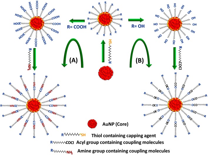
For the first time, Giersig and Mulvaney reported Mixed-monolayer mediated modification of AuNPs in 1993, where the AuNPs were stabilized using alkanethiols. This stabilization was introduced by mixing end-group functionalized organothiols (ω-functionalized organothiols) with pre-synthesized AuNPs in the presence of surfactants. Due to the soft character of gold and sulfur, thiol groups strongly bind to the gold through covalent interaction (Giersig and Mulvaney 1993) and hence the formed NPs show excellent stability and can be stored for years (Brust et al. 1995). Thus, to modify the surface of AuNPs, place-exchange methods are profoundly used where the substitution of existing thiol ligands from additive thiols are introduced. Templeton et al. and Hostetler et al. have shown method for the substitution of anchored thiol with the free thiol ligands (Hostetler et al. 1999; Templeton et al. 2000). The introduction of two or more functional ligands has also been reported for the synthesis of mixed-monolayer-protected AuNPs for synergistic applications. For example, hybrid AuNPs have been prepared using thiol-terminated ligands containing organic/inorganic dyes (Walter et al. 2002), smart polymers (Martin 1996), biomolecules (Giljohann et al. 2010), and drug molecules (Ghosh et al. 2008). These ligands on the surface of AuNPs interact amongst each other creating a rigid monolayer (Jadzinsky et al. 2007) which exhibits a certain level of intra-monolayer mobility as an optimal interaction with the analytes (Boal and Rotello 2000). Under the appropriate conditions, these ligands show intermolecular mobility by hopping between the NPs (Zachary and Chechik 2007) due to the effects of temperature and monolayer packing (Ionita et al. 2008). In general, the core of AuNPs contain hydrophobic groups and the secondary surface modifications with hydrophilic groups (viz. hydroxyl or carboxy moieties) helps to improve the dispersion of AuNPs preventing their aggregation. So far, a number of strategies have been reported following mixed-monolayer-based passivating approaches including the chemical coupling (Liu et al. 1998), polymerization (Mandal et al. 2002), electrostatic interaction (Chen et al. 2008), and selective intermolecular interaction (Giljohann et al. 2010; Braun et al. 2009). Among all, chemical coupling and polymerization are the most widely used methods, where the functionalization of AuNPs using carboxylic acid terminated was mostly used. In one of such examples, thiol ligands have also been used that forms amide linkage with linker molecules via. N-Ethyl-N-(3 dimethylaminopropyl)-carbodiimide coupling (Kamra et al. 2016). Apart from these, NPs with hydroxyl groups on their surface have also been reported to generate various functionalities by esterification reaction in presence of acyl moieties (Fig. 3) (Yoo et al. 2009).
Fig. 3.
Schematic representation of different types of secondary modification approaches for mixed-monolayer-passivated AuNP functionalization, (a) carbodiimide coupling at carboxylic end thiolated AuNPs (b) hydroxyl end thiol-stabilized AuNPs using RCOCl coupling reaction
Modification using other sulfur ligands
Sulfur-containing ligands viz. xanthates (Tzhayik et al. 2002), disulfides (Manna et al. 2003), di- and trithiols (Felidj et al. 2003), thioacetates, dithiocarbamates, trithiolates, thiolic acid, and resorcinarenetetrathiols (Balasubramanian et al. 2002) have also been used to functionalize AuNPs surface. In one such example, small-sized AuNPs (diameter 1.5 nm) have been prepared using a four-chained disulfide organic molecule. Since, the thiol moieties give better stabilization to AuNPs, these are most commonly used over other ligands for passivation. In addition, thio-ether- stabilized AuNPs have also been reported for AuNP surface functionalization, which shows relatively weaker passivating capacity (Shelley et al. 2002). Thus, to circumvent the limitations associated with the monomeric layer, poly-thio-ether passivating agents were used to stabilize the AuNPs (Li et al. 2001). Furthermore, metal-binding strengths between thiols and thio-ethers differ and hence alter the molecular arrangements, which contain multidentate thio-ethers that eventually lead to the unique morphologies and also allow the chemical reversibility. For instance, the tetradentate thio-ethers have been used to form reversible AuNP assembly (Maye et al. 2002). In another example, Sun et al. have described the synthesis of polycyclodextrin hollow gold spheres through oxidation-mediated thiol-stabilized AuNPs using iodine, resulting in decomposition to gold iodide with disulfide formation (Sun et al. 2001). However, the dispersion of these particles is major concerns especially in an organic solvent, thus to obtain improved dispersible particles, resorcinarene tetrathiol has been employed as a passivating agent, which is reported to yield mid-sized AuNPs (16–87 nm) (Yamamoto and Nakamoto 2003).
Surface modification using phosphine
The surface of AuNPs has also been stabilized and modified using phosphine, which is reported to act as an excellent precursor for functionalization with well-defined metallic cores (Weare et al. 2000). These functionalized particles have been used not only for catalysis (Schmid 1992) but also has been employed as the backbone for nanoscale electronic devices (Weare et al. 2000). Commonly, the approaches for synthesizing the phosphine- based AuNPs are carried out by the reduction of HAuCl4 followed by the treatment of phosphine derivatives for stabilizing the nanostructure such as PPh2(C6H4SO3Na–m) or P(C6H4SO3Na–m)3 (Schmid et al. 1996). In a report, the phosphine-mediated AuNPs were obtained, where the chloroaurate (AuCl4−) ions have been relocated after the reduction in aqueous NaBH4 to the organic phase (toluene) containing PPh3 (Weare et al. 2000). These uniquely synthesized NPs not only show the greater stability when stored in cold and dry conditions but also allow excellent surface functionalization. The process has also been tuned for larger particles synthesis by adjusting the important parameters of reaction time and temperature. In another strategy, AuNPs have been synthesized by following the Hutchison’s procedure where the rapid exchange of phosphine derivative of the different genre was employed, i.e., capping ligand and dissociated phosphines occur in dichloromethane at room temperature (Petroski et al. 2004). NPs from Hutchison’s preparation can also serve as the versatile AuNP precursors as the functionalization can be achieved with a wide range of ligands from ligand-exchange reactions to yield a diverse library of functional “nano-building blocks”, which can eventually lead to the unique nanostructures.
Surface modification using amine
Similar to the previously discussed strategies, there are a number of methods reported for the AuNPs surface functionalization. For this, Brust method is commonly adopted to modify the surface of NPs, where the present stabilizing thiol moieties are substituted by amine ligands. The major advantage of amine-capped AuNPs is that the surface chemistry of NPs can easily be probed by a variety of spectroscopic studies. Initially, the hydroxylamine has been used as a reducing agent for amine-capped AuNPs synthesis (Graf and van Blaaderen 2002). This paved the path towards extensive use of organic amines in a number of metal NPs, especially for AuNPs, due to their affinity for nitrogen. Amine-mediated synthesis of hydrophobic AuNPs has also been reported by Leff and co-workers using n-alkylamines (primary amines) (Leff et al. 1996). Similarly, aminomethyl pyrene (Thomas and Kamat 2000) and benzylamine (Thomas et al. 2002) have been reported to be used as stabilizing agents to obtain amine stabilized AuNPs. For simplifying the synthesis process, a direct one-pot synthesis of amine-stabilized AuNPs using 3-trimethoxysilylpropyl- diethylenetriamine has also been reported (Zhu et al. 2005). In addition to this, Aslam et al. demonstrated a one-step synthesis of oleylamine-capped water-soluble AuNPs (Aslam et al. 2004). Furthermore, a higher ratio of gold to amine has been used for the formation of products with mixed ligand shell, which suggests the ratio of the gold salt and amine ligands defines the dispersity index. Newmen et al. have reported an interesting study in this context, where the different ratios have tested for the AuNPs syntheses and found that in the ratio of 10:1(gold to amines) yields monodisperse nanoparticle, while the further lowering of amine ligands leads to the synthesis of polydispersed AuNPs. In addition to these, a variety of other amino compounds have also been employed to obtain amine-capped AuNPs including aromatic amines (Newman and Blanchard 2006), diamines (Selvakannan et al. 2004), tetraoctylammonium (Isaacs et al. 2005), laurylamine (Kumar et al. 2003), porphyrins (Kotiaho et al. 2010), and hyperbranched polyethylenimine (Duan and Nie 2007).
Apart from these, amino acids are also used for the amine passivation of AuNPs as these are capable of forming protective layers and the assembly of chloroaurate ions forming NPs. Functional groups of amino acids, viz. −SH and −NH2 possess high affinity for gold (Choudhary et al. 2016), thus provides the greater stability to the NPs. The physicochemical properties of the side groups present in the amino acids, such as polarity and hydrophilicity influence the reduction, stabilization, and conversion of gold ions (Au3+) to NPs (Huang et al. 2006b). So far, several amino acids have been reported for surface modification including tryptophan, lysine, aspartic acid (Shao et al. 2004), cysteine (Ma and Han 2008), and glutamic acid (Wangoo et al. 2008). In a report published by Selvakannan and co-workers, the synthesis and functionalization of water-soluble AuNPs have been mediated by amino acid lysine (Selvakannan et al. 2004). These lysine-capped aqueous AuNPs are electrostatically stabilized in solution and show excellent water-dispersibility. However, the AuNPs obtained from amine capped methods show pH dependent properties, due to which the particles aggregates to large superstructure on the fluctuation of pH.
Surface modification using polymers
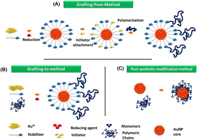
The polymers have also been used for capping the metallic NPs to provide greater stability. Initial attempt on polymer-stabilized AuNPs has been taken by Helcher in the presence of a polysaccharide in 1718, however, due to limited characterization availability, these were not properly and scientifically analyzed (Rac et al. 2014). Polymer-stabilized AuNPs have many advantages, which include longer stability, high surface density and ability to adjust solubility (Chandra et al. 2012; Zhu et al. 2012a; Chandra 2016). Few commonly used polymers for stabilization are: poly(N-vinylpyrrolidone) (PVP), poly(ethylene glycol) (PEG), poly(4-vinylpyridine), poly(vinyl alcohol) (PVA), poly-(vinyl methyl ether), chitosan, polyethyleneimine (PEI), poly(diallyl dimethyl ammonium chloride) (PDDA), polystyrene-block polymers, poly(methyl methacrylate) (PMMA), poly(dG)-poly(dC), and poly(N-isopropylacrylamide) (Mandal et al. 2002; Yilmaz and Suzer 2010). The adopted strategies for polymer stabilization follow (1) grafting from, (2) grafting to, and (3) post-synthetic modification approaches. A simple schematic representation of these methods has been shown in Fig. 4.
Fig. 4.
Schematic representation of the polymer-stabilized AuNPs synthesis using (a) grafting-from, (b) grafting-to, and (c) post-synthetic modification techniques
Grafting-from technique
The “grafting from” approach involves growing polymeric chains from scaffolds attached to the surface of AuNPs (Huo and Worden 2007; Shan and Tenhu 2007). Commonly, alkyl-thiol-passivated AuNPs are used as a precursor for capping the polymeric materials onto the nanoparticle’s surface. This technique provides various advantages such as precise control over the thickness, structure, and density of the polymeric layer. Apart from these, the AuNPs synthesized by this method possesses the chemically bonded scaffolds stabilization. These AuNPs are more robust as compared to those, which are synthesized by physiosorption using block copolymer micelles, water-soluble polymers, or star block copolymers. Using this method there are a number of strategies have been reported. For instance, the grafting of poly(methyl methacrylate) was established on to the AuNPs surface following the living radical polymerization (Mandal et al. 2002). Using similar approach, in another report, poly(N-butylacrylate) have been grown following atom transfer radical polymerization (Nuß et al. 2001). In addition to these, biopolymers viz. oligonucleotide, and peptides have also been reported on the surface of AuNPs followed by the propagation of the grafted molecules. For instance, a single-stranded oligonucleotide grafting was achieved onto the AuNPs with the help of thiol-linked primer-based passivation followed by DNA polymerization (Zhao et al. 2006). In another strategy, the AuNPs are stabilized using peptide chains, which were synthesized by the grafting of sulfhydryl amine groups onto the AuNPs followed by enzymatic peptide elongation. For instance, Higuchi and co-workers reported grafting of alpha-helical poly(gamma-methyl L-glutamate-co-L-glutamic acid) bio-polymeric moieties onto the surface of AuNPs (Higuchi et al. 2007).
Grafting-to technique
In “Grafting to” approach, gold cores are synthesized in polymer aggregates. The major advantage of this method is the availability of various polymers for functionalization. Since this follows a one-pot synthesis method, reacting materials are mixed in a single vessel thereby reducing a number of laborious steps involve in “grafting-from” technique eventually making the synthesis easy. In this method, two types of polymers are commonly used, one with the sulfur-containing group at the terminal end and other terminated with a sulfur-free group. Polymers terminated with a sulfur-containing group such as di-thioester, tri-thioester, thiol, thioether, and disulfide provide chemical-bonded shell layers around the gold cores (Liu et al. 2007; Aqil et al. 2008). Synthesis of these polymers was done by a radical polymerization using chain transfer reagents containing sulfur atoms (Wang et al. 2007). In polymeric aggregates without sulfur, evolution of gold cores results in gold nanocomposites in which the polymers interact with gold cores through multi-point physical adsorption (Sakai and Alexandridis 2004). However, these gold nanocomposites are unstable and the polymer has a high chance of getting detached from the AuNP surface because of the lack of stable chemical bonds. This can be overcome by cross-linking the polymer network or by the use of unimolecular micelles (Filali et al. 2005). This method has been used for the synthesis of AuNPs functionalized with artificial polymers, such as: PVP (Mohamed et al. 2017), poly(vinyl pyridine) (Zhang et al. 2018), PEG (Ocal et al. 2018), PVA (Kwiatkowska et al. 2018), PMMA (Zepon et al. 2015), PEI (Lazarus and Singh 2016), PDDA (Liu et al. 2016) as well as biopolymers (Chowdhury et al. 2018).
Post-synthetic modification techniques
In post-synthetic modification methods, AuNPs are first generated through conventional methods followed by functionalization (Kang and Taton 2005). AuNPs have been covalently conjugated with polymers having thiol groups, while the polymers devoid of thiol moieties were attached through physical adsorption onto the AuNPs surface. Similarly, biopolymers have also been used for the surface functionalization using the similar approach. Due to these distinctive properties and non-toxic nature, oligonucleotide-functionalized AuNPs have been used for highly sensitive and selective assays in detecting biomolecules and thus find several applications in biosensing, disease diagnostics, and gene expression studies. Moreover, due to their greater affinity towards DNA molecules, these are frequently used as non-viral vector for gene delivery (Biju 2014; Sahoo et al. 2017). In such examples, alkyl thiol-terminated oligonucleotide-functionalized AuNPs were synthesized with high stability in saline condition (Briley et al. 2015). Enzymes, peptides, antibodies, aptamer, etc., have also been used for the AuNPs functionalization in place of the chemical reagents (Baranwal et al. 2016; Kumar et al. 2008; Mahato et al. 2018b). So far, several applications have been reported using the AuNPs synthesized from these methods including DNA intercalation study (Wang et al. 2002), polynucleotides detection (Giljohann et al. 2010), and a number of protein detections.
Modification using silica
Silica has extensively been used for passivating the AuNPs, where a thin shell has been coated over NPs to prevent various detrimental interactions between proteins and other molecules with the nanoparticle’s surface. This step is followed by the binding of various molecules of interest, viz. spacer, target or coating molecule to the flanking linker on the surface of NPs. Thereafter, the functionalized NPs are sorted according to the number of bound molecules using various chromatographic techniques which also allow the selection of homo-functionalized NPs (Lévy et al. 2006). The silica coating onto the AuNPs greatly enhances its chemical stability against aggregation. These modifications are in general introduced using Stöber method, where tetraethylorthosilicate is used for passivation of gold core (Stöber et al. 1968). For instance, silica-coated AuNPs have been synthesized using (3-aminopropyl)trimethoxysilane configuring alkoxide flanking groups outwards from AuNPs (Liz-Marzán et al. 1996). Silica coating onto the AuNPs greatly enhances the chemical stability against aggregation and its solubility in different solvents. These modifications are also introduced using Stöber method, where the solution of tetra ethylorthosilicate is used in different alcohols such as methanol, ethanol, and isopropanol with ammonia. The resulting solution is then stirred to obtain the silica-coated NPs depending on the type of silicate ester and alcohol used and also on their volume ratios (Stöber et al. 1968). Thin silica shell-based AuNPs passivation has also been reported by Liu and Han, which has shown excellent biocompatibility and has been used in various colorimetric diagnostics, photothermal therapy, and SERS- based detections (Liu and Han 2010). In addition, functionalization of the silica-coated NPs surface with amino-, mercapto- and carboxy-terminated silanes allows the conjugation of other materials for further secondary modifications (Lee et al. 2008).
Applications of functionalized AuNPs
Functionalized AuNPs have extensively been used in various biomedical applications. So far, functionalized AuNPs have found in numerous applications of various fields such as electronics, photodynamic therapy, drug delivery and targeting, sensors, probes, diagnostics, and catalysis (Chandra et al. 2010, 2013b; Kumar et al. 2015). The potential applications of AuNPs in clinical and biomedical domains are described under various categories as follows.
Diagnostic applications
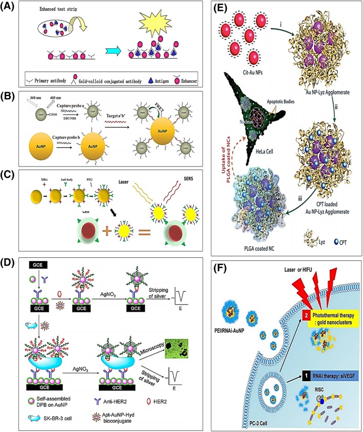
The unique optoelectronic properties, viz. SPR, Raman scattering, and fluorescence quenching led down the AuNPs in various diagnostic applications (Chandra 2016; Mahato et al. 2016a). Upon the exposure of electromagnetic radiation, the conduction electron (or plasmons) starts to oscillate on the surface of AuNPs. The coherent oscillation of the metal free electrons in resonance with the electromagnetic field also called as SPR develops strong electromagnetic fields onto the surface of particles. This subsequently enhances radiative properties such as absorption and scattering, as well as non-radiative properties, which eventually help to monitor the biomolecules. These, surface functionalities show the size-dependent SPR properties. For example, the thiol-stabilized AuNPs (diameter ≈ 5 nm) exhibit SPR at 530 nm, whereas amine-stabilized AuNPs (diameter ≈ 7 nm) exhibit SPR at 540 nm. These SPR properties of AuNPs are useful for biomolecular detection in real time with respect to the changes in refractive index upon binding to thin gold films (Liedberg et al. 1983). The shift in SPR due to the molecular interactions has been exploited for building various diagnostic strategies. For instance, Taton et al. has developed a technique that is capable to monitor these shifts upon nucleic acid interactions (Taton et al. 2000). The plasmonic effects of AuNPs have also led to the development of convenient colorimetric assays. These are based on the fact that the interacting electric fields of aggregating AuNPs have a tendency to lower the resonant frequency of plasmon oscillations, resulting in a visible color change with either bathochromic or hypsochromic shifting of the initial frequency (Srivastava et al. 2005). In such colorimetric assays, the color change is dependent on the action of target analyte, which either directly or indirectly triggers the further states of AuNPs aggregation or re-dispersion. In addition, AuNPs have been modified with linker molecules that can link them together in the presence of a molecule of interest. These AuNP-based colorimetric assays have been used in the detection of specific DNA strands, proteins, and small molecules. For instance, in dipstick-type pregnancy testing kits, AuNPs are modified with secondary antibodies against human gonadotropin hormone that subsequently binds to primary antibodies arranged over a small area to the dipstick (Fig. 5a) (Tanaka et al. 2006). In presence of the target analyte, the binding complex forms in a testing area consisting secondary antibody-conjugated AuNPs, analyte, and primary antibodies, resulting in the change in SPR properties of the AuNPs resulting the appearance of color (Stockman 2011; Tanaka et al. 2006).
Fig. 5.
Illustrations for different applications of functionalized AuNPs, where (a) shows the schematic of dipstick-type diagnostic device based on antibody-coated AuNPs for colorimetric detection (Reprinted with permission of Tanaka et al.; copyright Springer). b Shows functionalized AuNP–GDQ FRET pair-based detection of mecA gene sequence of Staphylococcus aureus (Reprinted with permission of Shi et al.; copyright Elsevier). c Shows functionalized AuNP-based SERS strategy for cell imaging (Reprinted with permission of Zhang et al.; copyright Springer). d Shows simultaneous detection and bio-imaging based on aptamer-based functionalized AuNPs (Reprinted with permission of Zhu et al.; copyright the American Chemical Society). e Shows co-functionalization of AuNPs and protein using polymers as nano-carrier for delivery of hydrophobic anticancer drug camptothecin (Reprinted with permission of Khandalia et al.; copyright the Royal Society of Chemistry). f Shows the anti-VEGF siRNA functionalized AuNP-based combinatorial theranostic strategies targeting early tumor cells (Reprinted with permission of Son et al.; copyright Theranostics)
Fluorescence resonance energy transfer (FRET) is a distance-dependent energy transfer-based technique, where a donor chromophore supplies emissive energy to the receptor counterpart after getting excited leading to the increased fluorescence intensity of the receptor counterpart of the FRET pair. For an excellent FRET-based optical quenching, AuNPs are a suitable candidate as it possesses high molar extinction coefficient and large energy bandwidth (Jain et al. 2007). Using AuNP-assisted FRET technique, Zhang et al. have reported a cholesterol sensor by functionalizing the AuNPs with β-cyclodextrin (Zhang et al. 2008). Additionally, researchers have developed several assays to monitor the cleavage of DNA by nucleases and reported the biomolecular detection up to femtomolar concentrations (Ray et al. 2006). In addition to this, accuracy has been enhanced using multicolored oligonucleotide-functionalized organic dye with AuNPs as nanoprobes (Ray et al. 2007). FRET-based assays have also been utilized to detect specific gene sequences. For instance, Shi et al. have developed novel FRET-based biosensor on graphene quantum dots and AuNPs for ultralow detection of a gene from Staphylococcus aureus, indicating immense application in medical diagnostics (Shi et al. 2015) (Fig. 5b).
Conventionally, Raman scattering has been used for various detections exploiting the intrinsic spectroscopic properties of analyte molecules but due to their poor intensities limits the usage in various diagnostic applications. Therefore, AuNP-assisted Raman spectroscopy has been adopted to overcome such limitations by employing AuNPs, which is also called surface enhanced Raman spectroscopy (SERS). These systems utilizes the intrinsic properties of the AuNPs for SERS phenomena (Talley et al. 2005; Bellamy and Garthwaite 2001). For instance, a tumor cell detection has been reported by Zhang et al. based on the AuNP-assisted SERS, where the antibody-tagged AuNPs were used (Zhang et al. 2016) and showed excellent analytical performance (Fig. 5c). SERS-based techniques have been found great attention in recent days due to their various advantages, viz. high quality, distinct, and noise-free signals over other diagnostic techniques. Furthermore, the tunability of these SERS-based properties have also been achieved with size, shape, orientation, and the aggregation of AuNPs, which makes the functionalization of AuNPs compatible for barcodes or recognition elements in developing various unique analytic tools (Zheng et al. 2012).
Sensors and biosensors
AuNP’s fascinating properties have extensively been utilized to design a number of bio/chemical sensors of optical and electrochemical formats (Kumar et al. 2015; Won et al. 2013; Zhu et al. 2012a; Chandra 2015). The smaller size and high aspect ratio of AuNPs provide stable immobilization of large numbers of biomolecules onto its surface while retaining their biological activities (Chandra et al. 2013b). Therefore, AuNPs are widely used as signal amplification tags in different types of biosensors because of its light-scattering, conducting, and local electromagnetic field enhancement properties (Mahato et al. 2016b; Mandal et al. 2018). Using these properties of AuNPs, various detection strategies have been employed for sensitive and selective detection of target DNA via. sequence-specific hybridization between the target and single-stranded oligonucleotide probe-conjugated AuNPs (Lyon et al. 1998; Spampinato et al. 2016; Bhatnagar et al. 2018; Chandra 2013). A prototype has been developed based on the oligonucleotide probe-conjugated AuNPs as SPR amplification tags that have satisfactorily enhanced the sensitivity by 1000-folds (He et al. 2000). Apart from these, a DNA fluoro-biosensor using a cerium complex Ce(QS)2Cl and thiolated probe-DNA with functionalized AuNPs have been reported based on DNA hybridization-mediated fluorescence quenching. Similarly, in another work, researchers used AuNPs as both nano-scaffolds for the attachment of capture sequences and as nano-quenchers of fluorophores attached to detect sequences, where 5′-thiolated 12-mer oligonucleotide-functionalized AuNPs were used for sequence-specific detection of target DNAs with a 2-nM detection limit (Wu et al. 2006). In another strategy, an ultra-selective rapid colorimetric biosensor for detecting the breast cancer gene BRAC1 using the DNA–AuNPs hybrid (Oh and Lee 2011). Apart from these, AuNP-based optical probes have also been used for clinical diagnosis of aminothiols, viz. cysteine, homo-cysteine, and glutathione (Xiao et al. 2012). Glycol-conjugated AuNPs have also been employed for selective and sensitive optical detection of a mannose-binding protein complex (Concanavalin A) based on surface plasmon absorption with reported excellent analytical performances, where the dynamic range of 12–45 nM was reported with the detection limit of 4.0 nM (Watanabe et al. 2012). Using AuNPs, there are a number of biosensing prototypes been reported based on the dual signal (optical/electrochemical) transduction. For instance, Boca et al. have demonstrated dual signal-based electrical/optical proof of concept biosensing prototype exploiting the nano-gaps formed during the self-assembly of AuNPs. This showed excellent SERS capabilities and chemi-resistivity of nano-gap AuNPs for the detection of 4-mercaptophenyl boronic acid (Boca et al. 2015). In another report, an aptamer-based AuNP-functionalized biosensor has been developed showing high sensitivity (20 nM) for biomolecular analysis using avidin as a model compound. The developed facile sensor was anticipated to be applied for in vitro analysis (Hernandez et al. 2009). Excellent electrical conductivity, high surface area, and catalytic properties of AuNPs are suitable for the electrochemical biosensing where the electrical responses generated are directly proportional to the analyte concentrations (Agrawal et al. 2013; Maurya et al. 2016). Until now, several electrochemical biosensors have been reported for detection of a number of analytes (Chandra 2016; Mahato et al. 2017). An electrochemical bifunctional nanosensor has been developed by Zhu et al. for the detection of HER2 positive breast cancer cells and HER2 protein biomarker using bioconjugate comprising hydrazine–AuNP–aptamer in a single sensor design (Zhu et al. 2012b) (Fig. 5d). This study elaborates the importance of AuNPs in nanobioconjugate development for biorecognition and also as a signal amplifier in a single experimental setting. In another study, AuNPs functionalized with thiol-modified receptor for glucose biosensing has been developed to monitor the glucose level of diabetic patients (Spampinato et al. 2016). In addition, functionalized AuNPs assisted strategies are constantly adding the qualities to various biosensors in terms of sensitivity, selectivity, limit of detection, dynamic range and compatible with numerous analytes including drugs, neurotransmitters, heavy metal ions, bacterial/cancer cells, and other metabolically active molecules (Zhu et al. 2012c; Chandra et al. 2011b; Kashish et al. 2017; Chung et al. 2018; Akhtar et al. 2018; Baranwal and Chandra 2018).
Bio-imaging
AuNPs and its composites have widely been used for bio-imaging applications exploiting their strong radiative properties. The AuNPs facilitate the biological features to visualize the surfaces under dark field microscopy. For instance, anti-epidermal growth factor receptor (anti-EGFR) antibody-functionalized AuNPs (40 nm) were used for the bio-imaging of overexpressed biomarker protein on cancer cells exploiting the SPR response of tagged AuNPs (El-Sayed et al. 2005). The SPR responses of these functionalized NPs irradiation appear in green light range (530 nm), which helps to visualize the immune interactions of tagged antibody to the present biomarker resulting the highly specific bio-imaging. Apart from this, the optimized geometries of AuNPs such as rods or cubes make them optimal tools for two-photon luminescence technique which uses a femtosecond near-infrared region laser excitation (Dreaden et al. 2012). This technique is known for its nonlinear optical imaging in near-infrared region where water and biomolecules show minimum absorption, thereby reducing background noises and increasing spatial resolution. These optical signatures have helped researchers to develop in vitro and in vivo imaging methods for detection of cancer and other living cells. For instance, Durr et al. used gold nanorods functionalized with anti-EGFR antibodies based on the above techniques for the molecular imaging of cancer cells at different depths within the tissues (Durr et al. 2007). Apart from these approaches, Chandra et al. have reported AuNPs mediated label free bio-imaging platform for cancer cells detection through the interaction between daunomycin and cell surface receptors. The bioimaging system, in this case, was also confirmed using electrochemical impedance spectroscopy (Chandra et al. 2011a).
Therapeutics
Functionalized AuNPs also show therapeutic effects including photothermal therapy (PTT) where the thermoablative properties of AuNPs are exploited to generate heat. The excitation of the AuNPs by near infrared or radio frequency radiations leads to the burning of the targeted cells (Huang et al. 2009; Jain et al. 2008). For example, a poly ethylene glycol-coated gold nanorods were used for tumor inhibition in an animal model using near-infrared PTT (Dreaden et al. 2012). Functionalization of AuNPs using DNA aptamers has been studied for various therapeutic studies including PTT and photodynamic therapies. In addition to this, these aptamer-functionalized AuNPs have also been studied for anticancer and antiviral therapies (Wang et al. 2016). Similarly, in a most recent study, AuNPs conjugated with DNA aptamers have found to be enhanced the therapeutic efficiency by inducing specificity, stability, uptake efficiencies of biomolecules. For instance, an aptamer–AuNP–graphene oxide nanocomposite developed by Wang et al. exhibited PTT effects even in an ultralow concentration of overexpressed MUC1 cancer cells (MCF-7) without affecting the healthy cells (Wang et al. 2016).
Drug delivery
Furthermore, AuNPs found use in the targeted delivery of various payloads ranging from small drug molecules to bio-macromolecules, owing to their low cytotoxicity, good cell permeability, and high drug-loading efficacies, stemming from their intrinsic greater surface-to-volume ratio. The attachment of payload can be achieved either by covalent chemical conjugation of small molecules, viz. drugs and neurotransmitters, or by non-covalent attachments, such as; electrostatic interactions as in the case of bio-macromolecules, e.g., peptides (Khandelia et al. 2014). The modes of attachment sometimes play an important role in delivering of species. In the case of covalent attachment of pro-drugs, they are delivered which are then processed in intracellular environment before assimilation, while the active drugs are generally attached with non-covalent means showing better release (Morgan et al. 2006). The release of the payloads can be triggered by various internal [e.g., glutathione (Hong et al. 2006), or pH (Polizzi et al. 2007)] or external stimuli (e.g., light) (Han et al. 2006). In general, AuNPs assisted delivery of molecules are optimized by tuning the particle size or by altering surface functionalization steps. For example, secondary coating with antibiofouling agents (viz. thiol-derivatized poly-ethylene glycol) was reported for delaying the response from reticuloendothelial system that acts as a biological barrier for drug delivery, which was reported the delayed delivery by 0.5–72 h in a mice model, claiming delivery clearance of approximately 150-fold than unmodified cetyltrimethylammonium bromide-capped AuNPs (Niidome et al. 2006). Different targeting moieties such as proteins, peptides, antibodies, and small molecules including folic acid and paclitaxel have also been attached to the AuNPs surface to facilitate selective cellular uptake and internalization of the drug-loaded nanocarriers. At the same time to achieve a highly specific targeted drug delivery to heterogeneous population of cancer cells in solid tumors, several receptor molecules (antibody, aptamer, peptide, etc.) are also attached onto the surface of AuNPs. In addition to this, several platforms have been demonstrated showing exciting results in delivering molecules into tumors (El-Sayed et al. 2005; Visaria et al. 2006; Huang et al. 2006a; Paciotti et al. 2006). For instance, Khandalia et al. have reported polymer-coated NP–protein conjugate to deliver the drug in the human cell lines and successfully induced the apoptosis to the target cells (Fig. 5e) (Khandelia et al. 2014). Some of the commonly FDA approved NPs-based drugs including paclitaxel, and doxorubicin were successfully cleared the clinical trial and used in a metastatic breast cancer phase III multiple-myeloma treatment in metastatic ovarian cancer, respectively (Singh et al. 2015).
Theranostics
Theranostics is an approach which provides the simultaneous benefits of therapeutics as well as imaging and diagnostics on a single platform (Chandra 2016). Since AuNPs exhibit several unique features such as SPR and absorption of near-infrared region of electrochemical radiation, by virtue of these the AuNPs show photothermal properties. Such properties have been exploited for targeted detections of cancerous/tumorigenic cells and their destruction simultaneously. The pioneering works on theranostics applications using the gold nanoshells were reported by Loo et al. (2005) and gold nano-spheres by Lapotko (2009). Following the trends, so far, a number of theranostic strategies have been reported using AuNPs for various types of cancers. For instance, AuNPs have been used for diagnosis and treatment of gastrointestinal (GI) cancers by targeting GI adenocarcinoma cells via. thermal induction (Singh et al. 2015). Here, the heating effects of AuNPs uncouple the heat sensitive chemical bonds facilitating a release of drugs directly at the target site (Singh et al. 2015). The detailed articles of this domain have been comprehensively reported elsewhere which includes theranostic applications of AuNPs for imaging, photothermal therapy (Curry et al. 2014), nano-cardiology (Spivak et al. 2013), single nanomaterial based treatment (Vinhas et al. 2015), and in theranostics oncology (Akhter et al. 2012) along with multifunctional composites (Khlebtsov et al. 2013). So far various strategies have been adopted including AuNP-based that have been developed by co-encapsulation of AuNPs with chemotherapeutic drugs including paclitaxel (Muthu et al. 2014), doxorubicin (Chen et al. 2013), and daunomycin (Chandra et al. 2013a) for efficient theranostic applications. In one of such examples, Shao et al. have developed an ellipsoidal NP–drug conjugate, which can harness the synergy of photothermally activated physical and biological effects for therapeutic goals. (Shao et al. 2013). In another example, the tumor necrosis factor-alpha coated gold nanospheres (Au-TNF-ɑ) have been used for the treatment followed by heating with laser pulses. In addition, higher efficacy has been obtained in pulsating laser-mediated treatment in in vivo studies carried out using mice models. In addition to these advancements, functionalized AuNPs have also reported to induce combinatorial theranostic effects to the targeted cells. In an article reported by Son et al. demonstrated that AuNPs functionalized with anti-VEGF siRNA for the cancer eradication in later stages (Fig. 5f) (Son et al. 2017).
Conclusions and future prospects
As per the above discussion on the synthesis, surface modification, and application potentials of AuNPs, it is evident that it is excellently compatible to the nanoplatforms for various biomedical applications. In particular, AuNPs are passivated with a variety of ligands, functional groups, imaging labels, therapeutic drugs, and carrier molecules before the actual applications. Commonly, the functionalization of the AuNPs is achieved by modifying its surface either with linker moieties followed by modification of capping agent of interest or by place-exchange reaction with originally stabilized ligand or by linking to the shell of stabilizing molecules. Thus, the functionalized AuNPs found an important place for serving a number of biomedical applications, viz. sensitive detection of biomolecules, cellular imaging, and drug delivery agent. In the recent era, tremendous researches on AuNP-based nano-platforms have been fabricated for the same purpose, yet there is an enormous scope of improvements to enhance their applicability in various biomedical applications. Few common instances to be considered as future prospects are as follows:
The AuNP-assisted optical imaging is possible, however, there are no models which can quantify the optical signals accurately particularly in deeper tissues to provide sufficient information about the target disease.
There are several reports that have used AuNP-based models for tumor/cancer therapy, but none of them has addressed the generalized case, which can target multiple closely related cells specifically without affecting surrounded healthy cells.
Several AuNPs-based sensing devices and many proof-of-concept prototypes have been reported for various clinical pathogens/samples, yet very few prototypes have been commercialized to provide personalized diagnostic platform. Therefore, more efforts should be given to improvise the existing strategies for better high throughput translational prototypes.
A number of AuNPs-based drug delivery systems have been reported, yet the therapeutic efficacy of these are limited with respect to dosage and target-specific non-toxic delivery.
AuNPs-based photothermal therapy has widely been reported for the treatment of cancerous cells, but suffers from the non-specific damage healthy cells due to the uncontrolled and uneven heating.
AuNPs-based theranostic strategies are fascinating in biomedical science; however, there are much remains to be done to achieve the real goals.
Therefore, the field remains open to many important discoveries to find new methods of synthesizing AuNPs with compounds that would promote excellent biocompatibility and would make them useful for diagnostics, bio-imaging, and therapeutics. Hence, the future work should be directed towards better understanding of the behavior of AuNPs in biological/real systems to overcome the limitations of conventionally developed AuNPs, which can assist the next-generation need of engineering and medicine by improving the clinical diagnostic platform.
Acknowledgements
This work is supported by Science and Engineering Research Board (SERB) project file no. ECR/2016/000100.
Compliance with ethical standards
Conflict of interest
Authors report no conflict of interest in this work.
References
- Abalde-Cela S, Taladriz-Blanco P, de Oliveira MG, Abell C. Droplet microfluidics for the highly controlled synthesis of branched gold nanoparticles. Sci Rep. 2018;8(1):2440. doi: 10.1038/s41598-018-20754-x. [DOI] [PMC free article] [PubMed] [Google Scholar]
- Agrawal B, Chandra P, Goyal RN, Shim Y-B. Detection of norfloxacin and monitoring its effect on caffeine catabolism in urine samples. Biosens Bioelectron. 2013;47:307–312. doi: 10.1016/j.bios.2013.03.025. [DOI] [PubMed] [Google Scholar]
- Akhtar MH, Hussain KK, Gurudatt NG, Chandra P, Shim Y-B. Ultrasensitive dual probe immunosensor for the monitoring of nicotine induced-brain derived neurotrophic factor released from cancer cells. Biosens Bioelectron. 2018;116:108–115. doi: 10.1016/j.bios.2018.05.049. [DOI] [PubMed] [Google Scholar]
- Akhter S, Ahmad MZ, Ahmad FJ, Storm G, Kok RJ. Gold nanoparticles in theranostic oncology: current state-of-the-art. Expert Opin Drug Deliv. 2012;9(10):1225–1243. doi: 10.1517/17425247.2012.716824. [DOI] [PubMed] [Google Scholar]
- Alex S, Tiwari A. Functionalized gold nanoparticles: synthesis, properties and applications—a review. J Nanosci Nanotechnol. 2015;15(3):1869–1894. doi: 10.1166/jnn.2015.9718. [DOI] [PubMed] [Google Scholar]
- Aqil A, Qiu H, Greisch J-F, Jérôme R, De Pauw E, Jérôme C. Coating of gold nanoparticles by thermosensitive poly (N-isopropylacrylamide) end-capped by biotin. Polymer. 2008;49(5):1145–1153. [Google Scholar]
- Ascencio JA, Pérez M, José-Yacamán M. A truncated icosahedral structure observed in gold nanoparticles. Sur Sci. 2000;447(1):73–80. [Google Scholar]
- Aslam M, Fu L, Su M, Vijayamohanan K, Dravid VP. Novel one-step synthesis of amine-stabilized aqueous colloidal gold nanoparticles. J Mater Chem. 2004;14(12):1795–1797. [Google Scholar]
- Balasubramanian R, Kim B, Tripp SL, Wang X, Lieberman M, Wei A. Dispersion and stability studies of resorcinarene-encapsulated gold nanoparticles. Langmuir. 2002;18(9):3676–3681. [Google Scholar]
- Baranwal A, Chandra P. Clinical implications and electrochemical biosensing of monoamine neurotransmitters in body fluids, in vitro, in vivo, and ex vivo models. Biosens Bioelectron. 2018;121:137–152. doi: 10.1016/j.bios.2018.09.002. [DOI] [PubMed] [Google Scholar]
- Baranwal A, Mahato K, Srivastava A, Maurya PK, Chandra P. Phytofabricated metallic nanoparticles and their clinical applications. RSC Adv. 2016;6(107):105996–106010. [Google Scholar]
- Baranwal A, Kumar A, Priyadharshini A, Oggu GS, Bhatnagar I, Srivastava A, Chandra P. Chitosan: an undisputed bio-fabrication material for tissue engineering and bio-sensing applications. Int J Biol Macromol. 2018;110:110–123. doi: 10.1016/j.ijbiomac.2018.01.006. [DOI] [PubMed] [Google Scholar]
- Baranwal A, Srivastava A, Kumar P, Bajpai VK, Maurya PK, Chandra P. Prospects of nanostructure materials and their composites as antimicrobial agents. Front Microbiol. 2018;9:422. doi: 10.3389/fmicb.2018.00422. [DOI] [PMC free article] [PubMed] [Google Scholar]
- Bellamy TC, Garthwaite J. Sub-second kinetics of the nitric oxide receptor, soluble guanylyl cyclase, in intact cerebellar cells. J Biol Chem. 2001;276(6):4287–4292. doi: 10.1074/jbc.M006677200. [DOI] [PubMed] [Google Scholar]
- Bhatnagar I, Mahato K, Ealla KKR, Asthana A, Chandra P. Chitosan stabilized gold nanoparticle mediated self-assembled gliP nanobiosensor for diagnosis of invasive aspergillosis. Int J Biol Macromol. 2018;110:449–456. doi: 10.1016/j.ijbiomac.2017.12.084. [DOI] [PubMed] [Google Scholar]
- Biju V. Chemical modifications and bioconjugate reactions of nanomaterials for sensing, imaging, drug delivery and therapy. Chem Soc Rev. 2014;43(3):744–764. doi: 10.1039/c3cs60273g. [DOI] [PubMed] [Google Scholar]
- Blanco E, Shen H, Ferrari M. Principles of nanoparticle design for overcoming biological barriers to drug delivery. Nat Biotechnol. 2015;33:941. doi: 10.1038/nbt.3330. [DOI] [PMC free article] [PubMed] [Google Scholar]
- Boal AK, Rotello VM. Fabrication and self-optimization of multivalent receptors on nanoparticle scaffolds. J Amer Chem Soc. 2000;122(4):734–735. [Google Scholar]
- Boca S, Leordean C, Astilean S, Farcau C. Chemiresistive/SERS dual sensor based on densely packed gold nanoparticles. Beilstein J Nanotechnol. 2015;6(1):2498–2503. doi: 10.3762/bjnano.6.259. [DOI] [PMC free article] [PubMed] [Google Scholar]
- Braun GB, Pallaoro A, Wu G, Missirlis D, Zasadzinski JA, Tirrell M, Reich NO. Laser-activated gene silencing via gold nanoshell—siRNA conjugates. ACS Nano. 2009;3(7):2007–2015. doi: 10.1021/nn900469q. [DOI] [PubMed] [Google Scholar]
- Briley WE, Bondy MH, Randeria PS, Dupper TJ, Mirkin CA. Quantification and real-time tracking of RNA in live cells using sticky-flares. Proc Natl Acad Sci. 2015;112(31):9591–9595. doi: 10.1073/pnas.1510581112. [DOI] [PMC free article] [PubMed] [Google Scholar]
- Brown HC, Kim S, Krishnamurthy S. Selective reductions. 26. Lithium triethylborohydride as an exceptionally powerful and selective reducing agent in organic synthesis. Exploration of the reactions with selected organic compounds containing representative functional groups. J Org Chem. 1980;45(1):1–12. [Google Scholar]
- Brust M, Fink J, Bethell D, Schiffrin D, Kiely C (1995) Synthesis and reactions of functionalised gold nanoparticles. J Chem Soc Chem Commun. (16):1655–1656
- Catherine L, Olivier P (2017) Gold nanoparticles for physics, chemistry and biology. World Scientific
- Chandra P. Advances in clinical diagnosis through electrochemical aptamer sensors. J Bioanal Biomed. 2013;5:e119. doi: 10.4172/1948-593X.1000e119. [DOI] [Google Scholar]
- Chandra P. Electrochemical nanobiosensors for cancer diagnosis. J Anal Bioanal Tech. 2015;6(1):e119. [Google Scholar]
- Chandra P. Nanobiosensors for personalized and onsite biomedical diagnosis. London: IET; 2016. [Google Scholar]
- Chandra P, Das D, Abdelwahab AA (2010) Gold nanoparticles in molecular diagnostics and therapeutics. Digest J Nanomater Biostructures (DJNB) 5(2)
- Chandra P, Noh H-B, Won M-S, Shim Y-B. Detection of daunomycin using phosphatidylserine and aptamer co-immobilized on Au nanoparticles deposited conducting polymer. Biosens Bioelectron. 2011;26(11):4442–4449. doi: 10.1016/j.bios.2011.04.060. [DOI] [PubMed] [Google Scholar]
- Chandra P, Zaidi SA, Noh H-B, Shim Y-B. Separation and simultaneous detection of anticancer drugs in a microfluidic device with an amperometric biosensor. Biosens Bioelectron. 2011;28(1):326–332. doi: 10.1016/j.bios.2011.07.038. [DOI] [PubMed] [Google Scholar]
- Chandra P, Koh WCA, Noh H-B, Shim Y-B. In vitro monitoring of i-NOS concentrations with an immunosensor: the inhibitory effect of endocrine disruptors on i-NOS release. Biosens Bioelectron. 2012;32(1):278–282. doi: 10.1016/j.bios.2011.11.027. [DOI] [PubMed] [Google Scholar]
- Chandra P, Noh H-B, Shim Y-B. Cancer cell detection based on the interaction between an anticancer drug and cell membrane components. Chem Commun. 2013;49(19):1900–1902. doi: 10.1039/c2cc38235k. [DOI] [PubMed] [Google Scholar]
- Chandra Pranjal, Singh Jai, Singh Amardeep, Srivastava Ananya, Goyal Rajendra N., Shim Yoon Bo. Gold Nanoparticles and Nanocomposites in Clinical Diagnostics Using Electrochemical Methods. Journal of Nanoparticles. 2013;2013:1–12. [Google Scholar]
- Chen S, Kimura K. Synthesis and characterization of carboxylate-modified gold nanoparticle powders dispersible in water. Langmuir. 1999;15(4):1075–1082. [Google Scholar]
- Chen Y-M, Yu C-J, Cheng T-L, Tseng W-L. Colorimetric detection of lysozyme based on electrostatic interaction with human serum albumin-modified gold nanoparticles. Langmuir. 2008;24(7):3654–3660. doi: 10.1021/la7034642. [DOI] [PubMed] [Google Scholar]
- Chen W-H, Xu X-D, Jia H-Z, Lei Q, Luo G-F, Cheng S-X, Zhuo R-X, Zhang X-Z. Therapeutic nanomedicine based on dual-intelligent functionalized gold nanoparticles for cancer imaging and therapy in vivo. Biomaterials. 2013;34(34):8798–8807. doi: 10.1016/j.biomaterials.2013.07.084. [DOI] [PubMed] [Google Scholar]
- Chen Y, Fan Z, Zhang Z, Niu W, Li C, Yang N, Chen B, Zhang H. Two-dimensional metal nanomaterials: synthesis, properties, and applications. Chem Rev. 2018;118(13):6409–6455. doi: 10.1021/acs.chemrev.7b00727. [DOI] [PubMed] [Google Scholar]
- Choudhary M, Yadav P, Singh A, Kaur S, Ramirez-Vick J, Chandra P, Arora K, Singh SP. CD 59 targeted ultrasensitive electrochemical immunosensor for fast and noninvasive diagnosis of oral cancer. Electroanalysis. 2016;28(10):2565–2574. doi: 10.1002/elan.201600238. [DOI] [Google Scholar]
- Chowdhury P, Roy B, Mukherjee N, Mukherjee S, Joardar N, Mondal MK, Roy D, Babu SPS (2018) Chitosan biopolymer functionalized gold nanoparticles with controlled cytotoxicity and improved antifilarial efficacy. Adv Compos Hybrid Mater 1–14
- Chung S, Chandra P, Koo JP, Shim Y-B. Development of a bifunctional nanobiosensor for screening and detection of chemokine ligand in colorectal cancer cell line. Biosens Bioelectron. 2018;100:396–403. doi: 10.1016/j.bios.2017.09.031. [DOI] [PubMed] [Google Scholar]
- Craig GA, Allen PJ, Mason MD (2010) Synthesis, characterization, and functionalization of gold nanoparticles for cancer imaging. Cancer Nanotechnol 177–193 [DOI] [PubMed]
- Curry T, Kopelman R, Shilo M, Popovtzer R. Multifunctional theranostic gold nanoparticles for targeted CT imaging and photothermal therapy. Contrast Media Mol Imaging. 2014;9(1):53–61. doi: 10.1002/cmmi.1563. [DOI] [PubMed] [Google Scholar]
- Daniel MC, Astruc D. Gold nanoparticles: assembly, supramolecular chemistry, quantum-size-related properties, and applications toward biology, catalysis, and nanotechnology. Chem Rev. 2004;104(1):293–346. doi: 10.1021/cr030698+. [DOI] [PubMed] [Google Scholar]
- Dhas TS, Kumar VG, Abraham LS, Karthick V, Govindaraju K. Sargassum myriocystum mediated biosynthesis of gold nanoparticles. Spectrochim Acta Part A Mol Biomol Spectrosc. 2012;99:97–101. doi: 10.1016/j.saa.2012.09.024. [DOI] [PubMed] [Google Scholar]
- Dreaden EC, Alkilany AM, Huang X, Murphy CJ, El-Sayed MA. The golden age: gold nanoparticles for biomedicine. Chem Soc Rev. 2012;41(7):2740–2779. doi: 10.1039/c1cs15237h. [DOI] [PMC free article] [PubMed] [Google Scholar]
- Duan H, Nie S. Etching colloidal gold nanocrystals with hyperbranched and multivalent polymers: a new route to fluorescent and water-soluble atomic clusters. J Amer Chem Soc. 2007;129(9):2412–2413. doi: 10.1021/ja067727t. [DOI] [PubMed] [Google Scholar]
- Durr NJ, Larson T, Smith DK, Korgel BA, Sokolov K, Ben-Yakar A. Two-photon luminescence imaging of cancer cells using molecularly targeted gold nanorods. Nano Lett. 2007;7(4):941–945. doi: 10.1021/nl062962v. [DOI] [PMC free article] [PubMed] [Google Scholar]
- Elahi N, Kamali M, Baghersad MH. Recent biomedical applications of gold nanoparticles: a review. Talanta. 2018;184:537–556. doi: 10.1016/j.talanta.2018.02.088. [DOI] [PubMed] [Google Scholar]
- El-Sayed IH, Huang X, El-Sayed MA. Surface plasmon resonance scattering and absorption of anti-EGFR antibody conjugated gold nanoparticles in cancer diagnostics: applications in oral cancer. Nano Lett. 2005;5(5):829–834. doi: 10.1021/nl050074e. [DOI] [PubMed] [Google Scholar]
- Faraday M. The Bakerian lecture: experimental relations of gold (and other metals) to light. Philos Trans R Soc Lond. 1857;147:145–181. [Google Scholar]
- Felidj N, Aubard J, Levi G, Krenn J, Hohenau A, Schider G, Leitner A, Aussenegg F. Optimized surface-enhanced Raman scattering on gold nanoparticle arrays. Appl Phys Lett. 2003;82(18):3095–3097. [Google Scholar]
- Filali M, Meier MA, Schubert US, Gohy J-F. Star-block copolymers as templates for the preparation of stable gold nanoparticles. Langmuir. 2005;21(17):7995–8000. doi: 10.1021/la050377o. [DOI] [PubMed] [Google Scholar]
- Ghosh P, Han G, De M, Kim CK, Rotello VM. Gold nanoparticles in delivery applications. Adv Drug Deliver Rev. 2008;60(11):1307–1315. doi: 10.1016/j.addr.2008.03.016. [DOI] [PubMed] [Google Scholar]
- Giersig M, Mulvaney P. Preparation of ordered colloid monolayers by electrophoretic deposition. Langmuir. 1993;9(12):3408–3413. [Google Scholar]
- Giljohann DA, Seferos DS, Daniel WL, Massich MD, Patel PC, Mirkin CA. Gold nanoparticles for biology and medicine. Angew Chem Int Ed. 2010;49(19):3280–3294. doi: 10.1002/anie.200904359. [DOI] [PMC free article] [PubMed] [Google Scholar]
- Graf C, van Blaaderen A. Metallodielectric colloidal core-shell particles for photonic applications. Langmuir. 2002;18(2):524–534. [Google Scholar]
- Han G, You CC, Kim BJ, Turingan RS, Forbes NS, Martin CT, Rotello VM. Light-regulated release of DNA and its delivery to nuclei by means of photolabile gold nanoparticles. Angew Chem. 2006;118(19):3237–3241. doi: 10.1002/anie.200600214. [DOI] [PubMed] [Google Scholar]
- He L, Musick MD, Nicewarner SR, Salinas FG, Benkovic SJ, Natan MJ, Keating CD. Colloidal Au-enhanced surface plasmon resonance for ultrasensitive detection of DNA hybridization. J Amer Chem Soc. 2000;122(38):9071–9077. [Google Scholar]
- Hernandez FJ, Dondapati SK, Ozalp VC, Pinto A, O’Sullivan CK, Klar TA, Katakis I. Label free optical sensor for avidin based on single gold nanoparticles functionalized with aptamers. J Biophotonics. 2009;2(4):227–231. doi: 10.1002/jbio.200910006. [DOI] [PubMed] [Google Scholar]
- Higuchi M, Ushiba K, Kawaguchi M. Structural control of peptide-coated gold nanoparticle assemblies by the conformational transition of surface peptides. J Colloid Interface Sci. 2007;308(2):356–363. doi: 10.1016/j.jcis.2006.12.069. [DOI] [PubMed] [Google Scholar]
- Hong R, Han G, Fernández JM, Kim B-j, Forbes NS, Rotello VM. Glutathione-mediated delivery and release using monolayer protected nanoparticle carriers. J Amer Chem Soc. 2006;128(4):1078–1079. doi: 10.1021/ja056726i. [DOI] [PubMed] [Google Scholar]
- Hostetler MJ, Templeton AC, Murray RW. Dynamics of place-exchange reactions on monolayer-protected gold cluster molecules. Langmuir. 1999;15(11):3782–3789. [Google Scholar]
- Huang X, El-Sayed IH, Qian W, El-Sayed MA. Cancer cell imaging and photothermal therapy in the near-infrared region using gold nanorods. J Amer Chem Soc. 2006;128(6):2115–2120. doi: 10.1021/ja057254a. [DOI] [PubMed] [Google Scholar]
- Huang Y-F, Lin Y-W, Chang H-T. Growth of various Au–Ag nanocomposites from gold seeds in amino acid solutions. Nanotechnology. 2006;17(19):4885. [Google Scholar]
- Huang X, Neretina S, El-Sayed MA. Gold nanorods: from synthesis and properties to biological and biomedical applications. Adv Mater. 2009;21(48):4880–4910. doi: 10.1002/adma.200802789. [DOI] [PubMed] [Google Scholar]
- Huo Q, Worden JG. Monofunctional gold nanoparticles: synthesis and applications. J Nanopart Res. 2007;9(6):1013–1025. [Google Scholar]
- Ionita P, Volkov A, Jeschke G, Chechik V. Lateral diffusion of thiol ligands on the surface of Au nanoparticles: an electron paramagnetic resonance study. Anal Chem. 2008;80(1):95–106. doi: 10.1021/ac071266s. [DOI] [PubMed] [Google Scholar]
- Isaacs SR, Cutler EC, Park J-S, Lee TR, Shon Y-S. Synthesis of tetraoctylammonium-protected gold nanoparticles with improved stability. Langmuir. 2005;21(13):5689–5692. doi: 10.1021/la050656b. [DOI] [PubMed] [Google Scholar]
- Jadzinsky PD, Calero G, Ackerson CJ, Bushnell DA, Kornberg RD. Structure of a thiol monolayer-protected gold nanoparticle at 1.1Å resolution. Science. 2007;318(5849):430–433. doi: 10.1126/science.1148624. [DOI] [PubMed] [Google Scholar]
- Jain PK, El-Sayed IH, El-Sayed MA. Au nanoparticles target cancer. Nano Today. 2007;2(1):18–29. [Google Scholar]
- Jain PK, Huang X, El-Sayed IH, El-Sayed MA. Noble metals on the nanoscale: optical and photothermal properties and some applications in imaging, sensing, biology, and medicine. Acc Chem Res. 2008;41(12):1578–1586. doi: 10.1021/ar7002804. [DOI] [PubMed] [Google Scholar]
- Kamra T, Chaudhary S, Xu C, Montelius L, Schnadt J, Ye L. Covalent immobilization of molecularly imprinted polymer nanoparticles on a gold surface using carbodiimide coupling for chemical sensing. J Colloid Interface Sci. 2016;461:1–8. doi: 10.1016/j.jcis.2015.09.009. [DOI] [PubMed] [Google Scholar]
- Kanaras AG, Kamounah FS, Schaumburg K, Kiely CJ, Brust M. Thioalkylated tetraethylene glycol: a new ligand for water-soluble monolayer protected gold clusters. Chem Commun. 2002;20:2294–2295. doi: 10.1039/b207838b. [DOI] [PubMed] [Google Scholar]
- Kang Y, Taton TA. Core/shell gold nanoparticles by self-assembly and crosslinking of micellar, block-copolymer shells. Angew Chem. 2005;117(3):413–416. doi: 10.1002/anie.200461119. [DOI] [PubMed] [Google Scholar]
- Kashish BS, Jyoti A, Mahato K, Chandra P, Prakash R. Highly sensitive in vitro biosensor for enterotoxigenic escherichia coli detection based on ssDNA anchored on PtNPs-chitosan nanocomposite. Electroanalysis. 2017;29(11):2665–2671. doi: 10.1002/elan.201600169. [DOI] [Google Scholar]
- Khandelia R, Jaiswal A, Ghosh SS, Chattopadhyay A. Polymer coated gold nanoparticle–protein agglomerates as nanocarriers for hydrophobic drug delivery. J Mater Chem B. 2014;2(38):6472–6477. doi: 10.1039/c4tb00800f. [DOI] [PubMed] [Google Scholar]
- Khlebtsov N, Bogatyrev V, Dykman L, Khlebtsov B, Staroverov S, Shirokov A, Matora L, Khanadeev V, Pylaev T, Tsyganova N. Analytical and theranostic applications of gold nanoparticles and multifunctional nanocomposites. Theranostics. 2013;3(3):167–180. doi: 10.7150/thno.5716. [DOI] [PMC free article] [PubMed] [Google Scholar]
- Koh WCA, Chandra P, Kim D-M, Shim Y-B. Electropolymerized self-assembled layer on gold nanoparticles: detection of inducible nitric oxide synthase in neuronal cell culture. Anal Chem. 2011;83(16):6177–6183. doi: 10.1021/ac2006558. [DOI] [PubMed] [Google Scholar]
- Kotiaho A, Lahtinen R, Efimov A, Lehtivuori H, Tkachenko NV, Kanerva T, Lemmetyinen H. Synthesis and time-resolved fluorescence study of porphyrin-functionalized gold nanoparticles. J Photochem Photobiol. 2010;212(2):129–134. [Google Scholar]
- Kumar A, Mandal S, Selvakannan P, Pasricha R, Mandale A, Sastry M. Investigation into the interaction between surface-bound alkylamines and gold nanoparticles. Langmuir. 2003;19(15):6277–6282. doi: 10.1021/la034209c. [DOI] [PubMed] [Google Scholar]
- Kumar SA, Peter Y-A, Nadeau JL. Facile biosynthesis, separation and conjugation of gold nanoparticles to doxorubicin. Nanotechnology. 2008;19(49):495101. doi: 10.1088/0957-4484/19/49/495101. [DOI] [PubMed] [Google Scholar]
- Kumar A, Hens A, Arun RK, Chatterjee M, Mahato K, Layek K, Chanda N. A paper based microfluidic device for easy detection of uric acid using positively charged gold nanoparticles. Analyst. 2015;140(6):1817–1821. doi: 10.1039/C4AN02333A. [DOI] [PubMed] [Google Scholar]
- Kumar A, Sharma S, Pandey LM, Chandra P. Nanoengineered material based biosensing electrodes for enzymatic biofuel cells applications. Mater Sci Energy Technol. 2018;1(1):38–48. doi: 10.1016/j.mset.2018.04.001. [DOI] [Google Scholar]
- Kwiatkowska A, Granicka LH, Grzeczkowicz A, Stachowiak R, Bącal P, Sobczak K, Darowski M, Kozarski M, Bielecki J. Gold nanoparticle-modified poly (vinyl chloride) surface with improved antimicrobial properties for medical devices. J Biomed Nanotechnol. 2018;14(5):922–932. doi: 10.1166/jbn.2018.2539. [DOI] [PubMed] [Google Scholar]
- Lapotko D. Therapy with gold nanoparticles and lasers: what really kills the cells? Nanomedicine. 2009;4(3):253–256. doi: 10.2217/nnm.09.2. [DOI] [PubMed] [Google Scholar]
- Lazarus GG, Singh M. In vitro cytotoxic activity and transfection efficiency of polyethyleneimine functionalized gold nanoparticles. Colloid Surf B. 2016;145:906–911. doi: 10.1016/j.colsurfb.2016.05.072. [DOI] [PubMed] [Google Scholar]
- Lee J, Park JC, Song H. A Nanoreactor framework of a Au@ SiO2 yolk/shell structure for catalytic reduction of p-nitrophenol. Adv Mater. 2008;20(8):1523–1528. [Google Scholar]
- Leff DV, Brandt L, Heath JR. Synthesis and characterization of hydrophobic, organically-soluble gold nanocrystals functionalized with primary amines. Langmuir. 1996;12(20):4723–4730. [Google Scholar]
- Lévy R, Wang Z, Duchesne L, Doty RC, Cooper AI, Brust M, Fernig DG. A generic approach to monofunctionalized protein-like gold nanoparticles based on immobilized metal ion affinity chromatography. ChemBioChem. 2006;7(4):592–594. doi: 10.1002/cbic.200500457. [DOI] [PubMed] [Google Scholar]
- Li X-M, de Jong MR, Inoue K, Shinkai S, Huskens J, Reinhoudt DN. Formation of gold colloids using thioether derivatives as stabilizing ligands. J Mater Chem. 2001;11(7):1919–1923. [Google Scholar]
- Liedberg B, Nylander C, Lunström I. Surface plasmon resonance for gas detection and biosensing. Sens Actuators. 1983;4:299–304. [Google Scholar]
- Liu S, Han MY. Silica-Coated Metal Nanoparticles. Chem An Asian J. 2010;5(1):36–45. doi: 10.1002/asia.200900228. [DOI] [PubMed] [Google Scholar]
- Liu J, Rinzler AG, Dai H, Hafner JH, Bradley RK, Boul PJ, Lu A, Iverson T, Shelimov K, Huffman CB. Fullerene pipes. Science. 1998;280(5367):1253–1256. doi: 10.1126/science.280.5367.1253. [DOI] [PubMed] [Google Scholar]
- Liu Y, Shipton MK, Ryan J, Kaufman ED, Franzen S, Feldheim DL. Synthesis, stability, and cellular internalization of gold nanoparticles containing mixed peptide-poly (ethylene glycol) monolayers. Anal Chem. 2007;79(6):2221–2229. doi: 10.1021/ac061578f. [DOI] [PubMed] [Google Scholar]
- Liu X-P, Tong J, Yuan Z, Yang Y, Mao C-J, Niu H-L, Jin B-K, Zhang S-Y. Highly sensitive electrochemical dopamine sensor from poly (diallyldimethylammonium chloride)-functionalized graphene nanoribbon/gold nanoparticle nanocomposite. J Nanosci Nanotechnol. 2016;16(2):1645–1649. doi: 10.1166/jnn.2016.10738. [DOI] [PubMed] [Google Scholar]
- Liz-Marzán LM, Giersig M, Mulvaney P. Synthesis of nanosized gold-silica core-shell particles. Langmuir. 1996;12(18):4329–4335. [Google Scholar]
- Lohse SE, Dahl JA, Hutchison JE. Direct synthesis of large water-soluble functionalized gold nanoparticles using Bunte salts as ligand precursors. Langmuir. 2010;26(10):7504–7511. doi: 10.1021/la904306a. [DOI] [PubMed] [Google Scholar]
- Loo C, Lowery A, Halas N, West J, Drezek R. Immunotargeted nanoshells for integrated cancer imaging and therapy. Nano Lett. 2005;5(4):709–711. doi: 10.1021/nl050127s. [DOI] [PubMed] [Google Scholar]
- Lyon LA, Musick MD, Natan MJ. Colloidal Au-enhanced surface plasmon resonance immunosensing. Anal Chem. 1998;70(24):5177–5183. doi: 10.1021/ac9809940. [DOI] [PubMed] [Google Scholar]
- Ma Z, Han H. One-step synthesis of cystine-coated gold nanoparticles in aqueous solution. Surf A. 2008;317(1):229–233. [Google Scholar]
- Mahato K, Baranwal A, Srivastava A, Maurya PK, Chandra P. Smart materials for biosensing applications. In: Pawar PM, Ronge BP, Balasubramaniam R, Seshabhattar S, editors. Techno-societal 2016, international conference on advanced technologies for societal applications. Berlin: Springer; 2016. pp. 421–431. [Google Scholar]
- Mahato K, Prasad A, Maurya P, Chandra P. Nanobiosensors: next generation point-of-care biomedical devices for personalized diagnosis. J Anal Bioanal Tech. 2016;7:e125. doi: 10.4172/2155-9872.1000e125. [DOI] [Google Scholar]
- Mahato K, Srivastava A, Chandra P. Paper based diagnostics for personalized health care: emerging technologies and commercial aspects. Biosens Bioelectron. 2017;96:246–259. doi: 10.1016/j.bios.2017.05.001. [DOI] [PubMed] [Google Scholar]
- Mahato K, Kumar A, Maurya PK, Chandra P. Shifting paradigm of cancer diagnoses in clinically relevant samples based on miniaturized electrochemical nanobiosensors and microfluidic devices. Biosens Bioelectron. 2018;100:411–428. doi: 10.1016/j.bios.2017.09.003. [DOI] [PubMed] [Google Scholar]
- Mahato K, Kumar S, Srivastava A, Maurya PK, Singh R, Chandra P. Electrochemical immunosensors: fundamentals and applications in clinical diagnostics. In: Vashist SK, Luong JHT, editors. Handbook of immunoassay technologies. New York: Elsevier; 2018. pp. 359–414. [Google Scholar]
- Mandal TK, Fleming MS, Walt DR. Preparation of polymer coated gold nanoparticles by surface-confined living radical polymerization at ambient temperature. Nano Lett. 2002;2(1):3–7. [Google Scholar]
- Mandal R, Baranwal A, Srivastava A, Chandra P. Evolving trends in bio/chemical sensors fabrication incorporating bimetallic nanoparticles. Biosens Bioelectron. 2018;117:546–561. doi: 10.1016/j.bios.2018.06.039. [DOI] [PubMed] [Google Scholar]
- Manna A, Chen P-L, Akiyama H, Wei T-X, Tamada K, Knoll W. Optimized photoisomerization on gold nanoparticles capped by unsymmetrical azobenzene disulfides. Chem Mater. 2003;15(1):20–28. [Google Scholar]
- Martin CR. Membrane-based synthesis of nanomaterials. Chem Mater. 1996;8(8):1739–1746. [Google Scholar]
- Maurya PK, Kumar P, Nagotu S, Chand S, Chandra P. Multi-target detection of oxidative stress biomarkers in quercetin and myricetin treated human red blood cells. RSC Adv. 2016;6(58):53195–53202. doi: 10.1039/C6RA05121A. [DOI] [Google Scholar]
- Maye MM, Chun SC, Han L, Rabinovich D, Zhong C-J. Novel spherical assembly of gold nanoparticles mediated by a tetradentate thioether. J Amer Chem Soc. 2002;124(18):4958–4959. doi: 10.1021/ja025724k. [DOI] [PubMed] [Google Scholar]
- Mohamed T, Matou-Nasri S, Farooq A, Whitehead D, Azzawi M. Polyvinylpyrrolidone-coated gold nanoparticles inhibit endothelial cell viability, proliferation, and ERK1/2 phosphorylation and reduce the magnitude of endothelial-independent dilator responses in isolated aortic vessels. Int J Nanomed. 2017;12:8813. doi: 10.2147/IJN.S133093. [DOI] [PMC free article] [PubMed] [Google Scholar]
- Morgan MT, Nakanishi Y, Kroll DJ, Griset AP, Carnahan MA, Wathier M, Oberlies NH, Manikumar G, Wani MC, Grinstaff MW. Dendrimer-encapsulated camptothecins: increased solubility, cellular uptake, and cellular retention affords enhanced anticancer activity in vitro. Cancer Res. 2006;66(24):11913–11921. doi: 10.1158/0008-5472.CAN-06-2066. [DOI] [PubMed] [Google Scholar]
- Muthu MS, Leong DT, Mei L, Feng S-S. Nanotheranostics-application and further development of nanomedicine strategies for advanced theranostics. Theranostics. 2014;4(6):660–677. doi: 10.7150/thno.8698. [DOI] [PMC free article] [PubMed] [Google Scholar]
- Nair B, Pradeep T. Coalescence of nanoclusters and formation of submicron crystallites assisted by Lactobacillus strains. Cryst Growth Des. 2002;2(4):293–298. [Google Scholar]
- Negishi Y, Takasugi Y, Sato S, Yao H, Kimura K, Tsukuda T. Magic-numbered Au n clusters protected by glutathione monolayers (n=18, 21, 25, 28, 32, 39): isolation and spectroscopic characterization. J Amer Chem Soc. 2004;126(21):6518–6519. doi: 10.1021/ja0483589. [DOI] [PubMed] [Google Scholar]
- Newman J, Blanchard G. Formation of gold nanoparticles using amine reducing agents. Langmuir. 2006;22(13):5882–5887. doi: 10.1021/la060045z. [DOI] [PubMed] [Google Scholar]
- Niidome T, Yamagata M, Okamoto Y, Akiyama Y, Takahashi H, Kawano T, Katayama Y, Niidome Y. PEG-modified gold nanorods with a stealth character for in vivo applications. J Control Release. 2006;114(3):343–347. doi: 10.1016/j.jconrel.2006.06.017. [DOI] [PubMed] [Google Scholar]
- Noh H-B, Chandra P, Moon JO, Shim Y-B. In vivo detection of glutathione disulfide and oxidative stress monitoring using a biosensor. Biomaterials. 2012;33(9):2600–2607. doi: 10.1016/j.biomaterials.2011.12.026. [DOI] [PubMed] [Google Scholar]
- Nuß S, Böttcher H, Wurm H, Hallensleben ML. Gold nanoparticles with covalently attached polymer chains. Angew Chem Int Ed. 2001;40(21):4016–4018. doi: 10.1002/1521-3773(20011105)40:21<4016::AID-ANIE4016>3.0.CO;2-J. [DOI] [PubMed] [Google Scholar]
- Ocal SK, Patarroyo J, Kiremitler NB, Pekdemir S, Puntes VF, Onses MS (2018) Plasmonic assemblies of gold nanorods on nanoscale patterns of poly (ethylene glycol): application in surface-enhanced raman spectroscopy. J Colloid Interface Sci 532:449–455 [DOI] [PubMed]
- Oh J-H, Lee J-S. Designed hybridization properties of DNA-gold nanoparticle conjugates for the ultraselective detection of a single-base mutation in the breast cancer gene BRCA1. Anal Chem. 2011;83(19):7364–7370. doi: 10.1021/ac201291y. [DOI] [PubMed] [Google Scholar]
- Paciotti GF, Kingston DG, Tamarkin L. Colloidal gold nanoparticles: a novel nanoparticle platform for developing multifunctional tumor-targeted drug delivery vectors. Drug Develop Res. 2006;67(1):47–54. [Google Scholar]
- Petroski J, Chou MH, Creutz C. Rapid phosphine exchange on 1.5-nm gold nanoparticles. Inorganic Chem. 2004;43(5):1597–1599. doi: 10.1021/ic035304b. [DOI] [PubMed] [Google Scholar]
- Polizzi MA, Stasko NA, Schoenfisch MH. Water-soluble nitric oxide-releasing gold nanoparticles. Langmuir. 2007;23(9):4938–4943. doi: 10.1021/la0633841. [DOI] [PubMed] [Google Scholar]
- Prasad A, Mahato K, Chandra P, Srivastava A, Joshi SN, Maurya PK. Bioinspired composite materials: applications in diagnostics and therapeutics. J Mol Eng Mater. 2016;04(01):1640004. doi: 10.1142/S2251237316400049. [DOI] [Google Scholar]
- Rac O, Suchorska-Woźniak P, Fiedot M, Teterycz H. Influence of stabilising agents and pH on the size of SnO2 nanoparticles. Beilstein J Nanotechnol. 2014;5(1):2192–2201. doi: 10.3762/bjnano.5.228. [DOI] [PMC free article] [PubMed] [Google Scholar]
- Ray PC, Fortner A, Darbha GK. Gold nanoparticle based FRET asssay for the detection of DNA cleavage. J Phys Chem B. 2006;110(42):20745–20748. doi: 10.1021/jp065121l. [DOI] [PubMed] [Google Scholar]
- Ray PC, Darbha GK, Ray A, Hardy W, Walker J. A gold-nanoparticle-based fluorescence resonance energy transfer probe for multiplexed hybridization detection: accurate identification of bio-agents DNA. Nanotechnology. 2007;18(37):375504. [Google Scholar]
- Sahoo SK, Misra R, Parveen S (2017) Nanoparticles: a boon to drug delivery, therapeutics, diagnostics and imaging. In: Balogh BP (ed) Nanomedicine in cancer. pan stanford, pp 73–124 [DOI] [PubMed]
- Sakai T, Alexandridis P. Single-step synthesis and stabilization of metal nanoparticles in aqueous pluronic block copolymer solutions at ambient temperature. Langmuir. 2004;20(20):8426–8430. doi: 10.1021/la049514s. [DOI] [PubMed] [Google Scholar]
- Sardar R, Shumaker-Parry JS. 9-BBN induced synthesis of nearly monodisperse ω-functionalized alkylthiol-stabilized gold nanoparticles. Chem Mater. 2009;21(7):1167–1169. [Google Scholar]
- Schaaff TG, Whetten RL. Giant gold-glutathione cluster compounds: intense optical activity in metal-based transitions. J Phys Chem B. 2000;104(12):2630–2641. [Google Scholar]
- Schmid G. Large clusters and colloids. Metals in the embryonic state. Chem Rev. 1992;92(8):1709–1727. [Google Scholar]
- Schmid G (2005) Nanoparticles: from theory to application. Wiley‐VCH Verlag GmbH & Co. KGaA
- Schmid G, Maihack V, Lantermann F, Peschel S (1996) Ligand-stabilized metal clusters and colloids: properties and applications. Dalton Trans (5):589–595
- Selvakannan P, Mandal S, Phadtare S, Gole A, Pasricha R, Adyanthaya S, Sastry M. Water-dispersible tryptophan-protected gold nanoparticles prepared by the spontaneous reduction of aqueous chloroaurate ions by the amino acid. J Colloid Interface Sci. 2004;269(1):97–102. doi: 10.1016/s0021-9797(03)00616-7. [DOI] [PubMed] [Google Scholar]
- Shan J, Tenhu H. Recent advances in polymer protected gold nanoparticles: synthesis, properties and applications. Chem Commun. 2007;44:4580–4598. doi: 10.1039/b707740h. [DOI] [PubMed] [Google Scholar]
- Shankar SS, Ahmad A, Pasricha R, Sastry M. Bioreduction of chloroaurate ions by geranium leaves and its endophytic fungus yields gold nanoparticles of different shapes. J Mater Chem. 2003;13(7):1822–1826. [Google Scholar]
- Shankar SS, Ahmad A, Sastry M. Geranium leaf assisted biosynthesis of silver nanoparticles. Biotechnol Progress. 2003;19(6):1627–1631. doi: 10.1021/bp034070w. [DOI] [PubMed] [Google Scholar]
- Shao Y, Jin Y, Dong S (2004) Synthesis of gold nanoplates by aspartate reduction of gold chloride. Chem Commun (9):1104–1105 [DOI] [PubMed]
- Shao J, Griffin RJ, Galanzha EI, Kim J-W, Koonce N, Webber J, Mustafa T, Biris AS, Nedosekin DA, Zharov VP (2013) Photothermal nanodrugs: potential of TNF-gold nanospheres for cancer theranostics. Sci Rep 3:1293. 10.1038/srep01293http://www.nature.com/articles/srep01293#supplementary-information [DOI] [PMC free article] [PubMed]
- Shelley EJ, Ryan D, Johnson SR, Couillard M, Fitzmaurice D, Nellist PD, Chen Y, Palmer RE, Preece JA. Dialkyl sulfides: novel passivating agents for gold nanoparticles. Langmuir. 2002;18(5):1791–1795. [Google Scholar]
- Shi J, Chan C, Pang Y, Ye W, Tian F, Lyu J, Zhang Y, Yang M. A fluorescence resonance energy transfer (FRET) biosensor based on graphene quantum dots (GQDs) and gold nanoparticles (AuNPs) for the detection of mecA gene sequence of Staphylococcus aureus. Biosens Bioelectron. 2015;67:595–600. doi: 10.1016/j.bios.2014.09.059. [DOI] [PubMed] [Google Scholar]
- Singh M, Harris-Birtill DC, Markar SR, Hanna GB, Elson DS. Application of gold nanoparticles for gastrointestinal cancer theranostics: a systematic review. Nanomed. 2015;11(8):2083–2098. doi: 10.1016/j.nano.2015.05.010. [DOI] [PubMed] [Google Scholar]
- Son S, Kim N, You DG, Hong YY, Yhee JY, Kim K, Kwon IC, Kim SH. Antitumor therapeutic application of self-assembled RNAi-AuNP nanoconstructs: combination of VEGF-RNAi and photothermal ablation. Theranostics. 2017;7(1):9–22. doi: 10.7150/thno.16042. [DOI] [PMC free article] [PubMed] [Google Scholar]
- Song J, Wang F, Yang X, Ning B, Harp MG, Culp SH, Hu S, Huang P, Nie L, Chen J. Gold nanoparticle coated carbon nanotube ring with enhanced raman scattering and photothermal conversion property for theranostic applications. J Amer Chem Soc. 2016;138(22):7005–7015. doi: 10.1021/jacs.5b13475. [DOI] [PMC free article] [PubMed] [Google Scholar]
- Spampinato V, Parracino MA, La Spina R, Rossi F, Ceccone G. Surface analysis of gold nanoparticles functionalized with thiol-modified glucose sams for biosensor applications. Front Chem. 2016;4:8. doi: 10.3389/fchem.2016.00008. [DOI] [PMC free article] [PubMed] [Google Scholar]
- Spivak MY, Bubnov RV, Yemets IM, Lazarenko LM, Tymoshok NO, Ulberg ZR. Gold nanoparticles-the theranostic challenge for PPPM: nanocardiology application. EPMA J. 2013;4(1):18. doi: 10.1186/1878-5085-4-18. [DOI] [PMC free article] [PubMed] [Google Scholar]
- Srivastava S, Frankamp BL, Rotello VM. Controlled plasmon resonance of gold nanoparticles self-assembled with PAMAM dendrimers. Chem Mater. 2005;17(3):487–490. [Google Scholar]
- Stöber W, Fink A, Bohn E. Controlled growth of monodisperse silica spheres in the micron size range. J Colloid Interface Sci. 1968;26(1):62–69. [Google Scholar]
- Stockman MI. Nanoplasmonics: past, present, and glimpse into future. Opt Express. 2011;19(22):22029–22106. doi: 10.1364/OE.19.022029. [DOI] [PubMed] [Google Scholar]
- Strozyk MS, Jimenez de Aberasturi D, Liz-Marzán LM. Composite polymer colloids for SERS-based applications. Chem Rec. 2018;18(7–8):807–818. doi: 10.1002/tcr.201700082. [DOI] [PubMed] [Google Scholar]
- Sugie A, Hatta T, Kanie K, Muramatsu A, Mori A. Synthesis of thiol-capped gold nanoparticles with organometallic reagents as a new class of reducing agent. Chem Lett. 2009;38(6):562–563. [Google Scholar]
- Sun L, Crooks RM, Chechik V (2001) Preparation of polycyclodextrin hollow spheres by templating gold nanoparticles. Chem Commun (4):359–360
- Talley CE, Jackson JB, Oubre C, Grady NK, Hollars CW, Lane SM, Huser TR, Nordlander P, Halas NJ. Surface-enhanced Raman scattering from individual Au nanoparticles and nanoparticle dimer substrates. Nano Lett. 2005;5(8):1569–1574. doi: 10.1021/nl050928v. [DOI] [PubMed] [Google Scholar]
- Tanaka R, Yuhi T, Nagatani N, Endo T, Kerman K, Takamura Y, Tamiya E. A novel enhancement assay for immunochromatographic test strips using gold nanoparticles. Anal Bioanal Chem. 2006;385(8):1414–1420. doi: 10.1007/s00216-006-0549-4. [DOI] [PubMed] [Google Scholar]
- Taton TA, Mirkin CA, Letsinger RL. Scanometric DNA array detection with nanoparticle probes. Science. 2000;289(5485):1757–1760. doi: 10.1126/science.289.5485.1757. [DOI] [PubMed] [Google Scholar]
- Teimouri M, Khosravi-Nejad F, Attar F, Saboury AA, Kostova I, Benelli G, Falahati M. Gold nanoparticles fabrication by plant extracts: synthesis, characterization, degradation of 4-nitrophenol from industrial wastewater, and insecticidal activity—a review. J Cleaner Prod. 2018;184:740–753. [Google Scholar]
- Templeton AC, Chen S, Gross SM, Murray RW. Water-soluble, isolable gold clusters protected by tiopronin and coenzyme a monolayers. Langmuir. 1999;15(1):66–76. [Google Scholar]
- Templeton AC, Wuelfing WP, Murray RW. Monolayer-protected cluster molecules. Acc Chem Res. 2000;33(1):27–36. doi: 10.1021/ar9602664. [DOI] [PubMed] [Google Scholar]
- Thomas KG, Kamat PV. Making gold nanoparticles glow: enhanced emission from a surface-bound fluoroprobe. J Amer Chem Soc. 2000;122(11):2655–2656. [Google Scholar]
- Thomas KG, Zajicek J, Kamat PV. Surface binding properties of tetraoctylammonium bromide-capped gold nanoparticles. Langmuir. 2002;18(9):3722–3727. [Google Scholar]
- Turkevich J, Stevenson PC, Hillier J. A study of the nucleation and growth processes in the synthesis of colloidal gold. Faraday Discuss. 1951;11(0):55–75. doi: 10.1039/DF9511100055. [DOI] [Google Scholar]
- Tzhayik O, Sawant P, Efrima S, Kovalev E, Klug J. Xanthate capping of silver, copper, and gold colloids. Langmuir. 2002;18(8):3364–3369. [Google Scholar]
- Vinhas R, Cordeiro M, Carlos F, Mendo S, Fernandes A, Figueiredo S, Baptista P. Gold nanoparticle-based theranostics: disease diagnostics and treatment using a single nanomaterial. Nanobiosensors Dis Diagn. 2015;4:11–23. [Google Scholar]
- Visaria RK, Griffin RJ, Williams BW, Ebbini ES, Paciotti GF, Song CW, Bischof JC. Enhancement of tumor thermal therapy using gold nanoparticle-assisted tumor necrosis factor-α delivery. Mol Cancer Ther. 2006;5(4):1014–1020. doi: 10.1158/1535-7163.MCT-05-0381. [DOI] [PubMed] [Google Scholar]
- Walter E, Murray B, Favier F, Kaltenpoth G, Grunze M, Penner R. Noble and coinage metal nanowires by electrochemical step edge decoration. J Phys Chem B. 2002;106(44):11407–11411. [Google Scholar]
- Wang G, Zhang J, Murray RW. DNA binding of an ethidium intercalator attached to a monolayer-protected gold cluster. Anal Chem. 2002;74(17):4320–4327. doi: 10.1021/ac0257804. [DOI] [PubMed] [Google Scholar]
- Wang Z, Tan B, Hussain I, Schaeffer N, Wyatt MF, Brust M, Cooper AI. Design of polymeric stabilizers for size-controlled synthesis of monodisperse gold nanoparticles in water. Langmuir. 2007;23(2):885–895. doi: 10.1021/la062623h. [DOI] [PubMed] [Google Scholar]
- Wang CC, Wu SM, Li HW, Chang HT. Biomedical applications of DNA-conjugated gold nanoparticles. ChemBioChem. 2016;17:1052–1062. doi: 10.1002/cbic.201600014. [DOI] [PubMed] [Google Scholar]
- Wangoo N, Bhasin K, Mehta S, Suri CR. Synthesis and capping of water-dispersed gold nanoparticles by an amino acid: bioconjugation and binding studies. J Colloid Interface Sci. 2008;323(2):247–254. doi: 10.1016/j.jcis.2008.04.043. [DOI] [PubMed] [Google Scholar]
- Watanabe S, Yamamoto S, Gouda A, Higashi Y. Highly sensitive optical sensing of carbohydrate-protein interactions using glycoconjugated gold nanoparticles. BUNSEKI KAGAKU. 2012;61(1):49–56. [Google Scholar]
- Waters CA, Mills AJ, Johnson KA, Schiffrin DJ (2003) Purification of dodecanethiol derivatised gold nanoparticles. Chem Commun (4):540–541 [DOI] [PubMed]
- Weare WW, Reed SM, Warner MG, Hutchison JE. Improved synthesis of small (d core≈1.5nm) phosphine-stabilized gold nanoparticles. J Amer Chem Soc. 2000;122(51):12890–12891. [Google Scholar]
- Won S-Y, Chandra P, Hee TS, Shim Y-B. Simultaneous detection of antibacterial sulfonamides in a microfluidic device with amperometry. Biosens Bioelectron. 2013;39(1):204–209. doi: 10.1016/j.bios.2012.07.043. [DOI] [PubMed] [Google Scholar]
- Wu Z-S, Jiang J-H, Fu L, Shen G-L, Yu R-Q. Optical detection of DNA hybridization based on fluorescence quenching of tagged oligonucleotide probes by gold nanoparticles. Anal Biochem. 2006;353(1):22–29. doi: 10.1016/j.ab.2006.01.018. [DOI] [PubMed] [Google Scholar]
- Wu Q, Chen L, Huang L, Wang J, Liu J, Hu C, Han H. Quantum dots decorated gold nanorod as fluorescent-plasmonic dual-modal contrasts agent for cancer imaging. Biosens Bioelectron. 2015;74:16–23. doi: 10.1016/j.bios.2015.06.010. [DOI] [PubMed] [Google Scholar]
- Xiao Q, Gao H, Lu C, Yuan Q. Gold nanoparticle-based optical probes for sensing aminothiols. TrAC Trends in Anal Chem. 2012;40:64–76. [Google Scholar]
- Yamamoto M, Nakamoto M. New type of monodispersed gold nanoparticles capped by myristate and PPh3 ligands prepared by controlled thermolysis of [Au (C13H27COO)(PPh3)] Chem Lett. 2003;32(5):452–453. [Google Scholar]
- Yee CK, Jordan R, Ulman A, White H, King A, Rafailovich M, Sokolov J. Novel one-phase synthesis of thiol-functionalized gold, palladium, and iridium nanoparticles using superhydride. Langmuir. 1999;15(10):3486–3491. [Google Scholar]
- Yeh Y-C, Creran B, Rotello VM. Gold nanoparticles: preparation, properties, and applications in bionanotechnology. Nanoscale. 2012;4(6):1871–1880. doi: 10.1039/C1NR11188D. [DOI] [PMC free article] [PubMed] [Google Scholar]
- Yilmaz E, Suzer S. Au nanoparticles in PMMA matrix: in situ synthesis and the effect of Au nanoparticles on PMMA conductivity. Appl Surf Sci. 2010;256(22):6630–6633. [Google Scholar]
- Yoo CI, Seo D, Chung BH, Chung IS, Song H. A facile one-pot synthesis of hydroxyl-functionalized gold polyhedrons by a surface regulating copolymer. Chem Mater. 2009;21(5):939–944. [Google Scholar]
- Zachary M, Chechik V. Hopping of thiolate ligands between Au nanoparticles revealed by EPR spectroscopy. Angew Chem Int Ed. 2007;46(18):3304–3307. doi: 10.1002/anie.200604070. [DOI] [PubMed] [Google Scholar]
- Zepon KM, Otsuka I, Bouilhac CC, Muniz EC, Soldi V, Borsali R. Glyco-nanoparticles made from self-assembly of maltoheptaose-block-Poly (methyl methacrylate): micelle, reverse micelle, and encapsulation. Biomacromol. 2015;16(7):2012–2024. doi: 10.1021/acs.biomac.5b00443. [DOI] [PubMed] [Google Scholar]
- Zhang N, Liu Y, Tong L, Xu K, Zhuo L, Tang B. A novel assembly of Au NPs–β-CDs–FL for the fluorescent probing of cholesterol and its application in blood serum. Analyst. 2008;133(9):1176–1181. doi: 10.1039/b803226b. [DOI] [PubMed] [Google Scholar]
- Zhang Q, Lu X, Tang P, Zhang D, Tian J, Zhong L. Gold Nanoparticle (AuNP)-based surface-enhanced raman scattering (SERS) probe of leukemic lymphocytes. Plasmonics. 2016;11(5):1361–1368. doi: 10.1007/s11468-016-0185-6. [DOI] [Google Scholar]
- Zhang Z, Sèbe G, Wang X, Tam KC. Gold nanoparticles stabilized by poly (4-vinylpyridine) grafted cellulose nanocrystals as efficient and recyclable catalysts. Carbohydr Polymers. 2018;182:61–68. doi: 10.1016/j.carbpol.2017.10.094. [DOI] [PubMed] [Google Scholar]
- Zhao W, Gao Y, Kandadai SA, Brook MA, Li Y. DNA polymerization on gold nanoparticles through rolling circle amplification: towards novel scaffolds for three-dimensional periodic nanoassemblies. Angew Chem Int Ed. 2006;45(15):2409–2413. doi: 10.1002/anie.200600061. [DOI] [PubMed] [Google Scholar]
- Zheng M, Li Z, Huang X. Ethylene glycol monolayer protected nanoparticles: synthesis, characterization, and interactions with biological molecules. Langmuir. 2004;20(10):4226–4235. doi: 10.1021/la035981i. [DOI] [PubMed] [Google Scholar]
- Zheng YB, Kiraly B, Weiss PS, Huang TJ. Molecular plasmonics for biology and nanomedicine. Nanomedicine. 2012;7(5):751–770. doi: 10.2217/nnm.12.30. [DOI] [PMC free article] [PubMed] [Google Scholar]
- Zhou J, Beattie DA, Sedev R, Ralston J. Synthesis and surface structure of thymine-functionalized, self-assembled monolayer-protected gold nanoparticles. Langmuir. 2007;23(18):9170–9177. doi: 10.1021/la700449f. [DOI] [PubMed] [Google Scholar]
- Zhu H, Pan Z, Hagaman EW, Liang C, Overbury SH, Dai S. Facile one-pot synthesis of gold nanoparticles stabilized with bifunctional amino/siloxy ligands. J Colloid Interface Sci. 2005;287(1):360–365. doi: 10.1016/j.jcis.2005.01.106. [DOI] [PubMed] [Google Scholar]
- Zhu Y, Chandra P, Ban C, Shim Y-B. Electrochemical evaluation of binding affinity for aptamer selection using the microarray chip. Electroanalysis. 2012;24(5):1057–1064. doi: 10.1002/elan.201100734. [DOI] [Google Scholar]
- Zhu Y, Chandra P, Shim Y-B. Ultrasensitive and selective electrochemical diagnosis of breast cancer based on a hydrazine–Au nanoparticle-aptamer bioconjugate. Anal Chem. 2012;85(2):1058–1064. doi: 10.1021/ac302923k. [DOI] [PubMed] [Google Scholar]
- Zhu Y, Chandra P, Song K-M, Ban C, Shim Y-B. Label-free detection of kanamycin based on the aptamer-functionalized conducting polymer/gold nanocomposite. Biosens Bioelectron. 2012;36(1):29–34. doi: 10.1016/j.bios.2012.03.034. [DOI] [PubMed] [Google Scholar]


