Abstract
Background
Infection of bones and joints remains one of the most commonly described complications of brucellosis in humans and is predominantly reported in all ages and sexes in high-risk regions, such as the Middle East, Asia, South and Central America, and Africa. We aimed to systematically review the literature and perform a meta-analysis to estimate the global prevalence of osteoarticular brucellosis (OAB).
Methodology
Major bibliographic databases were searched using keywords and suitable combinations. All studies reporting the incidence and clinical manifestations of osteoarticular brucellosis in humans, and demonstrated by two or more diagnostic methods (bacteriological, molecular, serological, and/or radiographic) were included. Random model was used, and statistical significance was set at 0.05%
Principal findings
A total of 56 studies met the inclusion criteria and were included in the systematic review and meta-analysis. There was an evidence of geographical variation in the prevalence of osteoarticular disease with estimates ranging from 27% in low-risk regions to 36% in high-risk regions. However, the difference was not significant. Thus, brucellosis patients have at least a 27% chance of developing osteoarticular disease.
Conclusions
The prevalence of OAB is not dependent on the endemicity of brucellosis in a particular region. Hence, further research should investigate the potential mechanisms of OAB, as well as the influence of age, gender, and other socioeconomic factor variations in its global prevalence, as this may provide insight into associated exposure risks and management of the disease.
Author summary
Brucellosis continues to be a global public health concern. It is caused by facultative, intracellular Brucella species. The most commonly described complication of brucellosis in humans is the infection of bones and joints, which is predominantly reported in all ages and sexes in high-risk regions, such as the Middle East, Asia, South and Central America, and Africa. In this current study, we systematically reviewed the literature and performed a meta-analysis to estimate the global prevalence of osteoarticular brucellosis. We demonstrated an evidence of geographical variation in the prevalence of osteoarticular brucellosis with estimates ranging from 27% in low-risk regions to 36% in high-risk regions. However, the difference was not significant. Therefore, the prevalence of osteoarticular brucellosis is not dependent on the endemicity of brucellosis in a particular region, and brucellosis patients have at least a 27% chance of developing osteoarticular disease.
Introduction
Brucellosis is a neglected disease worldwide and a growing public health concern in high-risk countries. It is caused by facultative, intracellular Brucella species. Brucella abortus (cattle), Brucella melitensis (goats and sheep), and Brucella suis (pigs) are known to be the most pathogenic to their target hosts as well as humans [1–5].
Humans are considered incidental hosts of brucellosis, and can acquire the disease via various routes, including oral, conjunctival, respiratory, cutaneous, transplancental, blood, and rarely by bone marrow transplantation [1,2,6–8]. However, infection is typically by direct exposure to contaminated animal products (e.g. consumption of unpasteurized milk), genital secretions, aborted fetuses, infectious aerosols, and accidental vaccine inoculations [5–7,9–13].
In humans, brucellosis manifests as a non-specific, flu-like illness characterized by undulant fever, headache, myalgia, arthralgia, lymphadenopathy, hepatomegaly and splenomegaly, among others. The risk of adverse pregnancy outcomes has also been reported in pregnant women infected with Brucella species [1,14–18]. Although brucellosis causes minimal mortality, the severe debilitating morbidity associated with the disease is of negative socioeconomic impact due to the time lost by patients and care-givers from normal daily productive activities, and the detrimental effects of antibiotic resistance resulting from prolonged use of antibiotics for treatment of the disease [3,19–22].
Infection of bones and joints remains one of the most commonly described complications of brucellosis in humans [13,21,23–26], and is predominantly reported in all ages and sexes in high-risk regions, such as the Middle East, Asia, South and Central America, and Africa [27–38]. Frequently, B. melitensis is isolated in cases of osteoarticular brucellosis (OAB) in high-risk regions. However, in low-risk regions, such as the United States, B. abortus is the most commonly encountered Brucella species, followed by B.suis [13,32,39–43].
Osteoarticular brucellosis (OAB) can be acute, subacute, or chronic. It is often diagnosed because of complaints of pain in joints or an evidence of infection at one or more locations of the musculoskeletal system [29,44,45]. These symptoms can present as inflammation (such as swelling, pain, functional disability, heat, tenderness, and redness) of bone and/or joints, or radiological evidence of bone anomalies [24,29,44–46]. Osteoarticular involvement can occur at any time during brucellosis infection and the main sites of the musculoskeletal system that are affected include the joints, spine, extraspinal tissues, tendon sheaths, as well as muscles [13,45,47–49].
Generally, OAB presents as sacroiliitis, peripheral arthritis, spondylitis, and osteomyelitis. Sacroiliitis is the inflammation of one or both sacroiliac joints. The onset of sacroiliitis may be preceded by non-specific flu-like symptoms such as fever, chills, sweats, and malaise [50], and is associated with severe pain in affected individuals [13,29,32,51]. The associated severe and acute pain has led to several misdiagnoses of this condition as leg monoplegia, fracture of the neck of femur, and prolapsed intervertebral discs [13,29,32,52]. The incidence of sacroiliitis varies widely (about 2% to 45%) depending on Brucella endemicity of the reporting region (14). Peripheral arthritis is one of the most common complications associated with brucellosis [13,23,32,45,48], and may affect patients of any age [24,29,46,53]. Arthritis may present as monoarticular, oligoarticular, or polyarticular distribution accompanied by pain and swelling of the affected region, especially in acute conditions [28,29,46,52,54,55]. The incidence of Brucella-induced arthritis is about 3% to 77% (13,31,38). Large joints such as the knees and hip are the most frequently involved peripheral joints, and less commonly, ankles, shoulders, elbows, wrists, and sternoclavicular joints are affected as well [23,32,45,46,48,56–59]. Clinical presentations of Brucella-induced arthritis are not specific, and should be differentiated from other types of arthritis by clinical history and a positive blood or synovial fluid culture of Brucella in infected individuals. Brucella-induced spondylitis is an inflammation of the spine and large joints that causes more serious complications than arthritis [54,60–63], and it typically begins at the disco-vertebral junction, but may spread to the whole vertebrae and to adjacent vertebral bodies [13]. The most commonly affected region is the lumbar spine, especially at the L4 and L5 levels. Other sites affected are the thoracic and cervical spine [26,54,62,63]. The diffuse form of spondylitis covers the entire vertebral body, and may extend to the adjacent disc, vertebrae and epidural space [51,64]. Destructive brucellar lesions of the spine are commonly reported in adults and can occur in any spinal region at single or multiple levels [13,30,32,65–68]. Apart from serology and culture, clinical history is valuable in the diagnosis of spondylitis since the presenting features are similar to other causes of spinal disease such as tuberculosis (13). Brucella-induced osteomyelitis is an infection of bone resulting in its inflammatory destruction and necrosis. It presents as motor weakness or paralysis and has been associated with a high rate of therapeutic failure and functional sequelae [69].
Several clinical reports suggest that individuals with Brucella infection commonly present with osteoarticular complication. Moreover, the prevalence of OAB is variably reported (2%-77%), depending on the virulence of Brucella species involved, age group and sex of the individuals affected, diagnostic methods, and endemicity of the reporting region [21,36,45,48,59,60,67,70,71].
Until this study, no attempt has been made to integrate all published studies and reports to derive a robust prevalence estimate of OAB. Therefore, the objective of this report was to systematically review the literature and perform a meta-analysis to estimate a well-grounded prevalence of OAB, which will help to establish disease awareness, facilitate early detection of the pathogen, facilitate development and validation of diagnostic tests, as well as demonstrate the need for vaccine development for prevention and control.
Methods
Eligibility criteria
All studies reporting the incidence and clinical manifestations of osteoarticular brucellosis in humans, or where prevalence of the disease could be calculated from available data were included in this current study. Studies reporting infection of the bones and/or joints, demonstrated by two or more diagnostic methods (bacteriological, molecular, serological, and/or radiographic) were included. Studies involving co-infection with other pathogens, evaluating therapeutic or surgical responses in osteoarticular brucellosis patients, as well as animal experimentations were excluded. Furthermore, review articles, case-control studies, conference proceedings, and book chapters were excluded.
Search strategy
Six databases were searched on March 6, 2018: Medline (Ovid), Global Health (Ovid), Northern Light Life Sciences (Ovid), CINAHL (Ebsco), Agricola (Ebsco), and Embase (Ovid). The searches included 3 concepts: brucellosis, prevalence or epidemiologic studies, and bone and joint infections or common manifestations of osteoarticular brucellosis such as arthritis, osteomyelitis, spondylitis, and sacroiliitis. (See S1 Text: Supplementary File for the details of the Medline (Ovid) search). The search was restricted to English Language reports and not restricted by year. In addition, references from the brucellosis entry from the Global Infectious Disease and Epidemiology Network (GIDEON) were collected. Cited and citing references of included and related reviews were retrieved using Scopus.
Screening
Citations were uploaded to Rayyan, an application designed for sorting citations [72]. Titles and abstract were screened. Those that seemed relevant were added to RefWorks and the full-text were reviewed.
Data extraction
Equivalent information was extracted from all included studies. This information comprised of the geographical region, sample size infected with brucellosis as well as those with osteoarticular involvement, age, sex, type of joints affected, and diagnostic methods (such as inflammatory signs, bacteriological culture, immunoassays, and radiographic imaging techniques). Prevalence and 95% confidence interval were calculated or extracted from the reported data.
Data analysis
The prevalence estimates for osteoarticular brucellosis in this review were based on the total number of individuals with confirmed brucellosis (denominator) and a proportion of these individuals with one or more osteoarticular disease manifestations. The meta-analytic integration of the individual study prevalence estimate was carried out using Stata15 and its “metaprop” and “galbr” commands. The “metaprop” command was developed specifically for meta-analysis of proportions and is based on the Freeman-Tukey double arcsine transformation for stabilizing variances. The “galbr” command produces a graphical display of the amount of heterogeneity among studies included in a meta-analysis. The “metaprop” command uses the numerator and denominator and carries out the Freeman-Tukey double arcsine transformation and then applied as fixed and/or random effects models using inverse variance weighting. The numerator and denominator data were used to estimate prevalence and these data were transformed into the Freeman-Tukey double arcsine equivalent with standard errors using Excel, and the data were then used to generate the galbraith plots.
Results
Study search
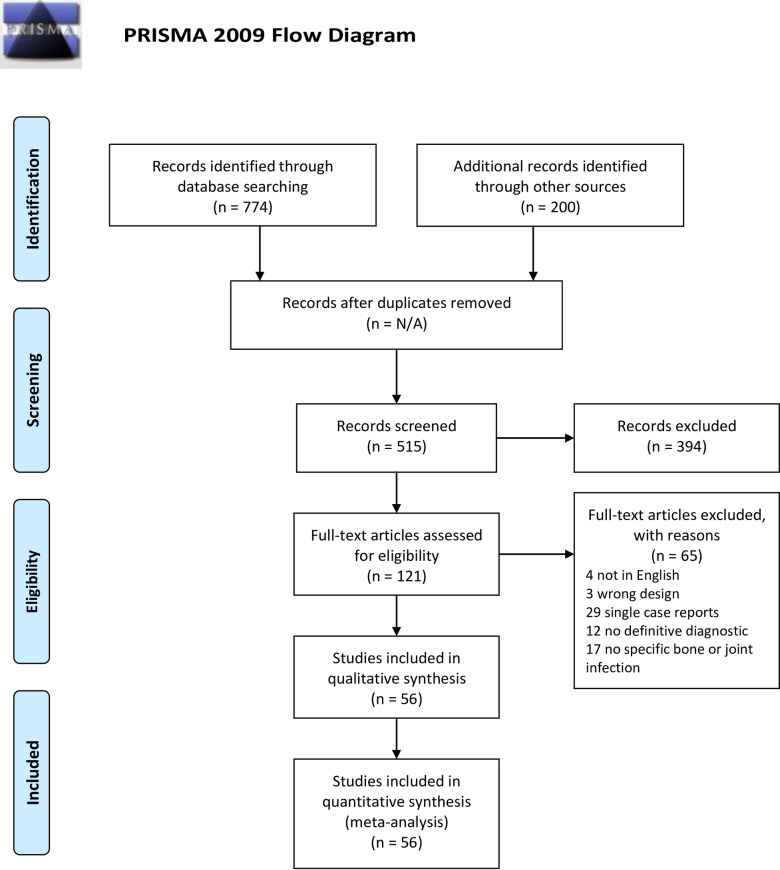
A total of 974 publications were identified, which led to 515 articles being analyzed for full-text review. After full-text review, 56 published studies met the inclusion criteria and were used in the meta-analysis. Fig 1 details the process of article screening and selection following the Preferred Reporting Items for Systematic Review and Meta-Analyses (PRISMA) statement guidelines [73].
Fig 1. Flow-chart of systematic review of osteoarticular brucellosis.
Included studies
All articles included in this study were either prospective (32%) or retrospective (64%), and the authors reported acute or chronic cases of human brucellosis and associated complications. Most of the included studies were from the Middle East, especially Turkey (37.5%), Iran (16%), Saudi Arabia (9%), Israel (3.5%), Kuwait (3.5%), Jordan (1.8%), and Iraq (1.8%). In Europe, studies were also reported from Spain (9%), Macedonia (7%), Germany (1.8%), Portugal (1.8%), and Kosovo (1.8%). Only one report was from South America, specifically Peru (1.8%). The most represented countries were Turkey, Iran, Spain, and Saudi Arabia, respectively, and B. melitensis was the predominant species isolated from either blood or bone marrow cultures of infected individuals. The age range of the study population was 0–88 years old. 25% of the included studies reported childhood OAB while 8% reported OAB in adults. Most studies reported varying proportion of osteoarticular brucellosis in both males and females. Table 1 details the characteristics of all the studies included in this review.
Table 1. Characteristics of included studies.
| Study | Country | Age | Sex ratio-Osteoarticular brucellosis (Male/Female) | Sample size (brucellosis) | Sample size (Osteoarticular brucellosis) | Prevalence/Proportion | Joints affected |
|---|---|---|---|---|---|---|---|
| Aktug-Demir et al., 2014 | Turkey | 18+ | N/A | 227 | 75 | 0.33 | Sacroiliac, spine |
| Al-Eissa et al., 1990 | Saudi Arabia | 0–14 | 15/25 (37.5/62.5%) | 102 | 40 | 0.39 | N/A |
| Ariza et al., 1993 | Spain | 7–83 | 42/20 (67.7/32.2%) | 530 | 62 | 0.12 | Spine, hip, bursa |
| Aydoslu et al., 2006 | Turkey | 17–76 | N/A | 47 | 14 | 0.3 | Sacroiliac, spine, peripheral arthritis |
| Benjamin et al., 1992 | Saudi Arabia | N/A | N/A | 157 | 57 | 0.36 | Hip, knee |
| Benjamin and Khan, 1994 | Saudi Arabia | 0–12 | N/A | 190 | 70 | 0.37 | Sacroiliac, spine, hip, knee, ankle, shoulder |
| Besharat et al., 2010 | Iran | N/A | 140 | 56 | 0.4 | N/A | |
| Bosilkovski et al., 2004a | Macedonia | 3–78 | N/A | 331 | 196 | 0.59 | Sacroiliac, spine, hip, bursa, sternochondral, costochondral |
| Bosilkovski et al., 2004b | Macedonia | 4–69 | 18/15 (54.5/45.5%) | 263 | 162 | 0.62 | Hip |
| Bosilkovski et al., 2010 | Macedonia | 1–82 | N/A | 550 | 299 | 0.54 | Peripheral arthritis, sacroilitis, spondylitis |
| Bosilkovski et al., 2013 | Macedonia | 0–14 | N/A | 317 | 133 | 0.42 | Sacroiliac, hip, knees, ankle, bursa, shoulder, elbow, wrist, interphalangeal, sternoclavicular |
| Bukharie, 2009 | Saudi Arabia | 13–81 | N/A | 84 | 54 | 0.64 | Spine |
| Bulut et al., 2011 | Turkey | 15–83 | N/A | 324 | 84 | 0.26 | Sacroiliac, spine |
| Cirakli et al., 2015 | Turkey | 2–17 | 42/10 (80.8/19.2%) | 52 | 11 | 0.21 | Hip, knee |
| Colmenero et al., 1991 | Spain | 14–73 | N/A | 263 | 65 | 0.25 | Sacroiliac, spine, ankle, olecranon bursa |
| Colmenero et al., 1992 | Spain | 14–82 | N/A | 593 | 58 | 0.1 | Spine |
| Colmenero et al., 1996 | Spain | 14+ | 2/- | 530 | 2 | 0.004 | Sacroiliac, spine |
| Colmenero et al., 2008 | Spain | >14 | 69/27 (72/28%) | 918 | 96 | 0.11 | Vertebral osteomyelitis |
| Dabbagh and Rasool 2009 | Iraq | <10 >60 | N/A | 80 | 45 | 0.56 | Knee, spine, sacroiliac |
| Dahouk et al., 2005 | Germany | 4–72 | 14/16 | 62 | 11 | 0.37 | Sacroiliac, sternoclavicular, spine, bursa |
| Demiroglu et al., 2007 | Turkey | 15–79 | N/A | 151 | 51 | 0.34 | Spine, sacroiliac, tendon |
| Ebrahimpour et al., 2017 | Iran | 15–80 | 299/165 (64.4/35.6%) | 1252 | 464 | 0.37 | Sacroiliac, hip, knee, ankle, elbow, shoulder |
| Eini et al., 2012 | Iran | 9–88 | N/A | 230 | 118 | 0.51 | Spine |
| Fanni et al., 2013 | Iran | 2–14 | N/A | 34 | 26 | 0.77 | Hip, knee, elbow, wrist, ankle, sacroiliac |
| Fruchtman et al., 2015 | Israel | 0–19 | N/A | 252 | 92 | 0.37 | N/A |
| Gonen et al., 2013 | Turkey | 15–88 | N/A | 201 | 50 | 0.25 | Sacroiliac, spine |
| Gotuzzo et al., 1987 | Peru | N/A | N/A | 92 | 22 | 0.24 | Sacroiliac, knee, ankle, spine |
| Guler et al., 2014 | Turkey | 3–82 | N/A | 370 | 178 | 0.48 | Sacroiliac, spine, bursa |
| Gur et al., 2003 | Turkey | N/A | 283 | 195 | 0.69 | Spine, sacroiliac | |
| Hizel et al., 2007 | Turkey | 15–81 | N/A | 163 | 72 | 0.44 | Spine, sacroiliac, paravertebral |
| Issa and Jamal, 1999 | Jordan | 3–14 | N/A | 68 | 38 | 0.56 | N/A |
| Jia et al., 2017 | China | 3–75 | N/A | 590 | 137 | 0.23 | Sacroiliac, knee, spine |
| Kazak et al., 2016 | Turkey | 15–85 | N/A | 164 | 87 | 0.53 | Sacroiliac, hip, ankle, knee, spine |
| Khateeb et al., 1990 | Kuwait | 13–75 | N/A | 400 | 104 | 0.46 | Sacroiliac, hip, knee, spine |
| Kokoglu et al., 2006 | Turkey | 15–69 | 67/71 (48.5/51.5%) | 138 | 64 | 0.14 | Sacroiliac, spine, peripheral arthritis |
| Kose et al., 2014 | Turkey | 14–83 | N/A | 72 | 10 | 0.31 | Sacroiliac, spine |
| Kouba et al., 2013 | Tunisia | 19–74 | 23/9 (72/28%) | 146 | 32 | 0.22 | Spine |
| Kursun et al, 2013 | Turkey | N/A | N/A | 447 | 137 | 0.31 | Spine |
| Lulu et al., 1988 | Kuwait | 10–60 | N/A | 400 | 105 | 0.26 | Sacroiliac, spine, hip, knee, shoulder, ankle, elbow |
| Memish et al., 2000 | Saudi Arabia | 0–40 | N/A | 160 | 68 | 0.42 | Sacroiliac, spine, hip, knee,ankle |
| Memut et al., 2012 | Turkey | 15–77 | N/A | 231 | 70 | 0.37 | Sacroiliac, spine, bursa |
| Mugahi et al., 2014 | Iran | 11–80 | N/A | 81 | 8 | 0.099 | N/A |
| Namiduru et al., 2003 | Turkey | 16–70 | 7/7/ (50/50%) | 186 | 14 | 0.08 | Spine |
| Okur et al., 2012 | Turkey | 2–16 | N/A | 147 | 20 | 0.14 | N/A |
| Parlak et al., 2015 | Turkey | 1–16 | N/A | 496 | 55 | 0.11 | Peripehral arthritis |
| Pourbagher et al., 2006 | Turkey | 2–77 | N/A | 251 | 114 | 0.45 | Sacroiliac, spine, hip, bursa |
| Qehaja-Bucaj et al., 2015 | Kosovo | 2–74 | N/A | 124 | 55 | 0.44 | Sacroiliac, spine, hip |
| Roushan et al., 2004 | Iran | 16–90 | N/A | 469 | 69 | 0.15 | Sacroiliac, spine, ankle, knee, hip, wrist, sternoclavicular |
| Roushan et al., 2005 | Iran | 3–15 | N/A | 111 | 35 | 0.32 | Sacroiliac, spine, ankle, knee, hip, wrist, shoulder |
| Santiago et al., 2011 | Portugal | N/A | 90 | 44 | 0.49 | N/A | |
| Sasan et al., 2012 | Iran | 0–16 | N/A | 82 | 52 | 0.63 | Knee and hip |
| Savas et al., 2007 | Turkey | 2–77 | N/A | 140 | 74 | 0.53 | Sacroiliac, spine |
| Tasova et al., 1999 | Turkey | 15–75 | 51/36 (58.6/41.4%) | 238 | 87 | 0.37 | Sacroiliac, spine, knee, ankle, bursa |
| Ulug et al., 2011 | Turkey | 4–15 | N/A | 22 | 5 | 0.23 | Sacroiliac, hip, spine, ankle, sternoclavicular |
| Zaks et al., 1995 | Israel | N/A | N/A | 90 | 40 | 0.41 | Sacroiliac, spine, hip, knee, ankle, shoulder, elbow |
| Zamani et al., 2011 | Iran | 2–12 | 14/10 (58.3/41.7%) | 96 | 24 | 0.25 | Knee, hip, ankle, wrist elbow |
For all individuals in the included studies, brucellosis was diagnosed based on the presence of inflammatory signs (pain, swelling, and tenderness) of the affected joints and one or more of other diagnostic methods including positive blood or synovial fluid culture; serology (using 2-mercapthoethanol-Standard Agglutination Test (1/160), Brucella Tube Agglutination Test (1:1280), Brucella Skin Test, Complement Fixation test, Rose Bengal test, Coomb’s test (1/320), Wright agglutination test, Immunofluorescence, or ELISA IgG and IgM); and anomalies of the bones and joints evident by varying imaging techniques. Participants in most of the included studies were diagnosed based on clinical signs and serology (70%), and only a few reported additional positive blood or synovial fluid culture (30%) (Table 2).
Table 2. Diagnostic methods of osteoarticular brucellosis used in included studies.
| Study | Country (n) | Diagnostic methods | |||
|---|---|---|---|---|---|
| Inflammatory signs | Culture | Immunoassays | Radiographs | ||
| Aktug-Demir et al., 2014 | Turkey (75) | Arthralgia | Blood culture | Standard tube agglutination test | Contrast enhanced MRI |
| Al-Eissa et al., 1990 | Saudi Arabia (40) | N/A | Blood culture | Brucella microagglutination test | N/A |
| Ariza et al., 1993 | Spain (62) | N/A | N/A | Standard tube agglutination test, Rose Bengal test, Coombs test | Plain radiographs, bone radionuclide scan |
| Aydoslu et al., 2006 | Turkey (14) | N/A | Blood culture | N/A | N/A |
| Benjamin et al., 1992 | Saudi Arabia (57) | N/A | Blood culture | N/A | N/A |
| Benjamin and Khan, 1994 | Saudi Arabia (70) | Pain, swelling, redness, functional disability | Blood and synovial fluid culture | Standard tube agglutination test | Plain radiographs |
| Besharat et al., 2010 | Iran (56) | Pain, fever, sweating | N/A | N/A | N/A |
| Bosilkovski et al., 2004a | Macedonia (196) | Pain, swelling, redness, functional disability | N/A | Standard tube agglutination test, Brucella Coombs test | Fabere test, plain radiograph, MRI, computed tomography, radionuclide bone scan, ultrasound |
| Bosilkovski et al., 2004b | Macedonia (162) | Pain, swelling, redness, functional disability | N/A | Standard tube agglutination test, Brucella Coombs test | Plain radiographs, bone radionuclide scan, ultrasound, MRI |
| Bosilkovski et al., 2010 | Macedonia (299) | Pain, swelling, redness, functional disability | N/A | Standard tube agglutination test, Brucella Coombs test, Brucellacapt test | MRI, radionuclide bone scans, computerized tomography |
| Bosilkovski et al., 2013 | Macedonia (133) | Pain, swelling, redness, functional disability | N/A | Standard tube agglutination test, Brucella Coombs test, Brucellacapt test | Stinchfield, Mennel test, abnormality on radiography, radionuclide bone scan or ultrasound examination, MRI, computed tomography |
| Bukharie, 2009 | Saudi Arabia (54) | Fever, pain | Blood and bone marrow culture | Standard tube agglutination test, ELISA (IgM and IgG) | N/A |
| Bulut et al., 2011 | Turkey (84) | Fever, sweating, arthralgia | Blood culture | Standard tube agglutination test | Plain radiographs, computed tomography scan, bone scan, MRI |
| Cirakli et al., 2015 | Turkey (11) | N/A | Body fluid culture | Standard tube agglutination test | N/A |
| Colmenero et al., 1991 | Spain (65) | Pain, swelling, redness, functional disability | N/A | Seroagglutination, Coombs, indirect immunofluorescence, Rose Bengal | Plain radiographs, radionuclide bone scan |
| Colmenero et al., 1992 | Spain (58) | Pain, swelling, redness, functional disability | N/A | Wright, Coombs, rose bengal test, indirect immunofluorescence | Plain radiographs, bone radionuclide scan |
| Colmenero et al., 1996 | Spain (2) | N/A | N/A | Wright, Coombs, indirect immunofluorescence | Computed tomography |
| Colmenero et al., 2008 | Spain (96) | Pain, swelling, redness, functional disability | N/A | Standard tube agglutination test, Coombs antibrucella or immunocapture agglutination test | Computed tomography |
| Dabbagh and Rasool 2009 | Iraq (45) | Pain, swelling, and restriction of movement | N/A | Brucella agglutination test and 2-Mercaptoethanol | Plain radiograph |
| Dahouk et al., 2005 | Germany (11) | N/A | Blood culture | Standard tube agglutination test, ELISA IgM, IgG | N/A |
| Demiroglu et al., 2007 | Turkey (51) | N/A | Bone marrow, sternoclavicular and psoas abscess culture | Standard tube agglutination test | N/A |
| Ebrahimpour et al., 2017 | Iran (464) | Swelling, effusion, restriction of movement | N/A | Standard tube agglutination test and 2-Mercaptoethanol | Plain radiographs, MRI, sonography |
| Eini et al., 2012 | Iran (118) | Arthralgia | Blood culture | Standard tube agglutination test, Brucella Coombs test, 2-Mercaptoethanol | N/A |
| Fanni et al., 2013 | Iran (26) | N/A | Blood and bone marrow culture | Serum agglutination test, Wright and Coombs test, 2-Mercaptoethanol | N/A |
| Fruchtman et al., 2015 | Israel (92) | Fever, myalgia, headache | Blood culture | Standard tube agglutination test | N/A |
| Gonen et al., 2013 | Turkey (50) | Blood and synovial fluid and bone marrow culture | Standard tube agglutination test, Coombs test | Plain radiographs, MRI, computed tomography, ultrasonography | |
| Gotuzzo et al., 1987 | Peru (22) | N/A | Blood, bone marrow, synovial culture | N/A | N/A |
| Guler et al., 2014 | Turkey (178) | Pain, swelling, redness, functional disability | Blood culture | Standard tube agglutination test | Radiological alterations and/or radionuclide uptake in any deep joint |
| Gur et al., 2003 | Turkey (195) | Pain, swelling, redness, functional disability | Blood and body fluid culture | Standard tube agglutination test | Plain radiographs, bone radionuclide scan, computed tomography, MRI |
| Hizel et al., 2007 | Turkey (72) | Pain, fever | Blood culture | Standard tube agglutination test | Bone Scintigrapghy |
| Issa and Jamal, 1999 | Jordan (38) | Arthralgia | Blood and bone marrow culture | Rose Bengal test, ELISA IgM and IgG, Wright test | N/A |
| Jia et al., 2017 | China (137) | Fatigue, fever, muscle and joint pain, headache | Blood culture | Standard tube agglutination test | N/A |
| Kazak et al., 2016 | Turkey (87) | Arthralgia | Blood and body fluid culture | Standard tube agglutination test and Rose bengal test, | N/A |
| Khateeb et al., 1990 | Kuwait (104) | Pain, swelling, restriction of movement | Culture negative | Microagglutination, slide agglutination test, ELISA (IgG, IgM, and IgA) | Plain radiographs- no apparent pathological changes |
| Kokoglu et al., 2006 | Turkey (64) | N/A | Blood and body fluids culture | Wright agglutination test | N/A |
| Kose et al., 2014 | Turkey (10) | Arthralgia | Blood and bone marrow culture | Standard tube agglutination test | N/A |
| Kouba et al., 2013 | Tunisia (32) | Pain | Blood and body fluids/tissue culture | Standard tube agglutination test | MRI, computed tomography |
| Kursun et al, 2013 | Turkey (137) | Arthralgia | Blood and bone marrow culture | Standard tube agglutination test | Whole-body bone scintigraphy (Technetium 99m) |
| Lulu et al., 1988 | Kuwait (105) | Pain and swelling | Culture negative | Microagglutination, slide agglutination test, ELISA (IgG, IgM, and IgA) | N/A |
| Memish et al., 2000 | Saudi Arabia (68) | N/A | Blood and body fluid culture | Microagglutination test | N/A |
| Memut et al., 2012 | Turkey (70) | Pain, swelling, redness, functional disability | Blood and bone marrow culture | Standard tube agglutination test, Rose Bengal test | Computed tomography, MRI |
| Mugahi et al., 2014 | Iran (8) | Arthralgia | N/A | Wright test and 2-Mecarptoethanol test | N/A |
| Namiduru et al., 2003 | Turkey (14) | Arthralgia | Blood and body fluid culture | Wright test | Plain radiograph, bone scintigraphy, MRI |
| Okur et al., 2012 | Turkey (20) | Arthralgia | Blood culture | Standard tube agglutination test | N/A |
| Parlak et al., 2015 | Turkey (55) | N/A | Blood culture | Brucella agglutination test, standard tube agglutination test | None |
| Pourbagher et al., 2006 | Turkey (114) | Arthralgia | Blood culture | Standard tube agglutination test | Joint sonography, radiography, radionuclide, bone scintigraphy, and MRI |
| Qehaja-Bucaj et al., 2015 | Kosovo (55) | Fever, arhtralgia, fatigue, sweating | N/A | Wright test | Plain radiographs |
| Roushan et al., 2004 | Iran (69) | Pain, swelling, redness, functional disability | N/A | Standard tube agglutination test, 2-Mercaptoethanol | Plain radiographs, bone radionuclide scan |
| Roushan et al., 2005 | Iran (35) | Pain, swelling, redness, functional disability | Blood culture | Standard tube agglutination test, 2-Mercaptoethanol | Radionuclide scan |
| Santiago et al., 2011 | Portugal (44) | N/A | N/A | ||
| Sasan et al., 2012 | Iran (52) | Arthralgia | Blood, urine and joint fluid culture | Wright test, Coombs test, Rose Bengal test, and 2-Mercaptoethanol | N/A |
| Savas et al., 2007 | Turkey (74) | Arthralgia | Blood culture | Standard tube agglutination test | N/A |
| Tasova et al., 1999 | Turkey (87) | Pain, swelling, redness, functional disability | Blood and body fluid culture | Standard tube agglutination test | Plain radiograph, radionuclide bone scan |
| Ulug et al., 2011 | Turkey (5) | Pain, swelling, redness, functional disability | N/A | Standard tube agglutination test | Computed tomography, MRI, plain Radiograph |
| Zaks et al., 1995 | Israel (40) | Pain, swelling, redness, functional disability | Positive culture | Standard tube agglutination test, Rose Bengal test, 2-Mercaptoethanol | Plain radiographs |
| Zamani et al., 2011 | Iran (24) | Pain, swelling, redness, functional disability | Blood culture | Standard tube agglutination test, 2-Mercaptoethanol | N/A |
Prevalence
The prevalence estimates ranged from 27% in low-risk regions (e.g. Europe and North America) to 36% in high-risk regions (e.g. Middle East and South America), with no significant difference between the two estimates, indicating that the prevalence of OAB is independent of the endemicity of a particular region. High-risk regions included those countries where high number of cases or incidence of brucellosis have been consistently reported, such as in the Middle East, Asia, South and Central America, and Africa [27–29,32–36].
Meta-analysis
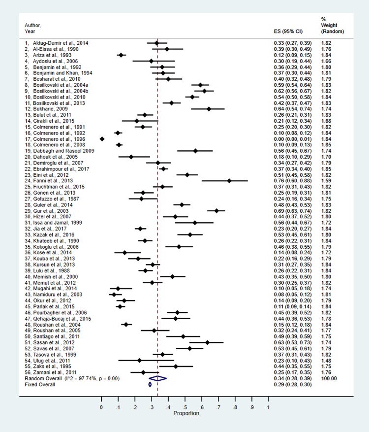
Fig 2 reflects the “metaprop” results of 56 prevalence estimates (converted back from the Freeman-Tukey transformations). The overall fixed effect estimate of prevalence was 0.29 (95%CI: 0.28 to 0.30). The fixed effect estimate was statistically heterogeneous with an I2 of 98.66%. The random effects estimate was 0.34 (95%CI: 0.28 to 0.39).
Fig 2. Metaprop results of prevalence estimates of osteoarticular brucellosis.
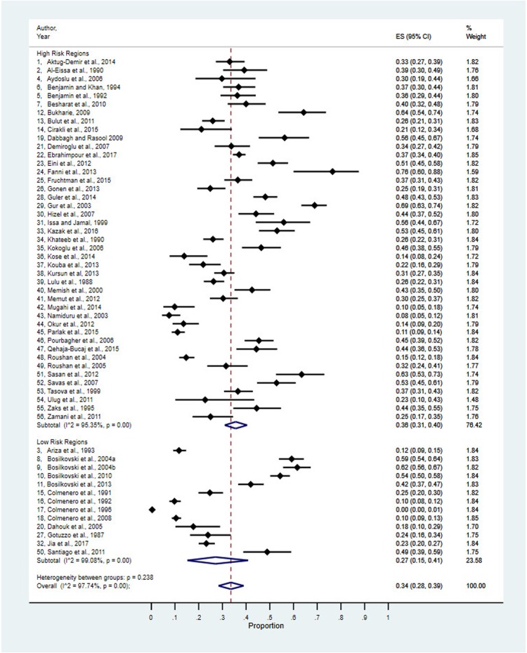
Fig 3 reflects the 56 prevalence estimates stratified by risk regions, which was determined based on previous reports of a high brucellosis incidence [27–29,32–36].
Fig 3. Prevalence estimates stratified by risk regions.
Both subgroups (high-risk and low-risk regions), as well as the overall result (Fig 3) were statistically heterogeneous. Stratification by risk regions alone was insufficient to explain the degree of heterogeneity. Random effects estimate for the two strata as prevalences were 0.36 (0.31 to 0.40) for high-risk countries and 0.27 (0.15 to 0.41) for low-risk countries. The estimates for the two strata were not statistically significantly different, suggesting that the prevalence of OAB is independent of the exposure risk of a particular region.
Sources of heterogeneity between studies can also be explored using meta-regression. To determine the source of heterogeneity in these studies, as well as influence of age and gender on the prevalence of OAB, we collected data on age and gender. However, these data were problematic, and thus, meta-regression could not be used. The age of individuals was typically reported as a range, for example, 16 to 75 years of age. Mean and/or median age of the population would have been more desirable for a meta-regression analysis. Additionally, some studies did not report any age data. Therefore, these studies were dropped out of the meta-regression analysis. Studies that did not report gender proportions were also excluded, and thus, meta-regression could not be used to further explore heterogeneity based on both age and gender.
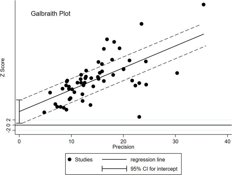
Outlier analysis has also been used to explain, and by removal of studies one at a time, explain heterogeneity. The degree of heterogeneity was such that outlier study removal would have left too few studies to calculate reasonable estimates. Galbraith plot is provided (Fig 4) using the Stata command “galbr” and the Excel computation of the Freeman-Tukey double arcsine transformation of prevalence values. Outlier studies are recognized as those outside the 2 parallel galbraith bands at values 2 and -2.
Fig 4. Galbraith plot analysis of all the included studies.
Discussion
The main objective of this study was to systematically review the literature and perform a meta-analysis to estimate the global prevalence of osteoarticular brucellosis (OAB). A total of 56 studies met the inclusion criteria and were included in the systematic review and meta-analysis. Although there was an evidence of geographical variation in the prevalence of OAB with estimates ranging from 27% in low-risk regions to 36% in high-risk regions, the difference was not significant. This result indicates that the prevalence of OAB is not dependent on the endemicity of brucellosis in a region, and that brucellosis patients have at least a 27% chance of developing osteoarticular disease. In addition, the result also suggests that brucellosis remains an important public health concern in both high-risk [74,75], and low-risk regions [42,43,76]. Therefore, early pathogen detection using sensitive and specific validated diagnostic techniques as well as treatment of the disease to stop disease manifestation are paramount in the control of OAB.
Classification of a region into high or low-risk was determined based on previous reports of consistently high incidence of brucellosis in different countries in Africa, Asia, Eastern Europe, Mediterranean Basin, Middle East, South and Central America, and The Caribbean, as signified by the Centers for Disease Control and Prevention [27–29,32–36]. Low-risk regions include those countries with little or no reports of incidence of brucellosis such as North and South America, and some parts of Europe. In low-risk regions, very few occasional cases of brucellosis occur, and are usually travel-related. For example, consumption of raw animal products (e.g. raw milk or cheese) while visiting high-risk countries [77]. Furthermore, OAB may be difficult to diagnose in low-risk regions, not due to a lack of appropriate infrastructure, but because the disease is less common and may be confused with other causes of arthritis in humans (e.g. Lyme disease), hence, an under-diagnosis or misdiagnosis of the disease.
In this current study, although not statistically significant, the higher prevalence of OAB in high-risk regions correlates with previous findings reporting a high incidence of brucellosis in Middle Eastern countries such as Iran and Saudi Arabia [75,78,79]. Moreover, prevalence variation in different parts of the world may be due to varying environmental and socioeconomic factors such as sanitation, availability of medical facilities for optimum treatment and care, brucellosis awareness in communities, diagnostic capabilities for prompt detection of the disease condition, amongst others [80–83]. Another important factor is under-diagnosis or misdiagnosis of brucellosis because the disease manifests as flu-like symptoms and may bear resemblance to other diseases with similar symptoms such as malaria, or dengue fever, which are common disease conditions in many parts of Africa, thus leading to delays in detection and appropriate treatment of the disease [81,82]. Also, Brucella-induced arthritis in older individuals is commonly overlooked as arthritis due to old age. Hence, an understanding of patients’ history and thorough clinical examinations are recommended.
Additionally, higher prevalence in high-risk regions may be due to close interactions with domestic animals such as raising animals in close proximity to human living areas, low public awareness of brucellosis as a serious debilitating disease, resistance to slaughtering infected animals, and the customary beliefs of raw milk ingestion [79,84].
Focal complications of brucellosis, such as osteoarticular involvement are frequently reported in children, especially in high-risk regions. For example, childhood OAB (age ≤ 18years) was reported in 25% of the studies included in this current meta-analysis study [85–91], while 8% of the studies reported OAB in adults only.
Clinical presentation of childhood brucellosis is similar to those observed in other flu-like illness such as malaria, influenza, or dengue and is often misdiagnosed and mistreated, especially in resource-limited settings [13,81,82,92–94]. In most reported cases, contact with contaminated animal products or consumption of raw unpasteurized milk has been shown as a risk factor for contracting the disease. For example, in some resource-limited settings, the available milk is rather given to children than adults, and if the milk is contaminated, it poses an increased risk of brucellosis infection. Most commonly, B. melitensis is the main causative agent in infected children, although other species such as B. abortus and B. suis have also been identified [13,36,38,91].
Most studies in this review reported age range for individuals presenting OAB, for example, an age range of 16–75 years, while some studies did not report any age data. Therefore, it was impossible to determine the actual variation in prevalence based on age of the study population. Thus meta-regression could not be used to further explore heterogeneity.
As regards gender, both sexes are affected equally, although some reports claim that the disease is more prevalent in males (80%) than in females (19%) because of the nature of the male job in such regions, which facilitates increased exposure to animals and their products, as observed in herdsmen, ranchers, pastoralists and abattoir workers [29,38,51]. In this current study, because of the limited information provided in the selected articles, it was impossible to determine variation in OAB prevalence based on gender of the study population.
There are several diagnostic tools for brucellosis. The gold standard of brucellosis diagnosis is the positive culture of Brucella from tissues and bodily fluids (e.g. blood, bone marrow, synovial, and cerebrospinal fluid) of infected patients, although culture yield is inversely related to the duration of illness [95–97]. For example, culture yield is greater during the acute stage of brucellosis while it is less in later stages of the disease or during occasional relapses [84]. Additionally, the likelihood of isolation in patients with chronic disease and focal complications can be improved by using sampling material from affected sites, such as synovial fluid in OAB cases [97].
Various Brucella culture systems including automated continuously monitored blood culture systems such as Bactec (BD Diagnostics, Sparks, MD, USA) and BacTAlert (bioMerieux, Durham, NC, USA) give higher yields than the conventional culture method and facilitate the detection of bacterial growth [84,98]. However, these culture systems are not routinely used in most high-risk regions because of insufficient infrastructure as well as trained personnel. Hence, classical bacteriological culture is a common diagnostic method for brucellosis in these regions because it is easily accessible [95–97].
Due to inconsistent yield of Brucella from culture systems, increased risk of personnel infection, as well as the lack of validated molecular-based diagnostic techniques, the common standard for diagnosis of brucellosis is serological assays, which include Serum Agglutination Test (SAT), Microagglutination Test (MAT), Enzyme Linked Immunosorbent Assay (ELISA), Indirect Coombs (Anti-Human Globulin) Test, Brucellacapt, Wright agglutination test, Rose Bengal Slide Agglutination Test (RB-SAT), Complement Fixation Test (CFT), Indirect immunofluorescence test (IF), and Immunochromatography Lateral Flow Assay. Among the serological assays, SAT is the most frequently used and standardized test. SAT is based on measuring an agglutination titer of different serum dilutions (1:20–1:1280) against a standardized concentration of whole Brucella cell suspension. The highest serum dilution showing more than 50% agglutination is considered the agglutination titre. A positive titre is 1:160 or more [84,98,99]. Multiple testing at 4–8 week intervals is recommended to overcome the drawback of inconsistent results. ELISA is another commonly used serological assay for diagnosing brucellosis. It is considered specific (95%) and sensitive (98%) and has been consistently shown to diagnose both focal and chronic brucellosis [98,100]. Generally, because of the variability in the specificity and sensitivity of the conventional serological tests routinely used in high-risk regions, a combination of varying serological tests (e.g. SAT and either indirect Coombs, Brucellacapt, or ELISA for IgG and IgM) is recommended for the definitive diagnosis of human brucellosis [97,98,100–102].
Correspondingly, all studies included in this systematic review and meta-analysis used a combination of serological tests including SAT (1/160), ELISA (IgG and IM), CFT, IF, Wright agglutination test, Brucella Tube Agglutination Test (1:1280), Brucella Skin Test, Coomb test (1/320), or RB-SAT (Table 2 shows the diagnostic tests used by the respective studies). All individuals presenting OAB in the articles selected for the current analyses were positive for two or more of the reported diagnostic methods (such as clinical signs of fever, inflamed joints, myalgia, arthralgia, and/or bacteriological culture, and serology) Table 2.
In addition to clinical and serological diagnosis of OAB, radiographic abnormalities of the bones and joints, which manifest as arthritis, sacroiliitis, and spondylitis, have been described using varying radiological techniques such as radionuclide bone scan, plain radiography, joint sonography, computed tomography, and contrast-enhanced magnetic resonance imaging, amongst others. The abnormalities in the affected osteoarticular regions included joint space narrowing or widening, subchondral erosion, subchondral sclerosis and/or soft tissue swelling [37,46,63,103]. For example, bone scans were considered positive for abnormalities when there was increased uptake of the compound in the respective osteoarticular regions [46]. Generally, radiological diagnosis of OAB in humans is nonspecific and inconsistent, but varying degrees of abnormalities of affected regions have been described [46,63]. In this current study, most of the individuals had a report of varying bone abnormalities evident by the respective imaging techniques.
Overall, the prospects of OAB diagnosis by a physician in high-risk versus low-risk regions differ. Brucella-induced osteoarticular involvement can be easily suspected in high-risk regions based on clinical signs and history of contact with animals and raw animal products, thereby facilitating rapid diagnosis and treatment. However, in low-risk regions, especially where brucellosis has been eradicated, a knowledge of patients’ clinical history (e.g. a travel-related exposure to and consumption of raw animal products such as milk and cheese) is particularly valuable to the diagnosis of OAB (13).
Since the clinical features of OAB are not specific and there is yet to be a single consistent definitive diagnostic technique, the clinical history of animal contact or consumption of raw animal products is especially important, as well as a combination of diagnostic methods (bacteriological culture, serology and imaging).
The purpose of the current study was to estimate the prevalence of OAB among brucellosis patients worldwide by performing a meta-analysis. For the first time, we have demonstrated that the prevalence of OAB is independent of brucellosis endemicity of a particular region, and that brucellosis patients have at least a 27% chance of developing an osteoarticular disease. Thus, brucellosis remains a public health concern in both high-risk and low-risk countries [104], although there are some limitations to this current study, such as incomplete data representation. For example, lack of vital demographics precluded the feasibility of estimating OAB prevalence based on age and gender. Nevertheless, the current review is still very valuable, and has contributed to our understanding of the global prevalence of Brucella-induced osteoarticular disease. Hence, this study has provided the basis for increased awareness of OAB, the need for the development and validation of diagnostic tests, and appropriate treatment regimen to reduce disease manifestation. Therefore, further research should investigate the potential mechanisms of OAB, as well as the influence of age, gender, and other socioeconomic factor variations in its global prevalence, as this may provide insight into associated exposure risks and management of the disease.
Supporting information
(DOCX)
(DOCX)
Data Availability
All relevant data are within the paper and its Supporting Information files.
Funding Statement
Funding for AAM has been provided by the National Institutes of Health (NIH) International Research Scientist Development Award (IRSDA/K01)(Award number: K01 TW009981-01). ASA has been funded by the CVM Graduate Diversity Fellowship from the College of Veterinary Medicine & Biomedical Sciences, Texas A&M University, College Station, Texas, USA. AAM and RG were also supported by the President's Excellence Funds (T3:Texas A&M Triads for Transformation), Texas A&M University, College Station, Texas, USA. The funders had no role in study design, data collection and analysis, decision to publish, or preparation of the manuscript.
References
- 1.Young EJ. Human brucellosis. Reviews of infectious diseases. 1983;5(5):821–842. [DOI] [PubMed] [Google Scholar]
- 2.Meltzer E, Sidi Y, Smolen G, Banai M, Bardenstein S, Schwartz E. Sexually transmitted brucellosis in humans. Clinical infectious diseases. 2010;51(2):e12–5. 10.1086/653608 [DOI] [PubMed] [Google Scholar]
- 3.Franco MP, Mulder M, Gilman RH, Smits HL. Human brucellosis. The Lancet infectious diseases. 2007;7(12):775–86 10.1016/S1473-3099(07)70286-4 [DOI] [PubMed] [Google Scholar]
- 4.Seleem MN, Boyle SM, Sriranganathan N. Brucellosis: a re-emerging zoonosis. Veterinary microbiology. 2010;140(3–4):392–8. 10.1016/j.vetmic.2009.06.021 [DOI] [PubMed] [Google Scholar]
- 5.de Figueiredo P, Ficht TA, Rice-Ficht A, Rossetti CA, Adams LG. Pathogenesis and immunobiology of brucellosis: review of Brucella–Host Interactions. The American journal of pathology. 2015;185(6):1505–1517. 10.1016/j.ajpath.2015.03.003 [DOI] [PMC free article] [PubMed] [Google Scholar]
- 6.Hashino M, Kim S, Tachibana M, Shimizu T, Watarai M. Vertical Transmission of Brucella abortus Causes Sterility in Pregnant Mice. Journal of Veterinary Medical Science. 2012;74(8):1075–1077. [DOI] [PubMed] [Google Scholar]
- 7.Kim S, Lee DS, Watanabe K, Furuoka H, Suzuki H, Watarai M. Interferon-γ promotes abortion due to Brucella infection in pregnant mice. BMC microbiology. 2005;5(1):22. [DOI] [PMC free article] [PubMed] [Google Scholar]
- 8.Wang Z, Wang SS, Wang GL, Wu TL, Lv YL, Wu QM. A pregnant mouse model for the vertical transmission of Brucella melitensis. The Veterinary Journal. 2014;200(1):116–21. 10.1016/j.tvjl.2013.12.021 [DOI] [PubMed] [Google Scholar]
- 9.Joffe B, Diamond MT. Brucellosis due to self-inoculation. Annals of internal medicine. 1966;65(3):564–5. [DOI] [PubMed] [Google Scholar]
- 10.Poole PM, Whitehouse DB, Gilchrist MM. A case of abortion consequent upon infection with Brucella abortus biotype 2. Journal of Clinical pathology. 1972;25(10):882–4. [DOI] [PMC free article] [PubMed] [Google Scholar]
- 11.McDermott JJ, Arimi SM. Brucellosis in sub-Saharan Africa: epidemiology, control and impact. Veterinary microbiology. 2000;90(1–4):111–34. [DOI] [PubMed] [Google Scholar]
- 12.Naparstek E, Block CS, Slavin S. Transmission of brucellosis by bone marrow transplantation. The Lancet. 1982;319(8271):574–5. [DOI] [PubMed] [Google Scholar]
- 13.Madkour MM. Osteoarticular Brucellosis: In: Madkour’s Brucellosis. Berlin, Heidelberg: Springer Berlin Heidelberg; 2001. pp. 74–84). [Google Scholar]
- 14.Sarram M, Feiz J, Foruzandeh M, Gazanfarpour P. Intrauterine fetal infection with Brucella melitensis as a possible cause of second-trimester abortion. American Journal of Obstetrics and Gynecology. 1974;119(5),657–660. [DOI] [PubMed] [Google Scholar]
- 15.Arenas-Gamboa AM, Rossetti CA, Chaki SP, Garcia-Gonzalez DG, Adams LG, Ficht TA. Human brucellosis and adverse pregnancy outcomes. Current tropical medicine reports. 2016;3(4):164–72. 10.1007/s40475-016-0092-0 [DOI] [PMC free article] [PubMed] [Google Scholar]
- 16.Nicoletti P. Brucellosis: past, present and future. Prilozi. 2010;31(1):21–32. [PubMed] [Google Scholar]
- 17.Vilchez G, Espinoza M, D’Onadio G, Saona P, Gotuzzo E. Brucellosis in pregnancy: clinical aspects and obstetric outcomes. International journal of infectious diseases. 2015;38:95–100. 10.1016/j.ijid.2015.06.027 [DOI] [PubMed] [Google Scholar]
- 18.Roushan MR, Ebrahimpour S. Human brucellosis: An overview. Caspian journal of internal medicine. 2015;6(1):46 [PMC free article] [PubMed] [Google Scholar]
- 19.Roth F, Zinsstag J, Orkhon D, Chimed-Ochir G, Hutton G, Cosivi O, Carrin G, Otte J. Human health benefits from livestock vaccination for brucellosis: case study. Bulletin of the World Health Organization, 2003;81(12);867–876. [PMC free article] [PubMed] [Google Scholar]
- 20.Pappas G, Memish ZA. Brucellosis in the Middle East: a persistent medical, socioeconomic and political issue. Journal of Chemotherapy. 2007;19(3):243–8. 10.1179/joc.2007.19.3.243 [DOI] [PubMed] [Google Scholar]
- 21.Dean AS, Crump L, Greter H, Hattendorf J, Schelling E, Zinsstag J. Clinical manifestations of human brucellosis: a systematic review and meta-analysis. PLoS Neglected Tropical Diseases. 2012;6(12):e1929 10.1371/journal.pntd.0001929 [DOI] [PMC free article] [PubMed] [Google Scholar]
- 22.Franc KA, Krecek RC, Häsler BN, Arenas-Gamboa AM. Brucellosis remains a neglected disease in the developing world: a call for interdisciplinary action. BMC public health. 2018;18(1):125 10.1186/s12889-017-5016-y [DOI] [PMC free article] [PubMed] [Google Scholar]
- 23.Gotuzzo E, Carrillo C, Guerra J, Llosa L. An evaluation of diagnostic methods for brucellosis—the value of bone marrow culture. Journal of infectious diseases. 1986;153(1):122–5. [DOI] [PubMed] [Google Scholar]
- 24.Aydin M, Yapar AF, Savas L, Reyhan M, Pourbagher A, Turunc TY, Demiroglu YZ, Yologlu NA, Aktas A. Scintigraphic findings in osteoarticular brucellosis. Nuclear medicine communications. 2005;26(7):639–47. [DOI] [PubMed] [Google Scholar]
- 25.Pourbagher A, Pourbagher MA, Savas L, Turunc T, Demiroglu YZ, Erol I, Yalcintas D. Epidemiologic, clinical, and imaging findings in brucellosis patients with osteoarticular involvement. American Journal of Roentgenology. 2006;187(4):873–80. 10.2214/AJR.05.1088 [DOI] [PubMed] [Google Scholar]
- 26.Bouaziz MC, Ladeb MF, Chakroun M, Chaabane S. Spinal brucellosis: a review. Skeletal radiology. 2008;37(9):785–90. 10.1007/s00256-007-0371-x [DOI] [PubMed] [Google Scholar]
- 27.Lulu AR, Araj GF, Khateeb MI, Mustafa MY, Yusuf AR, Fenech FF. Human brucellosis in Kuwait: a prospective study of 400 cases. QJM: An International Journal of Medicine. 1988;66(1):39–54. [PubMed] [Google Scholar]
- 28.Al-Rawi TI, Thewaini AJ, Shawket AR, Ahmed GM. Skeletal brucellosis in Iraqi patients. Annals of the rheumatic diseases. 1989;48(1):77 [DOI] [PMC free article] [PubMed] [Google Scholar]
- 29.Khateeb MI, Araj GF, Majeed SA, Lulu AR. Brucella arthritis: a study of 96 cases in Kuwait. Annals of the rheumatic diseases. 1990;49(12):994 [DOI] [PMC free article] [PubMed] [Google Scholar]
- 30.Ariza J, Pujol M, Valverde J, Nolla JM, Rufi G, Viladrich PF, Corredoira JM, Gudiol F. Brucellar sacroiliitis: findings in 63 episodes and current relevance. Clinical infectious diseases. 1993;16(6):761–5. [DOI] [PubMed] [Google Scholar]
- 31.Colmenero JD, Reguera J, Martos F, Sanchez-De-Mora D, Delgado M, Causse M, Martín-Farfán A, Juarez C. Complications associated with Brucella melitensis infection: a study of 530 cases. Medicine. 1996;75(4):195–211. [DOI] [PubMed] [Google Scholar]
- 32.Rajapakse CN. Bacterial infections: osteoarticular brucellosis. Bailliere's clinical rheumatology. 1995;9(1):161–77. [DOI] [PubMed] [Google Scholar]
- 33.Geyik MF, Gur A, Nas K, Cevik R, Sarac J, Dikici B, Ayaz C. Musculoskeletal involvement of brucellosis in different age groups: a study of 195 cases. Swiss Med Wkly. 2002;132(7–8):98–105. 2002/07/smw-09900 [DOI] [PubMed] [Google Scholar]
- 34.Guler S, Kokoglu OF, Ucmak H, Gul M, Ozden S, Ozkan F. Human brucellosis in Turkey: different clinical presentations. The Journal of Infection in Developing Countries. 2014;8(05):581–8. [DOI] [PubMed] [Google Scholar]
- 35.Fruchtman Y, Segev RW, Golan AA, Dalem Y, Tailakh MA, Novak V, Peled N, Craiu M, Leibovitz E. Epidemiological, diagnostic, clinical, and therapeutic aspects of Brucella bacteremia in children in southern Israel: A 7-year retrospective study (2005–2011). Vector-Borne and Zoonotic Diseases. 2015. March;15(3):195–201. 10.1089/vbz.2014.1726 [DOI] [PubMed] [Google Scholar]
- 36.Parlak M, Akbayram S, Doğan M, Tuncer O, Bayram Y, Ceylan N, Özlük S, Akbayram HT, Öner A. Clinical manifestations and laboratory findings of 496 children with brucellosis in Van, Turkey. Pediatrics International. 2015;57(4):586–9. 10.1111/ped.12598 [DOI] [PubMed] [Google Scholar]
- 37.Bosilkovski M, Kirova-Urosevic V, Cekovska Z, Labacevski N, Cvetanovska M, Rangelov G, Cana F, Bogoeva-Tasevska S. Osteoarticular involvement in childhood brucellosis: experience with 133 cases in an endemic region. The Pediatric infectious disease journal. 2013;32(8):815–9. 10.1097/INF.0b013e31828e9d15 [DOI] [PubMed] [Google Scholar]
- 38.Çıraklı S, Karlı A, Şensoy G, Belet N, Yanık K, Çıraklı A. Evaluation of childhood brucellosis in the central Black Sea region. Turk J Pediatr. 2015;57:123–28 [PubMed] [Google Scholar]
- 39.Evans AC. Brucellosis in the United States. American Journal of Public Health and the Nation’s Health. 1947;37(2):139–51. [PMC free article] [PubMed] [Google Scholar]
- 40.Bergsagel DE, Beamish RE, Wilt JC. Brucella arthritis of the hip joint: A review of the literature and report of a case treated with terramycin. Annals of internal medicine. 1952;37(4):767–76. [DOI] [PubMed] [Google Scholar]
- 41.Kaye D. Brucellosis In: Wilson JD, Braunwald E, Isselbacher KJ et al. Harrison's Principles of lnternal Medicine. 12th ed Volume 1 (eds). New York: McGraw-Hill Inc. Health Professions Division; 1991. Pp. 625–626. [Google Scholar]
- 42.Shoulder HK, Burden HL. News from the Centers for Disease Control and Prevention. Morbidity and Mortality Weekly Report. 2017;66(36):950–4. 10.15585/mmwr.mm6636a3 [DOI] [PMC free article] [PubMed] [Google Scholar]
- 43.Sfeir MM. Raw milk intake: beware of emerging brucellosis. Journal of Medical Microbiology. 2018. March 10.1099/jmm.0.000722 [DOI] [PubMed] [Google Scholar]
- 44.Buzgan T, Karahocagil MK, Irmak H, Baran AI, Karsen H, Evirgen O, Akdeniz H. Clinical manifestations and complications in 1028 cases of brucellosis: a retrospective evaluation and review of the literature. International journal of infectious diseases. 2010;14(6):e469–78. 10.1016/j.ijid.2009.06.031 [DOI] [PubMed] [Google Scholar]
- 45.Wong TM, Lou N, Jin W, Leung F, To M, Leung F. Septic arthritis caused by Brucella melitensis in urban Shenzhen, China: a case report. J Med Case Rep. 2014;8(1):367. [DOI] [PMC free article] [PubMed] [Google Scholar]
- 46.Bosilkovski M, Krteva L, Caparoska S, Dimzova M. Osteoarticular involvement in brucellosis: study of 196 cases in the Republic of Macedonia. Croat Med J. 2004;45(6):727–33. [PubMed] [Google Scholar]
- 47.Hashemi SH, Keramat F, Ranjbar M, Mamani M, Farzam A, Jamal-Omidi S. Osteoarticular complications of brucellosis in Hamedan, an endemic area in the west of Iran. Int J Inf Dis. 2007;11(6):496–500. [DOI] [PubMed] [Google Scholar]
- 48.Turan H, Serefhanoglu K, Karadeli E, Togan T, Arslan H. Osteoarticular involvement among 202 brucellosis cases identified in Central Anatolia region of Turkey. Int Med. 2011;50(5):421–8. [DOI] [PubMed] [Google Scholar]
- 49.Casalinuovo F, Ciambrone L, Cacia A, Rippa P. Contamination of bovine, sheep and goat meat with Brucella spp. Italian J food safety. 2016;5(3). [DOI] [PMC free article] [PubMed] [Google Scholar]
- 50.Miron D, Garty I, Tal I, Horovitz YO, Kedar AM. Sacroiliitis as a sole manifestation of Brucella melitensis infection in a child. Clinical nuclear medicine. 1987;12(6):466–7. [DOI] [PubMed] [Google Scholar]
- 51.Sharif HS, Aideyan OA, Clark DC, Madkour MM, Aabed MY, Mattsson TA, Al-Deeb SM, Moutaery KR. Brucellar and tuberculous spondylitis: comparative imaging features. Radiology. 1989;171(2):419–25. 10.1148/radiology.171.2.2704806 [DOI] [PubMed] [Google Scholar]
- 52.Ibero I, Vela P, Pascual E. Arthritis of shoulder and spinal cord compression due to Brucella disc infection. British journal of rheumatology. 1997;36(3):377–81. [DOI] [PubMed] [Google Scholar]
- 53.Gotuzzo E, Alarcón GS, Bocanegra TS, Carrillo C, Guerra JC, Rolando I, et al. Articular involvement in human brucellosis: a retrospective analysis of 304 cases In: Seminars in arthritis and rheumatism. Elsevier; 1982. pp. 245–255 [DOI] [PubMed] [Google Scholar]
- 54.Cobbaert K, Pieters A, Devinck MI, Devos M, Goethals I, Mielants H. Brucellar spondylodiscitis: case report. Acta Clinica Belgica. 2007. October 1;62(5):304–7. 10.1179/acb.2007.046 [DOI] [PubMed] [Google Scholar]
- 55.Köse Ş, Senger Ss, Akkoçlu G, Kuzucu L, Ulu Y, Ersan G, Oğuz F. Clinical manifestations, complications, and treatment of brucellosis: evaluation of 72 cases. Turkish journal of medical sciences. 2014;44(2):220–3. [DOI] [PubMed] [Google Scholar]
- 56.Roushan MR, Ahmadi SA, Gangi SM, Janmohammadi N, Amiri MJ. Childhood brucellosis in Babol, Iran. Tropical Doctor. 2005;35(4):229–31 10.1258/004947505774938693 [DOI] [PubMed] [Google Scholar]
- 57.Ulug M, Yaman Y, Yapici F, Can-Ulug N. Clinical and laboratory features, complications and treatment outcome of brucellosis in childhood and review of the literature. The Turkish journal of pediatrics. 2011;53(4):413 [PubMed] [Google Scholar]
- 58.Scian R, Barrionuevo P, Giambartolomei GH, De Simone EA, Vanzulli SI, Fossati CA, Baldi PC, Delpino MV. Potential role of fibroblast-like synoviocytes in joint damage induced by Brucella abortus infection through production and induction of matrix metalloproteinases. Infection and immunity. 2011;79(9):3619–32. 10.1128/IAI.05408-11 [DOI] [PMC free article] [PubMed] [Google Scholar]
- 59.Šiširak M, Hukić M. Osteoarticular complications of brucellosis: The diagnostic value and importance of detection matrix metalloproteinases. Acta medica academica. 2015;44(1):1 10.5644/ama2006-124.121 [DOI] [PubMed] [Google Scholar]
- 60.Colmenero JD, Reguera JM, Fernandez-Nebro A, Cabrera-Franquelo F. Osteoarticular complications of brucellosis. Annals of the rheumatic diseases. 1991;50(1):23 [DOI] [PMC free article] [PubMed] [Google Scholar]
- 61.Raptopoulou A, Karantanas AH, Poumboulidis K, Grollios G, Raptopoulou-Gigi M, Garyfallos A. Brucellar spondylodiscitis: noncontiguous multifocal involvement of the cervical, thoracic, and lumbar spine. Clinical imaging. 2006;30(3):214–7. 10.1016/j.clinimag.2005.10.006 [DOI] [PubMed] [Google Scholar]
- 62.Aktug-Demir N, Kolgelier S, Ozcimen S, Sumer S, Demir LS, Inkaya AC. Diagnostic clues for spondylitis in acute brucellosis. Saudi Medical Journal. 2014;35(8):816–20. [PubMed] [Google Scholar]
- 63.Lebre A, Velez J, Seixas D, Rabadão E, Oliveira J, da Cunha JS, Silvestre AM. Brucellar spondylodiscitis: case series of the last 25 years. Acta medica portuguesa. 2014;27(2):204–10. [PubMed] [Google Scholar]
- 64.Al-Shahed MS, Sharif HS, Haddad MC, Aabed MY, Sammak BM, Mutairi MA. Imaging features of musculoskeletal brucellosis. Radiographics. 1994;14(2):333–48. 10.1148/radiographics.14.2.8190957 [DOI] [PubMed] [Google Scholar]
- 65.Glasgow MM. Brucellosis of the spine. BJS. 1976;63(4):283–8. [DOI] [PubMed] [Google Scholar]
- 66.Marshall RW, Hall AJ. Brucellar spondylitis presenting as right hypochondrial pain. British medical journal (Clinical research ed.). 1983;287(6391):550. [DOI] [PMC free article] [PubMed] [Google Scholar]
- 67.Lifeso RM, Harder E, McCorkell SJ. Spinal brucellosis. Bone & Joint Journal. 1985;67(3):345–51. [DOI] [PubMed] [Google Scholar]
- 68.Özerbil ÖM, Ural O, Topatan HÌ, Erongun U. Lumbar spinal root compression caused by Brucella granuloma. Spine. 1998;23(4):491–3. [DOI] [PubMed] [Google Scholar]
- 69.Colmenero JD, Ruiz-Mesa JD, Plata A, Bermúdez P, Martín-Rico P, Queipo-Ortuño MI, Reguera JM. Clinical findings, therapeutic approach, and outcome of brucellar vertebral osteomyelitis. Clinical infectious diseases. 2008;46(3):426–33. 10.1086/525266 [DOI] [PubMed] [Google Scholar]
- 70.Gheita TA, Sayed S, Azkalany GS, El HF, Aboul-Ezz MA, Shaaban MH, Bassyouni RH. Subclinical sacroiliitis in brucellosis. Clinical presentation and MRI findings. Zeitschrift fur Rheumatologie. 2015. April;74(3):240–5. 10.1007/s00393-014-1465-1 [DOI] [PubMed] [Google Scholar]
- 71.Glick Y, Levin E, Saidel-Odes L, Schlaeffer F, Riesenberg K. Brucella melitensis (BM) bacteremia in hospitalized adult patients in southern Israel. Harefuah. 2016;155(2):88–91. [PubMed] [Google Scholar]
- 72.Ouzzani M, Hammady H, Fedorowicz Z, Elmagarmid A. Rayyan—a web and mobile app for systematic reviews. Syst Rev. 2016;5(1):210 10.1186/s13643-016-0384-4 [DOI] [PMC free article] [PubMed] [Google Scholar]
- 73.Moher D, Liberati A, Tetzlaff J, Altman DG, The PRISMA Group (2009). Preferred Reporting Items for Systematic Reviews and Meta-Analyses: The PRISMA Statement. PLoS Med 6(7): e1000097 10.1371/journal.pmed.1000097 [DOI] [PMC free article] [PubMed] [Google Scholar]
- 74.Pappas G, Papadimitriou P, Akritidis N, Christou L, Tsianos EV. The new global map of human brucellosis. The Lancet infectious diseases. 2006;6(2):91–9. 10.1016/S1473-3099(06)70382-6 [DOI] [PubMed] [Google Scholar]
- 75.Mirnejad R, Jazi FM, Mostafaei S, Sedighi M. Epidemiology of brucellosis in Iran: A comprehensive systematic review and meta-analysis study. Microbial pathogenesis. 2017;109:239–47. 10.1016/j.micpath.2017.06.005 [DOI] [PubMed] [Google Scholar]
- 76.Brown VR, Bowen RA, Bosco‐Lauth AM. Zoonotic pathogens from feral swine that pose a significant threat to public health. Transboundary and emerging diseases. 2018;65(3):649–59. 10.1111/tbed.12820 [DOI] [PubMed] [Google Scholar]
- 77.Al Dahouk S, Nöckler K, Hensel A, Tomaso H, Scholz HC, Hagen RM, Neubauer H. Human brucellosis in a nonendemic country: a report from Germany, 2002 and 2003. European Journal of Clinical Microbiology and Infectious Diseases. 2005;24(7):450–6. 10.1007/s10096-005-1349-z [DOI] [PubMed] [Google Scholar]
- 78.Al-Sekait MA. Epidemiology of brucellosis in Al medina region, saudi arabia. Journal of family & community medicine. 2000;7(1):47. [PMC free article] [PubMed] [Google Scholar]
- 79.Musallam II, Abo-Shehada MN, Hegazy YM, Holt HR, Guitian FJ. Systematic review of brucellosis in the Middle East: disease frequency in ruminants and humans and risk factors for human infection. Epidemiology & Infection. 2016;144(4):671–85. [DOI] [PubMed] [Google Scholar]
- 80.Minas M, Minas A, Gourgulianis K, Stournara A. Epidemiological and clinical aspects of human brucellosis in Central Greece. Japanese journal of infectious diseases. 2007;60(6):362 [PubMed] [Google Scholar]
- 81.Ducrotoy MJ, Bertu WJ, Ocholi RA, Gusi AM, Bryssinckx W, Welburn S, Moriyon I. Brucellosis as an emerging threat in developing economies: lessons from Nigeria. PLoS neglected tropical diseases. 2014;8(7):e3008 10.1371/journal.pntd.0003008 [DOI] [PMC free article] [PubMed] [Google Scholar]
- 82.Rossetti CA, Arenas-Gamboa AM, Maurizio E. Caprine brucellosis: A historically neglected disease with significant impact on public health. PLoS neglected tropical diseases. 2017;11(8):e0005692 10.1371/journal.pntd.0005692 [DOI] [PMC free article] [PubMed] [Google Scholar]
- 83.Kothalawala KA, Makita K, Kothalawala H, Jiffry AM, Kubota S, Kono H. Knowledge, attitudes, and practices (KAP) related to brucellosis and factors affecting knowledge sharing on animal diseases: a cross-sectional survey in the dry zone of Sri Lanka. Tropical animal health and production. 2018;50(5):983–9. 10.1007/s11250-018-1521-y [DOI] [PubMed] [Google Scholar]
- 84.Al Shaalan M, Memish ZA, Al Mahmoud S, Alomari A, Khan MY, Almuneef M, Alalola S. Brucellosis in children: clinical observations in 115 cases. International journal of infectious diseases. 2002;6(3):182–6. [DOI] [PubMed] [Google Scholar]
- 85.Adam A, Macdonald A, MacKenzie IG. Monarticular brucellar arthritis in children. Bone & Joint Journal. 1967;49(4):652–7. [PubMed] [Google Scholar]
- 86.Benjamin B, Annobil SH, Khan MR. Osteoarticular complications of childhood brucellosis: a study of 57 cases in Saudi Arabia. Journal of pediatric orthopedics. 1992;12(6):801–5. [DOI] [PubMed] [Google Scholar]
- 87.Benjamin B, Annobil SH. Childhood brucellosis in southwestern Saudi Arabia: a 5-year experience. Journal of tropical pediatrics. 1992;38(4):167–72. 10.1093/tropej/38.4.167 [DOI] [PubMed] [Google Scholar]
- 88.Benjamin B, Khan MR. Hip involvement in childhood brucellosis. Bone & Joint Journal. 1994. July 1;76(4):544–7. [PubMed] [Google Scholar]
- 89.Gur A, Geyik MF, Dikici B, Nas K, Çevik R, Saraç J, Hosoglu S. Complications of brucellosis in different age groups: a study of 283 cases in southeastern Anatolia of Turkey. Yonsei Medical Journal. 2003;44(1):33–44. 10.3349/ymj.2003.44.1.33 [DOI] [PubMed] [Google Scholar]
- 90.Sasan MS, Nateghi M, Bonyadi B, Aelami MH. Clinical features and long term prognosis of childhood brucellosis in northeast Iran. Iranian journal of pediatrics. 2012;22(3):319 [PMC free article] [PubMed] [Google Scholar]
- 91.Fanni F, Shahbaznejad L, Pourakbari B, Mahmoudi S, Mamishi S. Clinical manifestations, laboratory findings, and therapeutic regimen in hospitalized children with brucellosis in an Iranian Referral Children Medical Centre. Journal of health, population, and nutrition. 2013;31(2):218 [DOI] [PMC free article] [PubMed] [Google Scholar]
- 92.Feiz J, Sabbaghian H, Miralai M. Brucellosis due to B. melitensis in children: clinical and epidemiologic observations on 95 patients studied in central Iran. Clinical pediatrics. 1978;17(12):904–7. 10.1177/000992287801701210 [DOI] [PubMed] [Google Scholar]
- 93.Mantur BG, Akki AS, Mangalgi SS, Patil SV, Gobbur RH, Peerapur BV. Childhood brucellosis—a microbiological, epidemiological and clinical study. Journal of tropical pediatrics. 2004;50(3):153–7. 10.1093/tropej/50.3.153 [DOI] [PubMed] [Google Scholar]
- 94.Ducrotoy M, Bertu WJ, Matope G, Cadmus S, Conde-Álvarez R, Gusi AM, Welburn S, Ocholi R, Blasco JM, Moriyón I. Brucellosis in Sub-Saharan Africa: Current challenges for management, diagnosis and control. Acta tropica. 2017;165:179–93. 10.1016/j.actatropica.2015.10.023 [DOI] [PubMed] [Google Scholar]
- 95.Yagupsky P. Detection of Brucellae in blood cultures. Journal of clinical microbiology. 1999;37(11):3437–42. [DOI] [PMC free article] [PubMed] [Google Scholar]
- 96.Al Dahouk S, Sprague LD, Neubauer H. New developments in the diagnostic procedures for zoonotic brucellosis in humans. Rev Sci Tech. 2013;32(1):177–88. [DOI] [PubMed] [Google Scholar]
- 97.Al SDahouk, Tomaso H, Nöckler K, Neubauer H, Frangoulidis D. Laboratory-based diagnosis of brucellosis—a review of the literature. Part I: Techniques for direct detection and identification of Brucella spp. Clinical laboratory. 2003;49(9–10):487–505. [PubMed] [Google Scholar]
- 98.Araj GF. Update on laboratory diagnosis of human brucellosis. International journal of antimicrobial agents. 2010;36:S12–7. 10.1016/j.ijantimicag.2010.06.014 [DOI] [PubMed] [Google Scholar]
- 99.Gómez MC, Nieto JA, Rosa C, Geijo P, Escribano MA, Munoz A, López C. Evaluation of seven tests for diagnosis of human brucellosis in an area where the disease is endemic. Clinical and Vaccine Immunology. 2008;15(6):1031–3. 10.1128/CVI.00424-07 [DOI] [PMC free article] [PubMed] [Google Scholar]
- 100.Mohseni K, Mirnejad R, Piranfar V, Mirkalantari S. A Comparative Evaluation of ELISA, PCR, and Serum Agglutination Tests for Diagnosis of Brucella Using Human Serum. Iranian Journal of Pathology. 2017;12(4):371–6. [PMC free article] [PubMed] [Google Scholar]
- 101.Godfroid J, Nielsen K, Saegerman C. Diagnosis of brucellosis in livestock and wildlife. Croatian medical journal. 2010;51(4):296–305. 10.3325/cmj.2010.51.296 [DOI] [PMC free article] [PubMed] [Google Scholar]
- 102.Rubio M, Barrio B, Díaz R. Usefulness of Rose Bengal, Coombs and counter-immunoelectrophoresis for the diagnosis of human brucellosis cases with negative seroagglutination. Enfermedades infecciosas y microbiologia clinica. 2001;19(8):406–7. [DOI] [PubMed] [Google Scholar]
- 103.Namiduru M, Karaoglan I, Gursoy S, Bayazıt N, Sirikci A. Brucellosis of the spine: evaluation of the clinical, laboratory, and radiological findings of 14 patients. Rheumatology international. 2004;24(3):125–9. 10.1007/s00296-003-0339-7 [DOI] [PubMed] [Google Scholar]
- 104.https://www.cdc.gov/brucellosis/exposure/areas.html Centers for Disease Control and Prevention. Last visited, September 2018.
Associated Data
This section collects any data citations, data availability statements, or supplementary materials included in this article.
Supplementary Materials
(DOCX)
(DOCX)
Data Availability Statement
All relevant data are within the paper and its Supporting Information files.


