

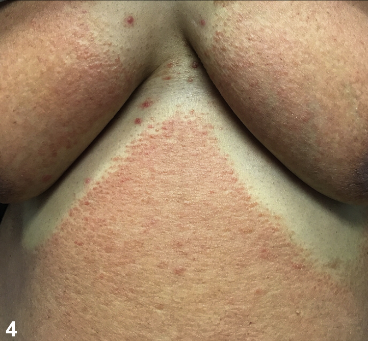
A 48-year-old Jamaican woman presented with several months of pruritus and a diffuse, erythematous scaly rash on the face, trunk, and extremities. At this time, there was no sparing of the folds. She was preemptively treated with triamcinolone 0.1% ointment and oral prednisone, 10 mg, for atopic dermatitis or contact dermatitis with minimal improvement. Laboratory values were significant for lymphocytosis and absolute eosinophilia. Biopsy found an atypical lymphoid infiltrate in the papillary dermis and epidermis (Fig 1). Nine months after initial presentation, the rash transformed into perifollicular red-brown papules that spared body folds and double-coverage areas (Fig 2, Fig 3, Fig 4). No palmoplantar hyperkeratosis or nail changes were noted. Repeat biopsy findings were again significant for an atypical lymphoid infiltrate in the papillary dermis and epidermis. Human T-cell lymphotropic virus-1 results were negative.
Fig 1.

Fig 2.

Fig 3.

Fig 4.
Question 1: What type of rash is manifested by this patient?
-
A.
Allergic contact dermatitis (ACD)
-
B.
Papuloerythroderma of Ofuji (PEO)
-
C.
Pityriasis rubra pilaris (PRP)
-
D.
Drug eruption
-
E.
Psoriasis
-
A.
ACD – Incorrect. Although contact dermatitis can spare skin folds, it is unlikely in a patient with no history of atopy. Furthermore, chronic ACD will have more scale and lichenification.1
-
B.
PEO – Correct. First described by Ofuji in 1984, PEO is characterized by a pruritic, diffuse erythroderma-like eruption formed by coalescence of flat-topped, red-to-brown papules with a cobblestone-like appearance.2 The eruption typically spares skin folds and creases, the “deck chair” sign. Other features of PEO seen in patients includes axillary and inguinal lymphadenopathy and palmoplantar hyperkeratosis and or nail bed infarction.3
-
C.
PRP – Incorrect. Although patients with PRP have follicular hyperaccentuation, such as our patient, the rash typically has a predominant scale and distinct orange-red color. The rash will also classically contain islands of sparing but will not spare the skin folds.1
-
D.
Drug eruption – Incorrect. Although drug eruptions have nonspecific rashes, they typically progress and resolve quickly. Our patient had a chronic rash and no history of new or changing medications.1
-
E.
Psoriasis – Incorrect. Although psoriasis is the most common underlying cause of erythroderma, it rarely spares the skin folds. Additionally, patients typically have a history of psoriasis, facial sparing, and nail changes, such as oil-drop spots and nail pits.1
Question 2: What cancer is most often associated with this rash?
-
A.
Cutaneous T-cell lymphoma (CTCL)
-
B.
Kidney cancer
-
C.
Gastric cancer
-
D.
Non-Hodgkin lymphoma
-
E.
Bladder cancer
-
A.
CTCL – Correct. At least 10 cases of CTCL manifesting as PEO have been reported in the literature, with an additional 8 to 10 cases beginning as PEO and later transforming into CTCL.3 Given that CTCL is difficult to diagnose because of variable presentation and histologic findings, patients with PEO should be monitored for transformation many years after their original diagnosis.3 Martinez-Barranca et al4 described a case of PEO that transformed into CTCL 7 years after the original diagnosis. A change in the rash size, texture, or pruritus should prompt an new biopsy to look for CTCL.3
-
B.
Kidney cancer – Incorrect. Kidney cancer was associated with 1 case of PEO in Japan, but no association has been made in the United States.3
-
C.
Gastric cancer – Incorrect. Gastric cancer is the second most common cancer associated with PEO. Most of these cases have been reported in older, Asian men.3 Given our patient is a Jamaican woman, this cancer is less likely.
-
D.
Non-Hodgkin lymphoma – Incorrect. Hematologic neoplasms are the third most common cancer associated with PEO; however, there are less than 5 cases of NHL associated with PEO in the literature.3
-
E.
Bladder cancer – Incorrect. One case of bladder cancer associated with PEO has been reported in Japan; however, no association has been made in the United States.3
Question 3: What histopathologic findings are most commonly associated with this rash?
-
A.
Hyperkeratosis and parakeratosis with a perivascular infiltrate of lymphocytes and neutrophils
-
B.
Foci of orthokeratosis alternating with parakeratosis in vertical and horizontal directions
-
C.
Dermal infiltration of lymphocytes, histiocytes, and eosinophils
-
D.
Spongiosis, acanthosis, and an eosinophilic infiltrate
-
E.
Nonspecific histopathologic changes, sometimes with eosinophils
-
A.
Hyperkeratosis and parakeratosis with a perivascular infiltrate of lymphocytes and neutrophils – Incorrect. This would be the histopathology expected in a biopsy of psoriasis.5
-
B.
Foci of orthokeratosis alternating with parakeratosis in vertical and horizontal directions – Incorrect. This is the histopathology of PRP.5
-
C.
Dermal infiltration of lymphocytes, histiocytes, and eosinophils – Correct. Patients with PEO classically have dermal infiltrates consisting of lymphocytes (100%), histiocytes (87%), and eosinophils (82%).3 Neutrophils and giant cells are present in less than 5% of cases.3 In patients with mycosis fungoides manifesting as PEO, the infiltrate will have a high percentage of atypical lymphocytes, as seen in our patient.3
-
D.
Spongiosis, acanthosis, and an eosinophilic infiltrate – Incorrect. This histopathology is usually seen in either atopic or contact dermatitis.5 The presence of eosinophils is more commonly seen in atopic dermatitis.
-
E.
Nonspecific histopathologic changes, sometimes with eosinophils – Incorrect. Drug eruptions will have nonspecific histopathology on biopsy.5 Eosinophils may be present in some cases. Biopsy will not help diagnose a drug eruption; however, it may help rule out other rashes that would have characteristic histopathologic findings.
Footnotes
Funding sources: None.
Conflicts of interest: None disclosed.
References
- 1.Mistry N., Gupta A., Alavi A. A Review of the diagnosis and management of erythroderma (generalized red skin) Adv Skin Wound Care. 2015;28(5):228–236. doi: 10.1097/01.ASW.0000463573.40637.73. [DOI] [PubMed] [Google Scholar]
- 2.Ofuji S., Furukawa F., Miyachi Y. Papuloerythroderma. Dermatologica. 1984;169:125–130. doi: 10.1159/000249586. [DOI] [PubMed] [Google Scholar]
- 3.Torchia D., Miteva M., Hu S. Papuloerythroderma 2009: two new cases and systematic review of the worldwide literature 25 years after its identification by Ofuji, et al. Dermatology (Basel) 2010;220(4):311–320. doi: 10.1159/000301915. [DOI] [PubMed] [Google Scholar]
- 4.Martinez-Barranca M.L., Munoz-Perez M.A., Garcia-Morales I. Ofuji Papuloerythroderma evolving to cutaneous T-cell lymphoma. J Eur Acad Dermatol Venereol. 2005;19(1):104–106. doi: 10.1111/j.1468-3083.2004.01081.x. [DOI] [PubMed] [Google Scholar]
- 5.Bolognia J., Jorizzo J., Schaffer J. 3rd ed. Elsevier Limited; London: 2012. Dermatology. [Google Scholar]