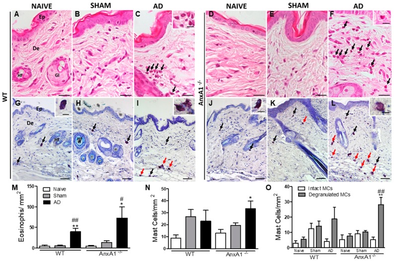
Figure 2.
Histopathology of the skin. (A,B,D,E,G,H,J,K): WT and AnxA1-/- control skins (Naïve and Sham). Atopic dermatitis (AD) characterized by intense influx of eosinophils (arrows) (C,F) and intact and degranulated mast cells (black and red arrows, respectively; I, L) into the dermis. Insets: detail of eosinophils (C,F), intact (G,J) and degranulated mast cells (I,L). Epidermis (Ep). Dermis (De). Hair follicle (Hf). Sebaceous gland (Gl). Stain: Hematoxylin-eosin (A–F) and toluidine blue (G–L). Bars: 20 μm (A–F), 50 μm (G–L), insets: 10 μm. (M): Quantification of eosinophils in the skin. (N): Quantification of the total number of mast cells in the skin. Data represent mean ± SEM of the number of cells per mm2 of the experimental groups (n = 3–5 animals/group). * p < 0.05; ** p < 0.01 vs. Naïve of the respective genotype; # p < 0.05; ## p < 0.01 vs. Sham of respective genotype (ANOVA, Bonferroni post test). (O): Quantification of intact and degranulated mast cells. ** p < 0.01 vs. intact mast cells of the AnxA1-/- AD group, ## p < 0.01 vs. degranulated mast cells from the AnxA1-/- Naïve and Sham groups (ANOVA, Bonferroni post test).

