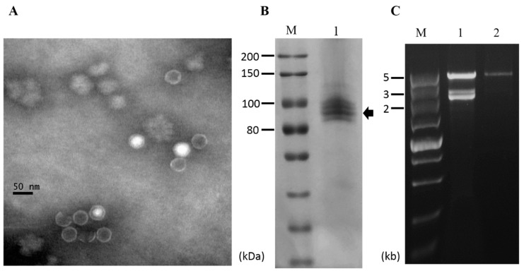
Figure 6.
Virus particles of NoRV2. (A) Electron micrograph of viral particles. The viral particles were uranyl acetate-stained and observed on a transmission electron microscope. The scale bar indicates 50 nm. (B) Protein components of virus particles analyzed by an 8% SDS-PAGE gel-electrophoresis; Lane M, Protein marker (200 kDa ladder, TransGen); Lane 1, the viral particles purified from the N. oryzae strain CS-7.5-4. (C) dsRNA extraction from the viral particles. The extracted nucleic acids were treated with DNase 1 and S1 nuclease as described above and electrophoresed in a 1% agarose gel. Lane M, DNA marker (5kb ladder, TaKaRa). Lane 1, dsRNA isolated from mycelia of the CS-7.5-4; Lane 2, dsRNA isolated from viral particles.

