Overview
Introduction
We describe a minimally invasive surgical technique for harvest of a quadriceps tendon autograft that reliably produces a graft suitable for anterior cruciate ligament (ACL) reconstruction while minimizing morbidity and complications classically associated with alternative autograft choices.
Step 1: Positioning
Position the patient supine on the operating room table.
Step 2: Marking of Landmarks
Palpate and mark soft-tissue and osseous landmarks on the involved extremity.
Step 3: Subcutaneous Dissection
Perform the incision and subcutaneous dissection.
Step 4: Graft Harvest
Harvest the graft.
Step 5: Graft Preparation
Prepare the graft.
Step 6: Closure
If a partial-thickness graft is harvested, no deep closure is needed.
Results
Since September of 2011, the quadriceps tendon has been our autograft of choice for ACL reconstruction.
Introduction
We describe a minimally invasive surgical technique for harvest of a quadriceps tendon autograft that reliably produces a graft suitable for anterior cruciate ligament (ACL) reconstruction while minimizing morbidity and complications classically associated with alternative autograft choices.
Recently there has been a surge of interest, biomechanical research, and reports of clinical outcomes regarding the quadriceps tendon and its utility as a graft option for ACL reconstruction. Advantages of quadriceps tendon autografts include excellent clinical results with low donor-site morbidity1-9, ease of harvest, and excellent biomechanical properties10-12. The quadriceps tendon is a versatile autograft choice that can be harvested with or without a bone block3,4,7,8,13-16; can be used for single or double-bundle reconstructions3,9,17,18; and can be employed for anatomic18, transtibial3,16, or all-inside reconstructions19.
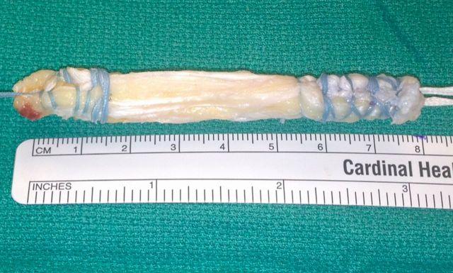
Traditionally, quadriceps tendon harvests have been performed through larger incisions, which allow visualization of the entire tendon. Our described technique is reproducible and consistently yields all-soft-tissue grafts of 9 to 10 mm in width, 6 to 7 mm deep, and 7 to 8 cm in length; longer grafts can be obtained with a patellar bone-block harvest. We demonstrate how this harvest is performed through a small (1.5-cm) incision.
Step 1: Positioning
Position the patient supine on the operating room table.
Position the patient supine on the operating room table, and perform an examination after successful general anesthesia has been administered.
If you wish to use a tourniquet, apply it to the involved extremity (but do not inflate it) and place the extremity into a circumferential arthroscopic leg-holder with the knee resting at 90° of flexion. Use of the tourniquet is optional but is recommended as it will assist with visualization through the small incision. Confirm the ability to hyperflex the knee to >120°.
Place the contralateral leg into a well-padded lithotomy leg-holder.
Prepare and drape the involved extremity in a sterile manner.
Perform a diagnostic arthroscopy either prior to or after quadriceps tendon graft harvest, depending on your preference.
Step 2: Marking of Landmarks
Palpate and mark soft-tissue and osseous landmarks on the involved extremity.
With the knee at 90° of flexion, mark the superior pole of the patella.
Mark the medial border of the vastus medialis obliquus and the lateral border of the patellar tendon above the patella.
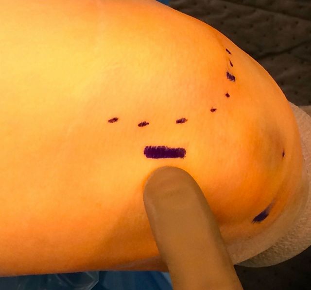
Then mark a longitudinal incision starting 0.5 cm above the patella at a point slightly lateral to the midpoint between the vastus medialis obliquus and the lateral quadriceps tendon extending proximally 1 to 2 cm (Fig. 1 and Video 1).
Fig. 1.
Preoperative marking of the vastus medialis obliquus and the planned incision 0.5 cm above the superior pole of the patella.
Video 1.
Landmarks and the skin incision are marked on the knee.
Step 3: Subcutaneous Dissection
Perform the incision and subcutaneous dissection.
If you wish to use a tourniquet, exsanguinate the extremity and inflate the tourniquet at this time.
Inject the planned incision site and subcutaneous tissue with local anesthetic with epinephrine solution.
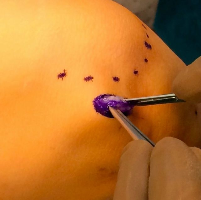
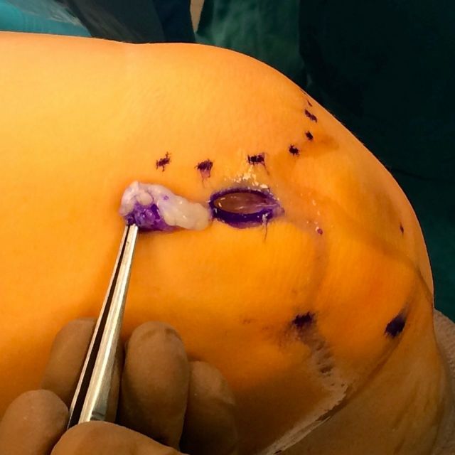
Make a 1.5-cm longitudinal incision and ellipse out the subcutaneous fat beneath the skin. This is key to allow for adequate visualization through the small incision (Figs. 2-A and 2-B).
Identify the fascia over the quadriceps tendon and excise it with Metzenbaum scissors or a scalpel.
Use a gauze sponge over a Key elevator to dissect bluntly over the anterior portion of the tendon both proximally and distally over the patella (Video 2).
Use a finger to ensure that all adhesions have been released proximally and distally.
Place an Army-Navy retractor into the proximal apex of the incision, and use the arthroscope (with the water off, and looking down at the tendon) to locate the vastus medialis obliquus, lateral tendon border, and proximal-most visualized portion of the rectus femoris tendon.
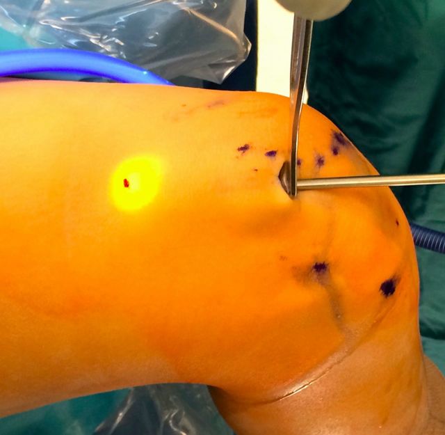
Rotate the arthroscope so that the light is pointing anteriorly (away from the tendon), and mark this point of light seen through the skin (Fig. 3).
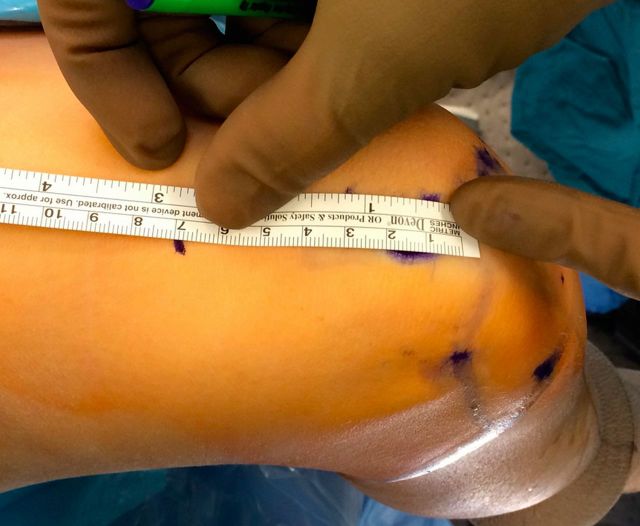
Measure this mark from the superior pole of the patella (Fig. 4 and Video 3). If an 8-cm graft is needed, this mark should be 8 cm from the superior pole.
Figs. 2-A and 2-B Ellipse and excision of subcutaneous fat is critical for visualization.
Fig. 2-A.
Before fat is excised.
Fig. 2-B.
After fat is excised.
Fig. 3.
The arthroscope is used to visualize the proximal rectus musculotendinous junction. The arthroscope is turned to look away from the tendon, and a mark is placed on the skin at the point of transillumination.
Fig. 4.
A second mark is made in line with this point and the superior pole of the patella at the length of planned graft harvest.
Video 2.
The skin incision and subcutaneous dissection.
Video 3.
The arthroscope is used to subcutaneously identify the vastus medialis obliquus and rectus femoris tendons.
Step 4: Graft Harvest
Harvest the graft.
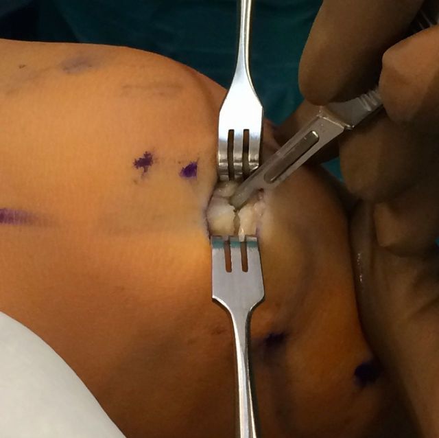
While using two Senn retractors to protect the skin, insert a double-bladed knife and incise the tendon longitudinally, using the previously made rectus femoris tendon marking for directional reference. Additionally, the length of the graft can be measured off of the knife handle (Video 4).
Continue the incision distally until the superior pole of the patella is encountered.
Slightly extend the knee and dissect the distal part of the quadriceps tendon off of the patella, taking care to taper the distal end of the tendon slightly to minimize the need for subsequent graft trimming, as 1 mm of thickness will be gained with later suture addition (Fig. 5).
We prefer to harvest a partial-thickness graft, although a full-thickness graft can be taken if needed. To harvest a partial-thickness graft, use a knife and/or Metzenbaum scissors to dissect proximally (Video 5).
If fat is encountered, do not dissect deeper as there is a risk of violating the capsule. Alternatively, if bone is desired use a saw to remove a bone block from the superior pole of the patella, taking care to avoid overpenetration into the articular surface.
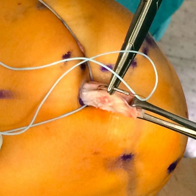
Once 3 cm of graft is freed distally, use a whipstitch or FiberLoop (Arthrex, Naples, Florida) to gain control of the distal part of the tendon, starting about 1.5 to 2 cm proximal to the distal extent of the graft. Grasping the distal tip of the tendon with an Allis clamp can aid with suture placement (Fig. 6 and Video 6).
While applying tension to the sutures, carry out additional dissection proximally with Metzenbaum scissors if necessary.
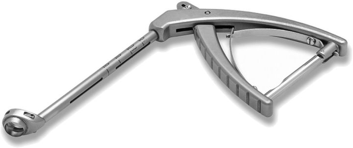
Use a closed-ended Arthrex graft gun to strip proximally until the desired length of graft is freed. The graft length is measured off of the graft gun.
Push the graft gun proximally and close the handle, cutting the graft proximally (Fig. 7 and Video 7)
After the graft is delivered from the wound, bring it to the back table for preparation (Fig. 8).
Fig. 5.
The quadriceps tendon is dissected off the superior pole of the patella.
Fig. 6.
Once 3 cm of graft has been dissected, the distal tip is grabbed with an Allis clamp, and a FiberLoop or whipstitch is used to gain control of the tendon.
Fig. 7.
The Arthrex graft gun with a combined tendon stripper and built-in tendon cutter. Markings on the shaft allow measurement of graft length prior to cutting.
Fig. 8.
Harvested graft after preparation.
Video 4.
The double-bladed knife is used to incise the quadriceps tendon in line with the previously identified rectus femoris tendon.
Video 5.
The tendon is dissected off the superior part of the patella.
Video 6.
A stitch is used to gain control of the distal part of the tendon.
Video 7.
The graft gun is used to strip the tendon proximally and cut the tendon at the desired graft length.
Step 5: Graft Preparation
Prepare the graft.
There are several methods of fixation for both the femoral and the tibial side.
For the femoral side, we prefer a cortical button, which is sewn to the graft with FiberWire (Arthrex) suture. Sew a second whipstitch or FiberLoop (Arthrex) to the other side of the graft.
We prefer to tie the tibial side over a screw and washer.
Step 6: Closure
If a partial-thickness graft is harvested, no deep closure is needed.
The arthroscope can be reintroduced into the wound to ensure that a partial-thickness graft was harvested (Video 8). If a full-thickness graft was harvested, either intentionally or unintentionally, close the capsular layer with absorbable suture to prevent fluid extravasation.
After irrigation, close the skin with absorbable suture and skin glue.
Video 8.
The arthroscope can be reintroduced into the harvest site to confirm partial-thickness harvest or identify capsular violation.
Results
Since September of 2011, the quadriceps tendon has been our autograft of choice for ACL reconstruction. We have prospectively collected data on 201 quadriceps tendon autograft ACL reconstructions (unpublished data), including primary (n = 181) and revision (n = 20) reconstructions and ACL reconstructions in elite athletes (Video 9).
We have seen no early failures with this surgical technique. Four patients have required revision reconstruction. All reinjuries requiring reconstruction were a result of sports or trauma. The only failure before six months occurred in a college athlete at five months postoperatively, after he jumped off several steps while running to catch a bus.
Video 9.
Interview with a collegiate soccer player who recently had two ACL reconstructions with bone-tendon-bone graft in one leg and a quadriceps tendon graft in the other.
What to Watch For
Indications
Primary ACL reconstruction.
Revision ACL reconstruction.
ACL reconstruction in a skeletally immature patient.
Contraindications
History of quadriceps tendon injury or surgery.
Preexisting quadriceps weakness or neuromuscular deficit.
Pitfalls & Challenges
Inadvertent capsular violation or full-thickness harvest.
If a large portion of the capsule is excised, deep closure can be challenging.
Failure to taper the graft end, necessitating graft trimming or a larger tunnel.
Clinical Comments
We prefer to use a free-tendon, all-soft-tissue graft because sufficient graft length can be reliably harvested without bone, and complications and morbidity with this technique are minimized with all-soft-tissue harvest. Additionally, harvesting the graft without bone is faster and technically easier than harvesting it with bone.
In our experience after a short learning curve, the quadriceps tendon harvest via this technique has been substantially faster than our bone-patellar tendon-bone or hamstring harvests.
We found no early failures related to suspensory fixation and all-soft-tissue grafts in our clinical follow-up.
Footnotes
Based on an original article: Arthroscopy. 2009 Dec;25(12):1408-14.
Disclosure: None of the authors received payments or services, either directly or indirectly (i.e., via his or her institution), from a third party in support of any aspect of this work. One or more of the authors, or his or her institution, has had a financial relationship, in the thirty-six months prior to submission of this work, with an entity in the biomedical arena that could be perceived to influence or have the potential to influence what is written in this work. In addition, one or more of the authors has a patent or patents, planned, pending, or issued, that is broadly relevant to the work. No author has had any other relationships, or has engaged in any other activities, that could be perceived to influence or have the potential to influence what is written in this work. The complete Disclosures of Potential Conflicts of Interest submitted by authors are always provided with the online version of the article.
References
- 1.Lee S, Seong SC, Jo H, Park YK, Lee MC. Outcome of anterior cruciate ligament reconstruction using quadriceps tendon autograft. Arthroscopy. 2004. October;20(8):795-802. [DOI] [PubMed] [Google Scholar]
- 2.Schulz AP, Lange V, Gille J, Voigt C, Fröhlich S, Stuhr M, Jürgens C. Anterior cruciate ligament reconstruction using bone plug-free quadriceps tendon autograft: intermediate-term clinical outcome after 24-36 months. Open Access J Sports Med. 2013;4:243-9. Epub 2013 Nov 19. [DOI] [PMC free article] [PubMed] [Google Scholar]
- 3.Geib TM, Shelton WR, Phelps RA, Clark L. Anterior cruciate ligament reconstruction using quadriceps tendon autograft: intermediate-term outcome. Arthroscopy. 2009. December;25(12):1408-14. [DOI] [PubMed] [Google Scholar]
- 4.Theut PC, Fulkerson JP, Armour EF, Joseph M. Anterior cruciate ligament reconstruction utilizing central quadriceps free tendon. Orthop Clin North Am. 2003. January;34(1):31-9. [DOI] [PubMed] [Google Scholar]
- 5.Stäubli HU. The quadriceps tendon-patellar bone construct for ACL reconstruction. Knieinstabilität und Knorpelschaden. 1998; p 129-39.
- 6.DeAngelis JP, Fulkerson JP. Quadriceps tendon—a reliable alternative for reconstruction of the anterior cruciate ligament. Clin Sports Med. 2007. October;26(4):587-96. [DOI] [PubMed] [Google Scholar]
- 7.Han HS, Seong SC, Lee S, Lee MC. Anterior cruciate ligament reconstruction: quadriceps versus patellar autograft. Clin Orthop Relat Res. 2008. January;466(1):198-204. Epub 2008 Jan 3. [DOI] [PMC free article] [PubMed] [Google Scholar]
- 8.Lee S, Seong SC, Jo CH, Han HS, An JH, Lee MC. Anterior cruciate ligament reconstruction with use of autologous quadriceps tendon graft. J Bone Joint Surg Am. 2007. October;89(Suppl 3):116-26. [DOI] [PubMed] [Google Scholar]
- 9.Shelton WR, Fagan BC. Autografts commonly used in anterior cruciate ligament reconstruction. J Am Acad Orthop Surg. 2011. May;19(5):259-64. [DOI] [PubMed] [Google Scholar]
- 10.Sasaki N, Farraro KF, Kim KE, Woo SLY. Biomechanical evaluation of the quadriceps tendon autograft for anterior cruciate ligament reconstruction: a cadaveric study. Am J Sports Med. 2014. March;42(3):723-30. Epub 2014 Jan 8. [DOI] [PMC free article] [PubMed] [Google Scholar]
- 11.Harris NL, Smith DA, Lamoreaux L, Purnell M. Central quadriceps tendon for anterior cruciate ligament reconstruction. Part I: Morphometric and biomechanical evaluation. Am J Sports Med. 1997. Jan-Feb;25(1):23-8. [DOI] [PubMed] [Google Scholar]
- 12.Stäubli HU, Schatzmann L, Brunner P, Rincón L, Nolte LP. Mechanical tensile properties of the quadriceps tendon and patellar ligament in young adults. Am J Sports Med. 1999. Jan-Feb;27(1):27-34. [DOI] [PubMed] [Google Scholar]
- 13.Joseph M, Fulkerson J, Nissen C, Sheehan TJ. Short-term recovery after anterior cruciate ligament reconstruction: a prospective comparison of three autografts. Orthopedics. 2006. March;29(3):243-8. [DOI] [PubMed] [Google Scholar]
- 14.Gorschewsky O, Klakow A, Pütz A, Mahn H, Neumann W. Clinical comparison of the autologous quadriceps tendon (BQT) and the autologous patella tendon (BPTB) for the reconstruction of the anterior cruciate ligament. Knee Surg Sports Traumatol Arthrosc. 2007. November;15(11):1284-92. Epub 2007 Aug 25. [DOI] [PubMed] [Google Scholar]
- 15.Chen CH, Chuang TY, Wang KC, Chen WJ, Shih CH. Arthroscopic anterior cruciate ligament reconstruction with quadriceps tendon autograft: clinical outcome in 4-7 years. Knee Surg Sports Traumatol Arthrosc. 2006. November;14(11):1077-85. Epub 2006 Jun 24. [DOI] [PubMed] [Google Scholar]
- 16.Kohl S, Stutz C, Decker S, Ziebarth K, Slongo T, Ahmad SS, Kohlhof H, Eggli S, Zumstein M, Evangelopoulos DS. Mid-term results of transphyseal anterior cruciate ligament reconstruction in children and adolescents. Knee. 2014. January;21(1):80-5. Epub 2013 Aug 21. [DOI] [PubMed] [Google Scholar]
- 17.Kim SJ, Jung KA, Song DH. Arthroscopic double-bundle anterior cruciate ligament reconstruction using autogenous quadriceps tendon. Arthroscopy. 2006. July;22(7):797.e1-5. [DOI] [PubMed] [Google Scholar]
- 18.Rabuck SJ, Musahl V, Fu FH, West RV. Anatomic anterior cruciate ligament reconstruction with quadriceps tendon autograft. Clin Sports Med. 2013. January;32(1):155-64. Epub 2012 Oct 13. [DOI] [PubMed] [Google Scholar]
- 19.Leitman EH, Morgan CD, Grawl DM. Quadriceps tendon anterior cruciate ligament reconstruction using the all-inside technique. Oper Tech Sports Med. 1999. October;7(4):179-88. [Google Scholar]


