Abstract
Objective:
To evaluate the safety and effectiveness of NOM (non-operative management) in the treatment of blunt liver trauma, following a standardized treatment protocol.
Methods:
All the hemodynamically stable patients with computed tomography (CT) diagnosis of blunt liver trauma underwent NOM. It included strict clinical and laboratory observation, 48-72h contrast enhanced ultrasonography (CEUS) or CT follow-up, a primary angioembolization in case of admission CT evidence of vascular injuries and a secondary angioembolization in presence of vascular injuries signs at follow-up CEUS.
Results:
181 patients (85.4%) [55 (30.4%) women and 126 (69.6%) men, median age 39 (range 14–71)] were included. Of these, 63 patients (34.8%) had grade I, 48 patients (26.5%) grade II, 39 patients (21.5%) grade III, 21 patients (11.6%) grade IV and 10 patients (5.5%) grade V liver injuries. The overall success rate of NOM was 96.7% (175/181). There was not significant difference in the success rate between the patients with different liver injuries grade. Morbidity rate was 7.4% (13/175). Major complications (2 bilomas, 1 liver hematoma and 2 liver abscesses) were successfully treated by CEUS or CT guided drainage. Eighteen (18/181) patients (9.9%) underwent angioembolization with successful results.
Conclusion:
Non-operative management of blunt liver trauma represents a safe and effective treatment for both minor and severe injuries, achieving an high success rate and an acceptable morbidity rate. The angiographic study with embolization, although required only in selected cases of vascular injuries, represents a fundamental therapeutic option in a significant percentage of patients.
Key Words: Hepatic trauma, Liver injury, Blunt trauma, Non-operative management, Angioembolization
Introduction
Trauma is the first cause of death and a significant cause of morbidity in people younger than 40 years of age in western countries, representing, therefore, a relevant clinical problem [1,2]. The liver and the spleen, despite relatively shielded by the inferior ribs, represent commonly injured organs during abdominal blunt trauma, accounting for about two-thirds of all visceral injuries [3-6]. Until three decades ago, the surgical treatment represented the most common therapeutic strategy for blunt trauma of abdominal parenchymatous organs [7-9]. Gradually, due to the advanced accuracy of diagnostic imaging, the improvement of interventional radiology techniques and the technical progress in intensive care, the conservative approach was encouraged and examined, showing satisfactory results [10-16]. At present, the nonoperative management (NOM) is the adopted strategy in hemodynamically stable patients with blunt abdominal trauma, representing the mainstay of treatment for minor splenic injuries (grades I-II according to the American Association for the Surgery of Trauma-AAST) [17], the first-line approach for severe splenic lesions (AAST grades III-V) and the standard of care for both minor and severe liver injuries [17-22].
Basing on the encouraging results reported in literature and on our own hospital resources, we developed and adopted, from January 2009, a treatment protocol for hemodynamically stable patients with blunt traumatic injuries of abdominal parenchymatous organs, in order to standardize the non-operative management. The results of this approach in treatment of splenic lesions were previously published [23]. In this study we aimed to evaluate the results of this management strategy in the treatment of blunt liver trauma, focusing on its safety, efficacy and complications.
Materials and Methods
Study population
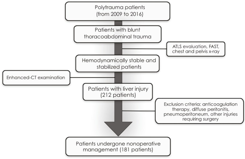
From January 2009 to January 2016, all the polytraumatized patients referring to the Emergency Department of the “A. Cardarelli” Hospital in Naples (Italy), were prospectively inserted into a database, including the demographic characteristics (age, gender, mechanism of injury), the Revised Trauma Score (RTS), the Glasgow Coma scale (GCS), the Injuries Severity Score (ISS) [24] and the results of clinical evaluation, diagnostic imaging and treatment strategy. Out of these, all the patients with blunt thoracoabdominal trauma were considered for the enrollment in this study. These last, after clinical evaluation according to the Advanced Trauma Life Support (ATLS®), underwent initial instrumental study with FAST (Focused Assessment with Sonography for Trauma) and chest and pelvis x-ray [25]. Then, the hemodynamically stable patients (systolic blood pressure ≥ 90 mmHg, heart rate < 100 bpm) and the hemodynamically stabilized patients (returned to normal vital signs after 1000 ml crystalloid infusion) were investigated with a total body computed tomography (CT) examination [26-30].
Basing on the clinical and instrumental evaluation, among all the enrolled patients, were included in this study the adult subjects (14 years or more), with initial hemodynamic stability or good response to 1000 ml crystalloid prompt infusion and CT evidence of AAST I-V grade liver injury. The patients with significant associated hemoperitoneum at CT (defined as intra-abdominal blood extended to at least two abdominal quadrants) were also included. All the patients receiving systemic anticoagulation, with diffuse peritonitis and with associated bowel injuries (pneumoperitoneum) or any other concomitant thoracoabdominal lesions requiring surgical procedure, were excluded. All the patients meeting these selection criteria were admitted to the Trauma Center and underwent NOM (Figure 1).
Fig. 1.
Flow-chart of the study design
ATLS: Advanced Trauma Life Support; FAST: Focused Assessment Sonography for Trauma; CT: Computer Tomogr a phy
Study protocol
The treatment protocol, previously described elsewhere [21], included arterial blood gas measurements every 12 hours, complete blood cell counts every 6 hours (until two stable hemoglobin examinations were obtained), an early contrast enhanced ultrasound (CEUS) or CT follow-up (48-72 hours after injury), a primary angioembolization in case of admission CT diagnosis of vascular injuries (contrast extravasation, pseudoaneurysm, arteriovenous fistula formation, vessel truncation), a secondary angioembolization in presence of active bleeding at follow-up CEUS or CT and a second look angiography in case of persistent falling of hematic hemoglobin level despite the primary or the secondary embolization. Failure of NOM was defined as the need of surgical abdominal exploration for hemodynamic instability, progressive fall of hemoglobin level despite two angioembolizations, clinical signs of diffuse peritonitis, missed abdominal injuries requiring surgery. NOM was considered successful when the patient was discharged without undergoing any abdominal surgical procedure. This study was not supported by any commercial company and had the approval of the institutional review board and of the ethical committee of our Institution; all the patients gave their informed written consent to take part in the study.
Statistical analysis
Statistical analysis was carried out using the program InStat Graph-Pad Prism® 5 (San Diego, California, USA). Values are expressed as means ± standard deviation (SD) or medians (range), according with distribution of data. Continuous data were compared between each group using the Mann-Whitney U-test. Prevalence data were compared between groups using the Fisher’s exact test. A probability value of less than 0.05 was considered significant.
Results
Study population
Among all the patients with blunt trauma observed during the study period, 212 were admitted with diagnosis of liver injury. Of these, 31 patients (14.6%) underwent operative management. The indications for surgery were the presence of persisting hemodynamic instability despite fluid therapy in 64.5% (20/31), diffuse peritonitis in 22.5% (7/31) and coexisting thoracoabdominal injuries requiring surgical procedure in 12.9% (4/31) of cases. Among the hemodynamically unstable patients, 7 (35%) were under systemic anticoagulation therapy and among the patients with diffuse peritonitis and hemoperitoneum, 4 (57.1%) had chronic liver disease. Out of patients with blunt liver trauma, 181 (85.4%) [55 (30.4%) women and 126 (69.6%) men, median age 39 (range 14–71)] satisfied selection criteria, were included in the study and constituted the object of analysis. The demographic characteristics and trauma severity of the included patients are showed in Table 1. The predominant cause of injury was vehicle traffic accident (56.9%), followed by aggression (20.9%), falling from height (12.1%) and pedestrian struck (9.9%).
Table 1.
Demographic and injury characteristics of the enrolled patients
| Features | |
|---|---|
| Sex (male) | 69.6% (126/181) |
| Age | 39 (12.3) |
| Ethnicity | Caucasian 93.3% (169/181) |
| African 6.6% (12/181) | |
| ISS a | 16 (3.2) |
| RTS b | 5.8 (1.2) |
| AAST c injury grade | 2 (1.8) |
| Associated injuries | 86.8% (157/181) |
| Mechanism of injury | Vehicle traffic accident (56.9%) |
| Aggression (20.9%) | |
| Pedestrian Struck (9.9%) | |
ISS: Injuries Severity Score;
RTS: Revised Trauma Score;
AAST: American Association for the Surgery of Trauma, Data are given as means with standard deviation in parenthesis, or percentages with row data in parenthesis.
According with AAST organ injury scale, 63 patients (34.8%) had grade I, 48 patients (26.5%) grade II, 39 patients (21.5%) grade III, 21 patients (11.6%) grade IV and 10 patients (5.5%) grade V injuries. Twenty-four patients (24/181 = 13.2%) presented isolated liver trauma whereas the remaining 157 patients (157/181 = 86.8%) showed multiple injuries. Among these last patients, the more frequent concomitant lesions were rib fractures, observed in 70.1% (127/181) of cases, followed by long bones fractures in 34.8% (63/181), head or maxillofacial injuries in 14.3% (26/181), hemothorax in 11.1% (24/181), renal injury in 8.8% (16/181), pelvic fractures in 9.3% (17/181), vertebral fractures in 6.6% (12/181), adrenal injury in 3.3% (6/181), splenic injury in 2.2% (4/181), small bowel injury and mesothelium hematoma in 1.1% (2/69) of cases.
Clinical outcome
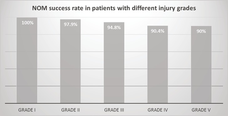
One hundred seventy-six patients (176/181 = 97.2%) [63 with grade I, 48 grade II, 38 grade III, 19 grade IV and 8 grade V injuries (mean injury grade = 2.21 ± 1.17)] didn’t show vascular injuries signs on admission CT and underwent, according with study protocol, observation with serial clinical, radiological and laboratory examination. Out of these, 1 patient (1/177 = 0.56%) with grade IV injuries showed, during hospitalization, progressive fall of hemoglobin level and liver active bleeding at follow-up CEUS; consequently, he underwent selective embolization of hepatic artery branches, with successful results. Eleven patients (11/176 = 6.2%) showed follow-up sonographic or CT findings of contained vascular injuries represented, in 7 cases, by isolated peripheral hepatic artery pseudoaneurysms and in 4 cases by pseudoaneurisms associated with arteriovenous fistulas. All these patients underwent angiography and were successfully treated with embolization. The non-operative management failed in 6 cases (6/176 = 3.4%). The indications for surgery were diffuse peritonitis due to bile peritoneal irritation in 2 patients, diffuse peritonitis due to concomitant small bowel injury in 1 patient, intra-abdominal hypertension in 1 patient and concomitant splenic injuries requiring delayed surgery in 2 patients. Six patients (6/181 = 3.3%) [1 with grade III, 3 grade IV, and 2 grade V injuries (mean injury grade = 4.2 ± 0.83)] showed admission CT findings of vascular injuries and underwent primary angioembolization. The indications for embolization were pseudoaneurysm and active bleeding in 33.3% (2/6) and 66.6% (4/6) of cases, respectively. No NOM failure was observed in this patients group. The overall success rate of NOM was 96.7% (175/181). There was not significant difference in the success rate between the patients with different liver injuries grade: particularly, the success rate ranged from 100% (63/63) in patients with grade I to 90% (9/10) in patients with grade V liver injuries grade (p>0.05: Fisher’s exact test) (Figure 2). The median of blood transfusions was significantly different between patients with minor (AAST grade I-II) and severe (AAST grade III-V) trauma [0.5 (0-2) vs 2 (0-4): p < 0.0001; Mann Whitney U-test].
Fig. 2.
Non-operative management (NOM) success rate in patients with different injury grades
Data are given as percentage; p>0.05: Fisher’s exact test; NOM: Nonoperative management
The overall median hospital stay was 11 days (7-17). No mortality was observed. The median follow-up period in the included patients was 24 months (6-36). In the patients successfully treated with NOM the morbidity rate was 7.4% (13/175). Minor complications included 3 cases of pleural effusions, 3 cases of endobronchitis and 2 cases of bladder catheter-related bacteremia. Major complications were all liver related and included 2 cases of biloma, 1 case of liver hematoma and 2 cases of liver abscesses. These complications were successfully treated by ultrasound (US) or CT guided drainage and did not require surgery. All the patients with major complications showed severe liver injuries (AAST grades III-V).
Discussion
During the last century, the surgical strategy has been widely adopted for the treatment of abdominal parenchimatous organs injuries, whereas from the ’80s, the standard of care for blunt liver and splenic trauma in hemodynamically stable patients, gradually shifted from operative to non-operative management. The surgical treatment, previously considered mandatory, remains, to date, still indicated in unstable patients not responder to fluid resuscitation and in case of failure of NOM [31-33]. Although we employed for many years the nonoperative management for treatment of blunt abdominal trauma, we recently adopted a standardized treatment protocol for the management of splenic and liver injuries in hemodynamically stable patients, developed on the base of existing literature, our experience and our own hospital resources.
This approach included strict clinical and laboratoristic observations, early CEUS follow-up [27], a primary angioembolization in case of admission CT diagnosis of vascular injuries and a secondary angioembolization in presence of vascular injuries signs at follow-up CEUS. Recently, we evaluated the effect of this approach on the outcome of patients with blunt splenic lesions, showing no mortality or major complications, a 95% success rate and a considerable impact of interventional radiology since more than a quarter of patients needed angioembolization [23].
In this study we reported the results of this management strategy in the treatment of minor and severe blunt liver trauma. Our data show, first of all, that the non-operative management, performed according with our protocol, represents an effective treatment option for blunt liver trauma. Indeed the overall success rate was higher than 96% and among the cases of unsuccessful NOM, 50% was related to concomitant bowel and splenic lesions and only 33% was due to liver related causes. According with other Authors [15, 34], although the success rate was lower in the subjects with high grade injury, no significant difference in this parameter was found between patients with different injury grade, suggesting that, in hemodynamically stable patients the nonoperative management could be a feasible strategy both in minor and in high grade blunt liver trauma, achieving high success rate even in treatment of severe liver injuries. Interestingly, a recent retrospective study showed that, using an established treatment protocol for nonoperative management of severe hepatic injuries, encouraging results can be achieved even in hemodinamically unstable patients [35]. However, the role of NOM in unstable patients with blunt liver trauma is still questionable and further researches with larger series and prospective design are needed to clarify this issue. Based on our results, the conservative approach represented a safe management strategy with a low morbidity rate. The major complications included 2 cases of biloma, 1 case of liver hematoma and 2 cases of liver abscesses, did not require surgery and were successfully treated by US or CT guided drainage.Compared to our experience with splenic trauma [23], the indication for angiography and angioembolization was less frequent in our series. Indeed, 3.3% of patients underwent a primary embolization for evidence of pseudo-aneurysm and active bleeding at admission CT and 6.6% of patients underwent a secondary embolization for the presence of liver active bleeding signs or contained vascular lesions (isolated pseudoaneurisms or associated with arteriovenous fistulas) at follow-up CEUS or CT. Overall, in our study, the indication to angiography and subsequent embolization occurred in 9.9% of the included patients.
According with some Authors [36] and in contrast with others [37], our results seem to suggest that embolization, although required only in selected cases of vascular lesions, represents a pivotal diagnostic and therapeutic tool in a significant number of patients with blunt liver injuries.
However, the onset of vascular injuries in patients with blunt liver trauma was less frequent than in patients with blunt splenic trauma [23]. Although the reasons of this are not clear, it can be argued that one of them may lie in the different friability between the hepatic and splenic parenchima that may result in a different incidence of vascular lesions.
In subjects with blunt liver trauma, the angiographic study with embolization, although showing a minor impact if compared with splenic lesions, represents a fundamental therapeutic option in a significant percentage of patients with associated contained vascular injuries. This study is a prospective observational study and the main limitation is represented by the absence of a control group to compare the effectiveness of our treatment protocol with other therapeutic approaches. Moreover, in this study, only patients with hemodynamic stability were included. The future research perspectives may be focused on the evaluation of safety and efficacy of an established treatment protocol for nonoperative management of severe liver injuries both in hemodynamically stable and hemodynamically unstable patients.
In conclusion, the nonoperative management of blunt hepatic trauma, according to our protocol, represents a safe and effective therapeutic approach for both minor and severe injuries, achieving an high success rate even in treatment of high-grade liver lesions and showing a low and acceptable morbidity rate.
Acknowledgments:
There are no acknowledgments
Funding/Support:
This research was not funded by any agency in the public, commercial, or not-for-profit sectors
Funding/Support:
All the Authors declare to not have financial interests related to the material in the manuscript.
Research involving human participants and/or animals :
The study involves Human Participants and the ethical committee of our Institution approved the study protocol.
Informed consent:
All the patients gave informed written consent.
Conflict of Interest:
None declared.
References
- 1.Gaines BA. Intra-abdominal solid organ injury in children: diagnosis and treatment. J Trauma. 2009;67(2 Suppl):S135–9. doi: 10.1097/TA.0b013e3181adc17a. [DOI] [PubMed] [Google Scholar]
- 2.Sauaia A, Moore FA, Moore EE, Moser KS, Brennan R, Read RA, et al. Epidemiology of trauma deaths: a reassessment. J Trauma. 1995;38(2):185–93. doi: 10.1097/00005373-199502000-00006. [DOI] [PubMed] [Google Scholar]
- 3.Smith J, Caldwell E, D'Amours S, Jalaludin B, Sugrue M. Abdominal trauma: a disease in evolution. ANZ J Surg. 2005;75(9):790–4. doi: 10.1111/j.1445-2197.2005.03524.x. [DOI] [PubMed] [Google Scholar]
- 4.Costa G, Tierno SM, Tomassini F, Venturini L, Frezza B, Cancrini G, et al. The epidemiology and clinical evaluation of abdominal trauma An analysis of a multidisciplinary trauma registry. Ann Ital Chir. 2010;81(2):95–102. [PubMed] [Google Scholar]
- 5.Hancock GE, Farquharson AL. Management of splenic injury. J R Army Med Corps. 2012;158(4):288–98. doi: 10.1136/jramc-158-04-03. [DOI] [PubMed] [Google Scholar]
- 6.Dupuy DE, Raptopoulos V, Fink MP. Current concepts in splenic trauma. J Intensive Care Med. 1995;10(2):76–90. doi: 10.1177/088506669501000204. [DOI] [PubMed] [Google Scholar]
- 7.Shackford SR, Molin M. Management of splenic injuries. Surg Clin North Am. 1990;70(3):595–620. doi: 10.1016/s0039-6109(16)45132-7. [DOI] [PubMed] [Google Scholar]
- 8.Morrison JJ, Bramley KE, Rizzo AG. Liver trauma--operative management. J R Army Med Corps. 2011;157(2):136–44. doi: 10.1136/jramc-157-02-03. [DOI] [PubMed] [Google Scholar]
- 9.Polanco P, Leon S, Pineda J, Puyana JC, Ochoa JB, Alarcon L, et al. Hepatic resection in the management of complex injury to the liver. J Trauma. 2008;65(6):1264–9; discussion 9-70. doi: 10.1097/TA.0b013e3181904749. [DOI] [PubMed] [Google Scholar]
- 10.Yanar H, Ertekin C, Taviloglu K, Kabay B, Bakkaloglu H, Guloglu R. Nonoperative treatment of multiple intra-abdominal solid organ injury after blunt abdominal trauma. J Trauma. 2008;64(4):943–8. doi: 10.1097/TA.0b013e3180342023. [DOI] [PubMed] [Google Scholar]
- 11.Tataria M, Nance ML, Holmes JHt, Miller CC, 3rd , Mattix KD, Brown RL, et al. Pediatric blunt abdominal injury: age is irrelevant and delayed operation is not detrimental. J Trauma. 2007;63(3):608–14. doi: 10.1097/TA.0b013e318142d2c2. [DOI] [PubMed] [Google Scholar]
- 12.Cirocchi R, Corsi A, Castellani E, Barberini F, Renzi C, Cagini L, et al. Case series of non-operative management vs operative management of splenic injury after blunt trauma. Ulus Travma Acil Cerrahi Derg. 2014;20(2):91–6. doi: 10.5505/tjtes.2014.99442. [DOI] [PubMed] [Google Scholar]
- 13.Pachter HL, Guth AA, Hofstetter SR, Spencer FC. Changing patterns in the management of splenic trauma: the impact of nonoperative management. Ann Surg. 1998;227(5):708–17; discussion 17-9. doi: 10.1097/00000658-199805000-00011. [DOI] [PMC free article] [PubMed] [Google Scholar]
- 14.Hsieh TM, Cheng Tsai T, Liang JL, Che Lin C. Non-operative management attempted for selective high grade blunt hepatosplenic trauma is a feasible strategy. World J Emerg Surg. 2014;9(1) doi: 10.1186/1749-7922-9-51. [DOI] [PMC free article] [PubMed] [Google Scholar]
- 15.Hommes M, Navsaria PH, Schipper IB, Krige JE, Kahn D, Nicol AJ. Management of blunt liver trauma in 134 severely injured patients. Injury. 2015;46(5):837–42. doi: 10.1016/j.injury.2014.11.019. [DOI] [PubMed] [Google Scholar]
- 16.Ochiai T, Igari K, Yagi M, Ito H, Kumagai Y, Iida M, et al. Treatment strategy for blunt hepatic trauma: analysis of 183 consecutive cases. Hepatogastroenterology. 2011;58(109):1312–5. doi: 10.5754/hge11042. [DOI] [PubMed] [Google Scholar]
- 17.Moore EE, Cogbill TH, Jurkovich GJ, Shackford SR, Malangoni MA, Champion HR. Organ injury scaling: spleen and liver (1994 revision) J Trauma. 1995;38(3):323–4. doi: 10.1097/00005373-199503000-00001. [DOI] [PubMed] [Google Scholar]
- 18.Franklin GA, Casos SR. Current advances in the surgical approach to abdominal trauma. Injury. 2006;37(12):1143–56. doi: 10.1016/j.injury.2006.07.018. [DOI] [PubMed] [Google Scholar]
- 19.Cirocchi R, Boselli C, Corsi A, Farinella E, Listorti C, Trastulli S, et al. Is non-operative management safe and effective for all splenic blunt trauma? A systematic review. Crit Care. 2013;17(5):R185. doi: 10.1186/cc12868. [DOI] [PMC free article] [PubMed] [Google Scholar]
- 20.Swift C, Garner JP. Non-operative management of liver trauma. J R Army Med Corps. 2012;158(2):85–95. doi: 10.1136/jramc-158-02-04. [DOI] [PubMed] [Google Scholar]
- 21.She WH, Cheung TT, Dai WC, Tsang SH, Chan AC, Tong DK, et al. Outcome analysis of management of liver trauma: A 10-year experience at a trauma center. World J Hepatol. 2016;8(15):644–8. doi: 10.4254/wjh.v8.i15.644. [DOI] [PMC free article] [PubMed] [Google Scholar]
- 22.Bouras AF, Truant S, Pruvot FR. Management of blunt hepatic trauma. J Visc Surg. 2010;147(6):e351–8. doi: 10.1016/j.jviscsurg.2010.10.004. [DOI] [PubMed] [Google Scholar]
- 23.Brillantino A, Iacobellis F, Robustelli U, Villamaina E, Maglione F, Colletti O, et al. Non operative management of blunt splenic trauma: a prospective evaluation of a standardized treatment protocol. Eur J Trauma Emerg Surg. 2016;42(5):593–8. doi: 10.1007/s00068-015-0575-z. [DOI] [PubMed] [Google Scholar]
- 24.Greenspan L, McLellan BA, Greig H. Abbreviated Injury Scale and Injury Severity Score: a scoring chart. J Trauma. 1985;25(1):60–4. doi: 10.1097/00005373-198501000-00010. [DOI] [PubMed] [Google Scholar]
- 25.Trauma ACoSCo. Advanced Trauma Life Suport ATLS: Student Course Manual. American College of Surgeons; 2012. [Google Scholar]
- 26.Iacobellis F, Ierardi AM, Mazzei MA, Magenta Biasina A, Carrafiello G, Nicola R, et al. Dual-phase CT for the assessment of acute vascular injuries in high-energy blunt trauma: the imaging findings and management implications. Br J Radiol. 2016;89(1061):20150952. doi: 10.1259/bjr.20150952. [DOI] [PMC free article] [PubMed] [Google Scholar]
- 27.Schueller G, Scaglione M, Linsenmaier U, Schueller-Weidekamm C, Andreoli C, De Vargas Macciucca M, et al. The key role of the radiologist in the management of polytrauma patients: indications for MDCT imaging in emergency radiology. Radiol Med. 2015;120(7):641–54. doi: 10.1007/s11547-015-0500-x. [DOI] [PubMed] [Google Scholar]
- 28.Romano S, Tortora G, Scaglione M, Lassandro F, Guidi G, Grassi R, et al. MDCT imaging of post interventional liver: a pictorial essay. Eur J Radiol. 2005;53(3):425–32. doi: 10.1016/j.ejrad.2004.12.019. [DOI] [PubMed] [Google Scholar]
- 29.Scaglione M, Iaselli F, Sica G, Feragalli B, Nicola R. Errors in imaging of traumatic injuries. Abdom Imaging. 2015;40(7):2091–8. doi: 10.1007/s00261-015-0494-9. [DOI] [PubMed] [Google Scholar]
- 30.Sica G, Guida F, Bocchini G, Codella U, Mainenti PP, Tanga M, et al. Errors in imaging assessment of polytrauma patients. Semin Ultrasound CT MR. 2012;33(4):337–46. doi: 10.1053/j.sult.2012.01.012. [DOI] [PubMed] [Google Scholar]
- 31.Ebrahimi S, Tahmasebi S, Rouhezamin MR, Mousavi SM, Abbasi HR, Bolandparvaz S, et al. Modified Perihepatic Packing; A Creative and Beneficial Method for Management of High Grade Liver Injury. Bull Emerg Trauma. 2013;1(1):22–7. [PMC free article] [PubMed] [Google Scholar]
- 32.Paydar S, Mahmoodi M, Jamshidi M, Niakan H, Keshavarz M, Moeenvaziri N, et al. Perihepatic Packing versus Primary Surgical Repair in Patients with Blunt Liver Trauma; an 8-year Experience. Bull Emerg Trauma. 2014;2(3):103–9. [PMC free article] [PubMed] [Google Scholar]
- 33.Banihashemi M, Safari A, Nezafat N, Tahamtan M, Negahdaripour M, Azarpira N, et al. Effect of Fibrin Packing on Managing Hepatic Hemorrhage and Liver Wound Healing in a Model of Liver Stab Wound in Rat. Bull Emerg Trauma. 2017;5(1):18–23. [PMC free article] [PubMed] [Google Scholar]
- 34.Tugnoli G, Cinquantini F, Coniglio C, Biscardi A, Piccinini A, Gordini G, et al. "The best is nothing": Non-operative management of hemodynamically stable grade V liver trauma. J Emerg Trauma Shock. 2015;8(4):239–40. doi: 10.4103/0974-2700.166756. [DOI] [PMC free article] [PubMed] [Google Scholar]
- 35.Inukai K, Uehara S, Furuta Y, Miura M. Nonoperative management of blunt liver injury in hemodynamically stable versus unstable patients: a retrospective study. Emerg Radiol. 2018;25(6):647–52. doi: 10.1007/s10140-018-1627-6. [DOI] [PubMed] [Google Scholar]
- 36.Letoublon C, Morra I, Chen Y, Monnin V, Voirin D, Arvieux C. Hepatic arterial embolization in the management of blunt hepatic trauma: indications and complications. J Trauma. 2011;70(5):1032–6; discussion 6-7. doi: 10.1097/TA.0b013e31820e7ca1. [DOI] [PubMed] [Google Scholar]
- 37.Bertens KA, Vogt KN, Hernandez-Alejandro R, Gray DK. Non-operative management of blunt hepatic trauma: Does angioembolization have a major impact? Eur J Trauma Emerg Surg. 2015;41(1):81–6. doi: 10.1007/s00068-014-0431-6. [DOI] [PubMed] [Google Scholar]


