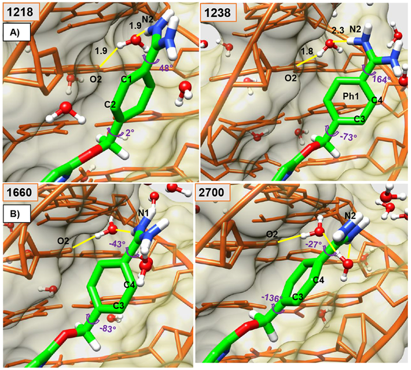
Figure 2.

Minor groove views of the DB2277-DNA complex are shown in panel A and B. Ø1 and α1 torsion angles are shown at several key times (frame-number on top-left) along with water-mediated H-bonds (yellow) (Å). Panel A shows larger twists for α1 and Ø1, compare 1218-frame with the orthogonal position of Ph1 relative to other aromatic groups of bound DB2277 in frame 1238. Starting at frame 1218 with C1 and C2 pointed out in panel A and end at the frame 2700 with C3 and C4 pointed out in Panel B. The DNA backbone is represented in orange-colored-stick with khaki-colored-space fill. The DB2277 molecule is shown in stick (green-C). Water molecules in ball and stick.
