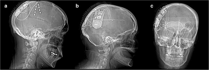
Fig. 1.
Computed tomography imaging of three patients post-implantation of the RNS System, demonstrating the range of cases in which the device may be used. (A) Two cortical strips targeting the right anterior and posterior premotor cortex. (B) Two depth electrodes targeting a heterotopia in the left frontal lobe. (C) Two depth electrodes targeting the left and right hippocampi

