

Abstract
Study Design:
Retrospective cohort study.
Objective:
To determine risk factors that may affect the rate of pedicle screws loosening in patients with degenerative diseases of the lumbar spine.
Methods:
A total of 250 patients with a low-grade spondylolisthesis and lumbar instability associated with degenerative diseases were enrolled. Preoperatively patients underwent computed tomography (CT) and cancellous bone radiodensity of a vertebral body was measured in Hounsfield units (HU). Pedicle screw fixation was used to treat patients either with a posterior fusion only or in combination with transforaminal lumbar interbody fusion (TLIF), anterior lumbar interbody fusion (ALIF), and direct lateral interbody fusion (D-LIF). Minimal follow-up period accounted for 18 months. Cases with screw loosening were registered assessing association with risk factors using logistic regression.
Results:
The rate of screw loosening was in positive correlation with the number fused levels and decreasing bone radiodensity. Fusion with a greater load-bearing surface cage was associated with the decrease in rate of pedicle screws loosening. Incomplete reduction in case of spondylolisthesis, bilateral facet joints removal, and laminectomy performed without anterior support favored pedicle screws loosening development. The estimated model classifies correctly 79% of cases with the specificity and sensitivity accounting for 87% and 66% respectively.
Conclusions:
The decreasing bone radiodensity in Hounsfield units has a considerable correlation with the rate of pedicle screws loosening. On the other hand, the length of fixation and applied surgical technique including fusion type also have a significant impact on complication rate. Spinal instrumentations should be planned by taking into account all potential risk factors and not characteristics relevant to bone quality assessment alone.
Keywords: lumbar spine, degenerative diseases, pedicle screws loosening, interbody fusion, Hounsfield units, decompression
Introduction
Degenerative diseases of the lumbar spine are frequently encountered in the older adult population and sometimes require surgical interventions using pedicle screw fixation. Despite the reported overall effectiveness, the applied interventions are associated with a certain rate of complications that frequently require repeated surgery. A typical complication that is associated with pedicle screw fixation is screw loosening with the reported rate ranging from 0.8% to 27% and even may exceed 50% in patients with osteoporosis.1,2,3,4 Taking into account great number of spinal instrumentations performed annually and complication related concern, factors that influence implant stability should be studied to predict complication and to work out an optimal surgical strategy.
The most frequently reported contributing factor to screws loosening is altered bone quality; however, the efficacy of different diagnostic modalities in bone quality assessment is still debated. Dual energy x-ray absorptiometry (DXA) is frequently used to assess bone mineral density (BMD), which is a part of bone quality assessment. On the other hand, the results of this examination can be strongly biased by degenerative changes in facet joints and even by aortic calcification.5-8 Being useful in the workup of a variety of spinal conditions, including degenerative diseases, computed tomography (CT) is also capable of accurately defining bone radiodensity using Hounsfield units (HU). Even though it has been reported that BMD has a strong relationship with radiodensity measured in HU, application of those parameters for implant failure prediction remains controversial.9-11
The additional factors that are supposed to influence pedicle screw instrumentation stability are lumbosacral fixation because of sacral anatomy and multilevel fusion because of an increased load on pedicle screws.1,3,4 It has been shown that the resection of ligaments, facet joints, and laminectomy are associated with the increased range of movements in a spinal segment and as a consequence, an increase in the stress on screw-bone interface that may lead to pedicle screws loosening.12,13 It has been reported that the lack of the anterior support is a significant factor for pedicle screws loosening; however, the extent of the influence and the role of the applied fusion type remain undetermined.14-16
Despite a considerable number of studies that were published on screws loosening, the reported data remains inconclusive because of different criteria used as indicator for implant loosening; the enrolled groups were heterogeneous regarding pathology without assessment of potential bias related. Furthermore, the majority of studies investigated the only single factor influence without assessment of other potential effects.3
The objective of this study is to determine risk factors that may affect the rate of pedicle screws loosening, unique contribution, and their interference in patients with degenerative diseases of the lumbar spine.
Patients and Methods
This study is a retrospective evaluation of 250 patients with degenerative diseases of the lumbar spine and evident instability of spinal segments including 80 males and 170 females with a male to female ratio of 0.47. The average age of participants at the time of operation was 52 years (SD = 12.11; range 28-74 years). Patients with only axial pain and those who presented with neurorological symptoms associated with spinal stenosis were enrolled. Participants underwent spinal instrumentations employing pedicle screws fixation during the period from 2012 to 2015. The duration of follow-up period was 18 months. The collected data during the follow-up period was analyzed retrospectively and finally radiographic criterion was used to assess outcomes. This study was reviewed and approved by local institutional review board committee, as long as all applied methods were conventional, and no additional risks were found; the informed written consent was received from all patients.
The inclusion criterion for participation in this study was presence of a degenerative disease of the lumbar spine with unstable spinal segments, which was confirmed by functional radiograms or having low-grade symptomatic spondylolisthesis. Indications for spinal instrumentations were
neurological deficit
claudication
axial and radicular pain syndromes with visual analog scale (VAS) over 4 and Oswestry Disability Index (ODI) over 40% resistant to repeated conservative treatment
The exclusion criteria were
patients with high-grade spondylolisthesis (grades 3 and 4)
patients with degenerative deformities that required more than fixation of 5 segments or spinopelvic fixation
evidence of tumor-related lesions of the lumbar spine
patients with sagittal and frontal imbalance and spinopelvic parameters mismatches that require more than 5 segment fixation and spinopelvic fixation
patients hospitalized for revision surgery
patients with screw malposition and redirection detected on postoperative CT images
patients with different types of fusion applied on different levels
Before the procedure, patients underwent a functional X-ray imaging and CT examination, the criterion for spinal instability were anterior translation greater than 3 mm and rotation more than 10°.17 CT was used as a part of preoperative work. The CT scans were performed from the T12-L5 levels using a single CT scanner (Aquilion 32, Toshiba Corporation). The scans used a slice thickness of 0.5 mm, covering a scan area of 50 cm. The scan parameters included tube voltage 120 kV, tube current 300 mA, auto mAċs range 180 to 400; 1.0 s/3.0mm/0.5 × 32, helical-pitch 21.0. Integrated software was utilized for calculations of bone density (Vitrea Version 5.2.497.5523) incorporating a window width/window level ratio of 2000/500. During CT examinations, measurements of a vertebral body cancellous bone radiodensity in HU were obtained at standard level of L3 in the sagittal, axial, and coronal planes. Measurements in the axial plane were taken at the level of the middle of the pedicles while those in the sagittal and coronal planes were taken along the geometric center of vertebra body. Oval-shaped trabecular bone samples were selected using the maximal achievable diameters without traversing into cortical bone to calculate bone density in each plane. Then out of those figures, an average radiodensity was calculated for each case.
Pedicle screw fixation with 6 mm polyaxial screws was used to treat patients either as a stand-alone technique with the posterior fusion or in combination with interbody fusion. The applied technique was standard. Pedicle screws were introduced at least to the anterior third of a vertebral body, bicortical screw placement was not used in the enrolled patients. Transforaminal lumbar interbody fusion (TLIF) with a single cage or anterior lumbar interbody fusion (ALIF) and direct lateral interbody fusion (D-LIF) applying cage with a greater loadbearing surface were used for interbody fusion (cages of approximately the same properties were used for transpsoas and anterior approaches). Cages that were used to perform interbody fusion are present in Figure 1. Autograft of a locally harvested bone was used to perform TLIF while solid allografts were used to perform ALIF and D-LIF. If indicated, a decompression of the nerve roots and spinal cord was performed. The extensiveness of the applied decompression was categorized as unilateral facet joints removal, bilateral facet joints with interspinous ligament removal, and laminectomy.
Figure 1.
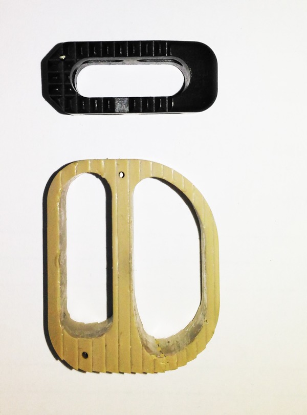
Cages that were used to perform transforaminal lumbar interbody fusion (TLIF) and either anterior lumbar interbody fusion (ALIF) or direct lateral interbody fusion (D-LIF) in patients enrolled in this study.
The duration of the follow-up was 18 months. Patients underwent examination using VAS scale, ODI questionnaire, and CT examinations at the period of 6, 12, and 18 months after interventions. Radiologic method was used to assess surgical outcomes in this study. Patients with pedicle screws loosening detected on CT images were registered, the criterion for screw loosening was at least 1-mm radiolucent zone around the screw and double halo sign.3 Figure 2 demonstrates the CT image in patient with pedicle screws loosening signs. Finally, surgical outcomes were classified in dichotomized scale either as presented with complication signs regardless the number of screws loosened or without screws loosening detected.
Figure 2.
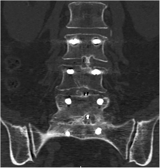
Computed tomographic image of a lumbar spine in a coronal plane. Bilateral double halo sign is evident on L3 level (radiolucent zone surrounded by sclerotic bone).
Statistical Analysis
A power analysis was performed to estimate the required sample size and it has been estimated that a sample of 180 patients was required to achieve 80% power. The association between screws loosening rate and potential risk factors was assessed using logistic regression analysis. Somers’ D correlation was used as a part of logistic regression analysis to assess the degree to which a dependent variable is associated with a particular predictor.
Results
A heterogeneous group of 250 patients with degenerative diseases of the lumbar spine who underwent spinal instrumentations with pedicle screws fixation was enrolled in this study, the characteristics of the enrolled group are provided in Table 1.
Table 1.
Characteristics of the Enrolled Group of Patients.
| Characteristic | No. of patients (%) |
|---|---|
| No. of levels fused | |
| 1 | 153 (61.2) |
| 2 | 70 (28.0) |
| 3 | 21 (8.4) |
| 4 | 5 (2.0) |
| 5 | 1 (0.4) |
| Out of those with lumbosacral fixation | 122 (48.8) |
| The applied fusion type | |
| Posterior fusion only | 38 (15.2) |
| TLIF | 162 (64.8) |
| Cage with a greater load surface (ALIF, D-LIF) | 50 (20.0) |
| The amount of decompression | |
| No resection of posterior structures | 74 (29.6) |
| Laminectomy | 62 (24.8) |
| Unilateral total facet joints removal | 118 (47.2) |
| Bilateral total facet joints removal on at least 1 level | 56 (22.4) |
| Residual slip of vertebra in case of spondylolisthesis >3 mm | 49 (19.6) |
| Characteristics of bone | |
| Radiodensity (HU) | Mean = 127.94; SD = 41.12; range 41,13-282.00 |
Abbreviations: ALIF, anterior lumbar interbody fusion; D-LIF, direct lateral interbody fusion; TLIF, transforaminal lumbar interbody fusion.
During the follow-up period, 97 patients had presented with CT signs of pedicle screws loosening (1-mm radiolucent zone around at least 1 screw and double halo sign); however, out of those only 39 complained of axial pain with ODI values over 40; those patients underwent revision surgery.
Using logistic regression analysis, the relationship between potential risk factors and the rate of pedicle screws loosening was estimated. The following factors were taken into account assessing risk for pedicle screws loosening development: bone density measured in HU, number of fused levels (the extension of fixation), posterior fusion performed without anterior support, type of interbody fusion, the extensiveness of decompression, unrestored alignment with anterior translation of more than 3 mm in case of spondylolisthesis and lumbosacral fusion. The parameters of estimated logistic regression model with highest explanatory value are presented in Table 2. According to the regression analysis results presented in Table 2, the most significant contributing factors to pedicle screw loosening were the number of fused levels and radiodensity. The rate of screws loosening was in positive correlation with the number fused levels and decreasing bone density. Application of cage with greater loadbearing surface applying ALIF and transpsoas approaches was associated with the decrease in rate of pedicle screws loosening (22% vs 43%). Incomplete reduction in case of spondylolisthesis was associated with a moderate rise in the rate of pedicle screws loosening (49% vs 36%). The influence of factors related to the amount of performed decompression was either small or moderate. Bilateral facet joints removal and laminectomy performed without anterior support favored pedicle screws loosening development (55% vs 34% and 91% vs 33% respectively). The influence of other factors including higher order interactions between predictors was insignificant. The overall goodness of fit of estimated model was χ2 = 97.771, P < .0001. The estimated model classifies correctly 79% of cases with the specificity and sensitivity accounting for 87% and 66% respectively.
Table 2.
The Estimated Parameters of Logistic Regression Model.a
| Components of Regression Model | Regression Coefficient | Odds Ratio per Unit Change [95% CI] | Somers’ D r |
|---|---|---|---|
| Intercept | −0.1438 P = .8758 | — | — |
| No. of fused levels | 0.8905 P = .0198 | 2.4364 [1.1534, 5.1464] | 0.6402 |
| Radiodensity, HU | −0.0264 P < .0001 | 0.9739 [0.9640, 0.9840] | −0.5735 |
| TLIF cage versus ALIF and D-LIF cage used | 1.0591 P = .0272 | 2.8838 [1.1282, 7.3719] | 0.5305 |
| Bilateral total facet joints removal (at least one level) | 1.0186 P = .0471 | 2.7694 [1.0132, 7.5694] | 0.0350 |
| Residual slip over 3 mm in case of spondylolisthesis | 0.9374 P = .0184 | 2.5534 [1.1726, 5.5604] | 0.1122 |
| Laminectomy performed without interbody fusion | 0.9374 P = .0013 | 36.3566 [4.1372, 319.4933] | 0.0190 |
| Lumbosacral fusion | 0.4803 P = .6002 | 1,5042 [0.3249, 6.9633] | 0.3717 |
| Laminectomy | −0.7075 P = .2124 | 0.4928 [0.1616, 1.5027] | 0.1186 |
| Posterior fusion performed without interbody fusion | −0.6161 P = .1074 | 0.540 [0.2548, 1.1445] | 0.0513 |
Abbreviations: ALIF, anterior lumbar interbody fusion; D-LIF, direct lateral interbody fusion; TLIF, transforaminal lumbar interbody fusion.
aP values in boldface indicate statistical significance (P < .05).
Discussion
Being the only way to improve the quality of life in certain cases, pedicle screws fixation is broadly used in patients with degenerative diseases of the lumbar spine and number of interventions performed annually is on the rise across the world. Taking into account the observed tendency, even a small rate of complications can cause considerable negative social effects and over expenses. Screws loosening is one of the most frequently reported typical complication of pedicle screws fixation that is associated with the necessity of the revision surgery. Despite a considerable number of articles covering this topic, the reported data remains, to some extent, inconclusive, because of different criteria used for pedicle screw loosening identification and because of different study designs.3 The majority of studies were focused on impact of BMD on the rate of screws loosening; however, it has been proven that bone quality is not the only contributing factor.1,3,4
The most spoken predictor favoring pedicle screws loosening development is altered bone quality with osteoporosis in severe cases. Osteoporosis is defined as a disease with a generalized decrease in bone mass and associated decline in the architectural makeup of bone tissue, with a result of a decrease in bone strength and an increased risk in the incidence of bone low-energy fractures.5 BMD is the most frequently used criterion for bone quality assessment. On the other hand, it has been proven that BMD is not the ultimate parameter associated with the mechanical properties of bone.5,18 As a consequence, the controversies on validity of diagnostic modalities used for bone quality assessment are still present. The vast majority of trials, which are focused on implant studies, are based on the assumption that figures of BMD that correspond to the osteoporosis criterion are also associated with a decline in capability to sustain loads without failure in patients who were operated on with pedicle screw fixation. On the other hand, there is still inconclusive evidence that those figures are the best predictive value in relation to the loosening of pedicle screws. DXA is frequently used as a screening tool for patients with osteoporosis and osteopenia detection, although, the preciseness of this option remains controversial because of a potential bias caused by degenerative changes in posterior structures and even by calcified aorta.19-22 In contrast, according to the findings reported in recent articles, there is growing evidence that radiodensity measured in HU has a strong relationship with bone mineral density and potentially has a predictive value in relation to the rate of low-energy fractures, implant instability, and pseudoarthrosis.9,23-26 The results of our data analysis show that the rate of pedicle screws loosening was in positive relation with the decreasing radiodensity and the statistical significance of regression coefficient was strong; however, the impact of other detected factors were also considerable.
The second most contributing factor in the estimated regression model was the length of fixation. The significance of this factor was discussed in several articles and the explanation of observed effect is an increase in cantilever bending moments loaded by longer level instrumentation.1,3,4 According to the results of the analysis the impact of fixation length on the pedicle screws loosening rate was considerable and comparable to the one associated with bone radiodensity in HU.
The least discussed factors with potential relevance to the pedicle screws loosening development, are the type of fusion and the extensiveness of decompression. Biomechanical studies have shown that inadequate anterior support may increase stress to the bone-screw interface finally resulting in a screw loosening.3 The biomechanical properties and outcomes of various types of fusion were studied in several articles and it has been reported that interbody fusion from the anterior approach does not provide considerable benefits over transforaminal interbody fusion, nonetheless, the stability provided by ALIF cage was slightly higher.14-16 According to the results of the regression analysis, application of a broad cage used for ALIF was associated with a significant decrease in the rate of screws loosening. A possible explanation of the observed effect is that cages applied for TLIF procedure provides support in the central part of the endplate, conversely broad cage provides load on periphery of endplate with greater compressive strength and distribution of load on greater surface, finally providing tolerance to the applied forces.27
Lumbosacral fusion was reported as a potential risk factor for screws loosening development because of the anatomy specificity and greater loads on pedicle screws.4,28 According to the results of our study, lumbosacral fusion turned out to be insignificant factor while the sample size of 250 patients with the proportion of those with lumbosacral fusion accounted for 48.8% is sufficient to assess the potential influence. The explanation for the observed result is that predictors of a stronger impact override the influence of lumbosacral fixation on pedicle screws stability.
It has been clearly determined that gradual resection of facet joints result in an increased range of motion in rotation, while laminectomy increases the range of motion in bending and extension.12,13 On the other hand, the most reliable mechanisms of screws loosening were considered cyclic caudocephalad toggling and rotational stress causing micromovements between vertebral body and screws that may finally resulted in the screws loosening.29 After the bilateral facet joints removal an additional rotational stress at the interface of bone and screw was expected, finally resulting in an increased rate of loosening. Even though the effect of bilateral facet joints removal was statistically significant, the relative influence was small. It was expected that laminectomy and posterior fusion without anterior support would cause a considerable increase in pedicle screws loosening rate; however, neither a former nor a latter caused any significant effect independently. In contrast, a combination of those factors noticeably favored the development of pedicle screws loosening and strongly outweighed the expected first order effects.
Overall impact of altered biomechanics related to an incompletely reduced vertebral body in the case of spondylolisthesis remains unclear because no additional benefits associated with complete reduction versus fusion in situ were found in patients with a low-grade spondylolisthesis.30 On the other hand, it has been proven that even biomechanical properties like spinopelvic parameters may influence loosening rate.4 According the results of our study the residual slip of over 3 mm in case of spondylolisthesis is a significant factor for the development of pedicle screws loosening; furthermore, a considerable unique contribution in the estimated model cannot be denied because no significant correlation with any other factors was found.
Finally, the results of our study provide some evidence that bone radiodensity measured in HU has relevance to pedicle screws loosening prediction. On the other hand, studied characteristic of cancellous bone is not the only contributing factor to pedicle screws loosening development. The results of the regression analysis demonstrate that pedicle screws loosening rate can be strongly influenced by the number of levels fused and applied surgical technique.
This study has certain limitations that must be acknowledged. The proportion of the enrolled patients with altered bone quality was relatively high so that it is plausible that screw loosening would have an overall high prevalence. In addition, the number of participants in this study was relatively small, however, the power analysis confirmed that the sample size was sufficient to support the reached conclusions.
Conclusion
The decreasing bone radiodensity in Hounsfield units has a considerable correlation with the rate of pedicle screws loosening. On the other hand, the length of fixation and applied surgical technique, including fusion type, also have a considerable impact on complication rate. Spinal instrumentations should be planned by taking into account all potential risk factors and not characteristics relevant to bone quality assessment alone.
Footnotes
Declaration of Conflicting Interests: The author(s) declared no potential conflicts of interest with respect to the research, authorship, and/or publication of this article.
Funding: The author(s) received no financial support for the research, authorship, and/or publication of this article.
References
- 1. Röllinghoff M, Schlüter-Brust K, Groos D, et al. Mid-range outcomes in 64 consecutive cases of multilevel fusion for degenerative diseases of the lumbar spine. Orthop Rev (Pavia). 2010;2:e3 doi:10.4081/or.2010.e3. [DOI] [PMC free article] [PubMed] [Google Scholar]
- 2. Wu ZX, Gong FT, Liu L, et al. A comparative study on screw loosening in osteoporotic lumbar spine fusion between expandable and conventional pedicle screws. Arch Orthop Trauma Surg. 2012;132:471–476. doi:10.1007/s00402-011-1439-6. [DOI] [PubMed] [Google Scholar]
- 3. Galbusera F, Volkheimer D, Reitmaier S, Berger-Roscher N, Kienle A, Wilke HJ. Pedicle screw loosening: a clinically relevant complication? Eur Spine J. 2015;24:1005–1016. doi:10.1007/s00586-015-3768-6. [DOI] [PubMed] [Google Scholar]
- 4. Kim JB, Park SW, Lee YS, Nam TK, Park YS, Kim YB. The effects of spinopelvic parameters and paraspinal muscle degeneration on S1 screw loosening. J Korean Neurosurg Soc. 2015;58:357–362. doi:10.3340/jkns.2015.58.4.357. [DOI] [PMC free article] [PubMed] [Google Scholar]
- 5. Kanis JA, McCloskey EV, Johansson H, Cooper C, Rizzoli R, Reginster JY; Scientific Advisory Board of the European Society for Clinical and Economic Aspects of Osteoporosis and Osteoarthritis (ESCEO) and the Committee of Scientific Advisors of the International Osteoporosis Foundation (IOF). European guidance for the diagnosis and management of osteoporosis in postmenopausal women. Osteoporos Int. 2013;24:23–57. doi:10.1007/s00198-012-2074-y. [DOI] [PMC free article] [PubMed] [Google Scholar]
- 6. Mallard F, Bouvard B, Mercier P, Bizot P, Cronier P, Chappard D. Trabecular microarchitecture in established osteoporosis: relationship between vertebrae, distal radius and calcaneus by X-ray imaging texture analysis. Orthop Traumatol Surg Res. 2013;99:52–59. doi:10.1016/j.otsr.2012.08.004. [DOI] [PubMed] [Google Scholar]
- 7. Burke CJ, Didolkar MM, Barnhart HX, Vinson EN. The use of routine non density calibrated clinical computed tomography data as a potentially useful screening tool for identifying patients with osteoporosis. Clin Cases Miner Bone Metab. 2016;13:135–140. doi:10.11138/ccmbm/2016.13.2.135. [DOI] [PMC free article] [PubMed] [Google Scholar]
- 8. Fountoulis G, Kerenidi T, Kokkinis C, et al. Assessment of bone mineral density in male patients with chronic obstructive pulmonary disease by DXA and quantitative computed tomography. Int J Endocrinol. 2016;2016:6169721 doi:10.1155/2016/6169721. [DOI] [PMC free article] [PubMed] [Google Scholar]
- 9. Schreiber JJ, Anderson PA, Rosas HG, Buchholz AL, Au AG. Hounsfield units for assessing bone mineral density and strength: a tool for osteoporosis management. J Bone Joint Surg Am. 2011;93:1057–1063. doi:10.2106/JBJS.J.00160. [DOI] [PubMed] [Google Scholar]
- 10. Choi MK, Kim SM, Lim JK. Diagnostic efficacy of Hounsfield units in spine CT for the assessment of real bone mineral density of degenerative spine: correlation study between T-scores determined by DEXA scan and Hounsfield units from CT. Acta Neurochir (Wien). 2016;158:1421–1427. doi:10.1007/s00701-016-2821-5. [DOI] [PubMed] [Google Scholar]
- 11. Kohan EM, Nemani VM, Hershman S, Kang DG, Kelly MP. Lumbar computed tomography scans are not appropriate surrogates for bone mineral density scans in primary adult spinal deformity. Neurosurg Focus. 2017;43:E4 doi:10.3171/2017.9.FOCUS17476. [DOI] [PubMed] [Google Scholar]
- 12. Bisschop A, van Engelen SJ, Kingma I, et al. Single level lumbar laminectomy alters segmental biomechanical behavior without affecting adjacent segments. Clin Biomech (Bristol, Avon). 2014;29:912–917. doi:10.1016/j.clinbiomech.2014.06.016. [DOI] [PubMed] [Google Scholar]
- 13. Lee KK, Teo EC. Effects of laminectomy and facetectomy on the stability of the lumbar motion segment. Med Eng Phys. 2004;26:183–192. doi:10.1016/j.medengphy.2003.11.006. [DOI] [PubMed] [Google Scholar]
- 14. Mobbs RJ, Phan K, Malham G, Seex K, Rao PJ. Lumbar interbody fusion: techniques, indications and comparison of interbody fusion options including PLIF, TLIF, MI-TLIF, OLIF/ATP, LLIF and ALIF. J Spine Surg. 2015;1:2–18. doi:10.3978/j.issn.2414-469X.2015.10.05. [DOI] [PMC free article] [PubMed] [Google Scholar]
- 15. Jägersberg M, Schneider K, Schaller C, Richter M. ALIF versus TLIF for post-discectomy syndrome. J Neurol Surg A Cent Eur Neurosurg. 2014;75:329–335. doi:10.1055/s-0034-1372432. [DOI] [PubMed] [Google Scholar]
- 16. Niemeyer TK, Koriller M, Claes L, Kettler A, Werner K, Wilke HJ. In vitro study of biomechanical behavior of anterior and transforaminal lumbar interbody instrumentation techniques. Neurosurgery. 2006;59:1271–1276. doi:10.1227/01.NEU.0000245609.01732.E4. [DOI] [PubMed] [Google Scholar]
- 17. Leone A, Guglielmi G, Cassar-Pullicino VN, Bonomo L. Lumbar intervertebral instability: a review. Radiology. 2007;245:62–77. doi:10.1148/radiol.2451051359. [DOI] [PubMed] [Google Scholar]
- 18. Cheung AM, Adachi JD, Hanley DA, et al. High-resolution peripheral quantitative computed tomography for the assessment of bone strength and structure: a review by the Canadian Bone Strength Working Group. Curr Osteoporos Rep. 2013;11:136–146. doi:10.1007/s11914-013-0140-9. [DOI] [PMC free article] [PubMed] [Google Scholar]
- 19. Tenne M, McGuigan F, Besjakov J, Gerdhem P, Åkesson K. Degenerative changes at the lumbar spine—implications for bone mineral density measurement in elderly women. Osteoporos Int. 2013;24:1419–1428. doi:10.1007/s00198-012-2048-0. [DOI] [PubMed] [Google Scholar]
- 20. Rehman Q, Lang T, Modin G, Lane NE. Quantitative computed tomography of the lumbar spine, not dual x-ray absorptiometry, is an independent predictor of prevalent vertebral fractures in postmenopausal women with osteopenia receiving long-term glucocorticoid and hormone-replacement therapy. Arthritis Rheum. 2002;46:1292–1297. doi:10.1002/art.10277. [DOI] [PubMed] [Google Scholar]
- 21. Smith JA, Vento JA, Spencer RP, Tendler BE. Aortic calcification contributing to bone densitometry measurement. J Clin Densitom. 1999;2:181–183. [DOI] [PubMed] [Google Scholar]
- 22. Atalay A, Kozakcioglu M, Cubuk R, Tasali N, Guney S. Degeneration of the lumbar spine and dual-energy X-ray absorptiometry measurements in patients without osteoporosis. Clin Imaging. 2009;33:374–378. doi:10.1016/j.clinimag.2008.12.005. [DOI] [PubMed] [Google Scholar]
- 23. Bredow J, Boese CK, Werner CM, et al. Predictive validity of preoperative CT scans and the risk of pedicle screw loosening in spinal surgery. Arch Orthop Trauma Surg. 2016;136:1063–1067. doi:10.1007/s00402-016-2487-8. [DOI] [PubMed] [Google Scholar]
- 24. Schwaiger BJ, Gersing AS, Baum T, Noël PB, Zimmer C, Bauer JS. Bone mineral density values derived from routine lumbar spine multidetector row CT predict osteoporotic vertebral fractures and screw loosening. AJNR Am J Neuroradiol. 2014;35:1628–1633. doi:10.3174/ajnr.A3893. [DOI] [PMC free article] [PubMed] [Google Scholar]
- 25. Matsukawa K, Abe Y, Yanai Y, Yato Y. Regional Hounsfield unit measurement of screw trajectory for predicting pedicle screw fixation using cortical bone trajectory: a retrospective cohort study. Acta Neurochir (Wien). 2018;160:405–411. doi:10.1007/s00701-017-3424-5. [DOI] [PubMed] [Google Scholar]
- 26. Wagner SC, Formby PM, Helgeson MD, Kang DG. Diagnosing the undiagnosed: osteoporosis in patients undergoing lumbar fusion. Spine (Phila Pa 1976). 2016;41:E1279–E1283. doi:10.1097/BRS.0000000000001612. [DOI] [PubMed] [Google Scholar]
- 27. Cadman J, Sutterlin C, 3rd, Dabirrahmani D, Appleyard R. The importance of loading the periphery of the vertebral endplate. J Spine Surg. 2016;2:178–184. doi:10.21037/jss.2016.09.08. [DOI] [PMC free article] [PubMed] [Google Scholar]
- 28. Yu BS, Zhuang XM, Zheng ZM, Zhang JF, Li ZM, Lu WW. Biomechanical comparison of 4 fixation techniques of sacral pedicle screw in osteoporotic condition. J Spinal Disord Tech. 2010;23:404–409. doi:10.1097/BSD.0b013e3181b63f4d. [DOI] [PubMed] [Google Scholar]
- 29. Mizuno T, Kasai Y, Sakakibara T, Yoshikawa T, Inaba T. Biomechanical study of rotational micromovement of the pedicle screw. Springerplus. 2016;5:1016 doi:10.1186/s40064-016-2694-3. [DOI] [PMC free article] [PubMed] [Google Scholar]
- 30. Bai X, Chen J, Liu L, et al. Is reduction better than arthrodesis in situ in surgical management of low-grade spondylolisthesis? A system review and meta analysis. Eur Spine J. 2017;26:606–618. doi:10.1007/s00586-016-4810-z. [DOI] [PubMed] [Google Scholar]