Chikungunya virus is an important human pathogen which now circulates in both the Old and New Worlds. As in the case of other Old World alphaviruses, CHIKV nsP2 not only has enzymatic functions in viral RNA replication but also is a critical inhibitor of the antiviral response and one of the determinants of CHIKV pathogenesis. In this study, we have applied a new strategy to select a variety of CHIKV nsP2 mutants that no longer exhibited transcription-inhibitory functions. The designed CHIKV variants became potent type I interferon inducers and acquired a less cytopathic phenotype. Importantly, they demonstrated the same replication rates as the parental CHIKV. Mutations in the same identified peptide of nsP2 proteins derived from other Old World alphaviruses also abolished their nuclear functions. Such mutations can be further exploited for development of new attenuated alphaviruses.
KEYWORDS: alphaviruses, chikungunya virus, noncytopathic replication, transcription inhibition, viral RNA replication, innate immunity, nsP2, replicon, vaccines, virus-host interactions
ABSTRACT
Alphavirus infections are characterized by global inhibition of cellular transcription and rapid induction of a cytopathic effect (CPE) in cells of vertebrate origin. Transcriptional shutoff impedes the cellular response to alphavirus replication and prevents establishment of an antiviral state. Chikungunya virus (CHIKV) is a highly pathogenic alphavirus representative, and its nonstructural protein 2 (nsP2) plays critical roles in both inhibition of transcription and CPE development. Previously, we have identified a small peptide in Sindbis virus (SINV) nsP2 (VLoop) that determined the protein’s transcriptional inhibition function. It is located in the surface-exposed loop of the carboxy-terminal domain of nsP2 and exhibits high variability between members of different alphavirus serocomplexes. In this study, we found that SINV-specific mutations could not be directly applied to CHIKV. However, by using a new selection approach, we identified a variety of new VLoop variants that made CHIKV and its replicons incapable of inhibiting cellular transcription and dramatically less cytopathic. Importantly, the mutations had no negative effect on RNA and viral replication rates. In contrast to parental CHIKV, the developed VLoop mutants were unable to block induction of type I interferon. Consequently, they were cleared from interferon (IFN)-competent cells without CPE development. Alternatively, in murine cells that have defects in type I IFN production or signaling, the VLoop mutants established persistent, noncytopathic replication. The mutations in nsP2 VLoop may be used for development of new vaccine candidates against alphavirus infections and vectors for expression of heterologous proteins.
IMPORTANCE Chikungunya virus is an important human pathogen which now circulates in both the Old and New Worlds. As in the case of other Old World alphaviruses, CHIKV nsP2 not only has enzymatic functions in viral RNA replication but also is a critical inhibitor of the antiviral response and one of the determinants of CHIKV pathogenesis. In this study, we have applied a new strategy to select a variety of CHIKV nsP2 mutants that no longer exhibited transcription-inhibitory functions. The designed CHIKV variants became potent type I interferon inducers and acquired a less cytopathic phenotype. Importantly, they demonstrated the same replication rates as the parental CHIKV. Mutations in the same identified peptide of nsP2 proteins derived from other Old World alphaviruses also abolished their nuclear functions. Such mutations can be further exploited for development of new attenuated alphaviruses.
INTRODUCTION
The Alphavirus genus in the Togaviridae family contains a variety of human and animal pathogens, which are widely distributed on all continents (1). Based on the geographical circulation, they can be divided into the New World (NW) and Old World (OW) alphaviruses. However, the recent spread of the OW chikungunya virus (CHIKV) to Central and South America and the Caribbean islands suggested that such division no longer reflects the current viral distribution and renders some flexibility (2, 3). Under natural conditions, alphaviruses are transmitted by persistently infected mosquito vectors between amplifying vertebrate hosts. In vertebrates, they develop a variety of diseases with different clinical symptoms (1). The NW alphaviruses induce highly debilitating disease that results in meningoencephalitis with a frequent lethal outcome or neurological sequelae (4). The OW representatives, exemplified by Sindbis virus (SINV), Semliki Forest virus (SFV), and CHIKV, are generally less pathogenic than those prevalent in the New World, and their human-associated diseases are usually limited to rash, fever, and arthritis. However, within recent years, CHIKV has become a viral pathogen of particular concern because of its spread to new areas and the severity of symptoms induced in humans (5–9). In many cases, CHIKV-specific polyarthritis is characterized by excruciating joint pain that can continue for years (5–9).
As for other alphaviruses, the CHIKV genome (G RNA) is represented by a single-stranded RNA of positive polarity of ∼11.5 kb in length (9). It mimics the structure of cellular mRNAs in that it contains a cap at the 5ʹ terminus and a poly(A) tail at the 3ʹ terminus. The G RNA encodes only a few proteins. The nonstructural viral proteins (nsPs) are translated directly from G RNAs as polyprotein precursor P123 or P1234 (1). Together with a CHIKV-specific set of host factors (10, 11), they form viral replication complexes (RCs) that initially contain P123 plus nsP4 (12, 13). At later times postinfection (p.i.), after complete polyprotein processing by nsP2-associated protease activity, the mature RCs include individual nsP1, nsP2, nsP3, and nsP4 (14, 15). These RCs are efficient in the synthesis of viral G RNA and subgenomic (SG) RNA, which serves as a template for translation of the precursor of viral structural proteins. After a few steps of processing, the latter proteins form infectious viral particles (16).
Despite recent progress in understanding the functions of nsPs in viral replication and interactions with host cells, many processes mediated by viral nsPs remain to be characterized. Alphavirus nsP2 has numerous known enzymatic activities, which include its function as a helicase (17–19) during RNA replication, protease function in ns polyprotein processing (15, 20), and RNA 5ʹ triphosphatase function during capping of viral G and SG RNAs (21). nsP2 can also acquire mutations that compensate the negative effects of modifications introduced into the promoter elements of viral genomes, nsP3, or capsid proteins (22–25). Another critically important function of nsP2, which is specific for the OW alphaviruses, including CHIKV, is its ability to translocate into the nuclei of vertebrate cells (26–28). In this compartment, nsP2 induces polyubiquitination of the catalytic subunit of cellular DNA-dependent RNA polymerase II, RPB1 (29). This ultimately leads to proteasomal degradation of RPB1 and abrogates synthesis of cellular mRNAs. The resulting global transcriptional shutoff makes cells incapable of activating the transcription-dependent antiviral response and cell signaling and induces cell death (29–31). Expression of nsP2 alone without viral replication is also highly cytotoxic for vertebrate cells (29), suggesting critical activity of this protein in viral pathogenesis on molecular and cellular levels. CHIKV nsP2 was also proposed to inhibit IFN-stimulated JAK-STAT signaling by interfering with STAT1 phosphorylation and its translocation into the (32, 33). However, in SINV-infected cells, this function directly depends on the nsP2-induced inhibition of cellular transcription (34).
Previous studies showed that the carboxy-terminal S-adenosyl-l-methionine (SAM)-dependent RNA methyltransferase-like (SAM MTase-like) domain of SINV and SFV nsP2 is not directly involved in protease and helicase functions of the protein (35–37), despite the fact that its presence strongly stimulates both functions. However, defined point mutations in this domain had deleterious effects on the ability of nsP2 to induce transcriptional shutoff (26, 30, 38, 39). The designed viral mutants and/or mutated alphavirus replicons that expressed no structural proteins were dramatically less cytopathic than their wild-type (wt) counterparts and capable of inducing an antiviral response in infected cells (40–42). These results suggested that the SAM MTase-like domain plays a critical role in the nuclear function of the OW alphavirus nsP2 proteins. However, these mutations in SINV and SFV nsP2 also strongly affected RNA and viral replication rates. Therefore, their effects on viral cytopathogenicity were difficult to unambiguously interpret (26, 41).
Previously, the protocol for the development of noncytopathic alphavirus genome-based constructs has been successfully applied to SINV and SFV (26, 41). The defective, self-replicating genomes (replicons) of the latter viruses had all of the structural genes in the SG RNA replaced by selectable markers. Despite lacking the structural genes, such replicons remained cytopathic. However, rare spontaneous mutations in the nsP2-coding sequence could produce a noncytopathic phenotype and make the replicons capable of persistent replication in some vertebrate cells lines that had defects in type I interferon (IFN) response. Development of attenuated CHIKV constructs was found to be more challenging, and multiple mutations in nsP2 were required for making CHIKV replicons less cytopathic (43). These mutations also had a deleterious effect on RNA replication rates and thus were likely not applicable for the development of stable, replication-competent viruses.
Alphaviruses with altered nuclear functions represent important systems for further understanding of the molecular mechanism of their pathogenesis and virus-host interactions. Thus, if they remain capable of efficient replication, CHIKV mutants that lack host inhibitory function could provide an opportunity for generating attenuated viruses. The results of our prior study on SINV strongly suggested a critical function of a short, highly variable peptide loop (termed VLoop) located on the surface of nsP2 SAM MTase-like domain in the protein’s nuclear functions (44). However, the SINV-related data were not directly applicable to CHIKV, which is a more important human pathogen. Therefore, we applied a new selection approach to identify a variety of CHIKV variants and CHIKV replicons with a mutated VLoop. The developed viruses retained high levels of replication but no longer exhibited transcription inhibitory functions. The designed CHIKV replicons and viruses became noncytopathic in murine cells and can be used in a wide range of applications. The selected mutations can be also further applied in development of vaccine candidates against CHIKV infection.
RESULTS
Introduction of attenuating, SINV-specific mutations into CHIKV nsP2 makes the virus less cytopathic.
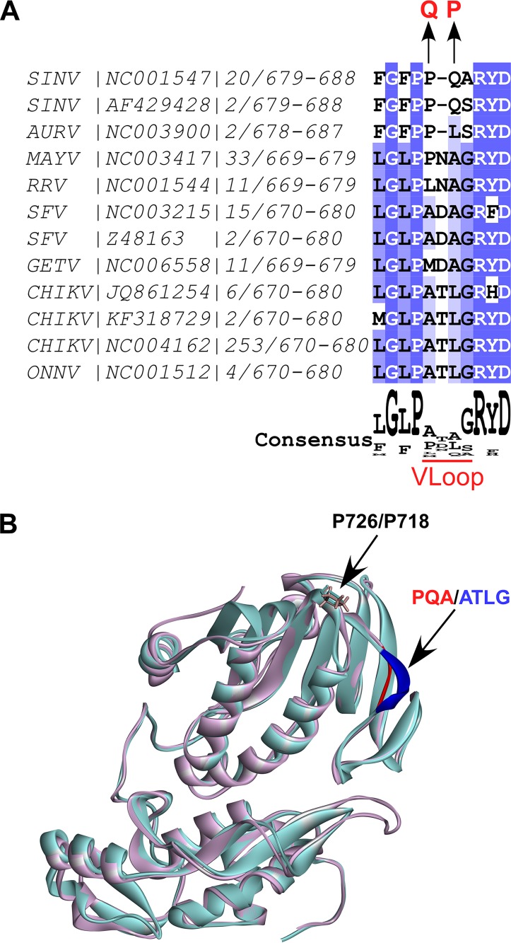
Recently, we selected a variety of SINV variants with mutated P683 or Q684 in the carboxy-terminal SAM MTase-like domain of nsP2 (44). These mutations strongly affected the ability of SINV nsP2 to induce RPB1 degradation and consequently made the virus incapable of efficient inhibition of the antiviral response. Importantly, these mutations did not affect SINV replication rates in the tested cell lines of vertebrate origin. Therefore, in this new study, we initially made an attempt to apply these SINV-based data for the development of attenuated mutants of the most pathogenic OW alphavirus, CHIKV. The alignment of the available sequences of the OW alphavirus nsP2 proteins demonstrated that the SINV-specific P683 and Q684 mutations are located in the short surface-exposed loop between two conserved amino acid sequences (Fig. 1A). This loop exhibited a very low level of identity between the members of SINV and SFV serocomplexes (Fig. 1A), and its overall length varied between 3 and 4 amino acids (aa). Thus, it was termed VLoop. Structural alignment of the carboxy-terminal domains of SINV and CHIKV nsP2 proteins demonstrated that for both viruses, VLoop was located in close proximity to P726 (SINV), which was previously shown to be a critical determinant of protein’s function in the degradation of RPB1 subunit of RNA polymerase II (29).
FIG 1.
CHIKV nsP2-specific VLoop demonstrates high variability and is located on the surface of the carboxy-terminal SAM MTase-like domain. (A) The nsP2 protein sequences of 365 OW alphaviruses were downloaded from Virus Pathogen Resources (ViPR). The sequences were aligned using Muscle in Jalview. The fragments of nsP2 corresponding to aa 679 to 688 of SINV were aligned, and then the redundant sequences and those present in a single strain were deleted. The GenBank accession number for the representative strain, numbers of the strains included into alignment for each group, and amino acid positions in nsP2 are indicated. Mutations identified in attenuated SINV are shown above the alignment. (B) The 3D structures of CHIKV (PDB code 3TRK) and SINV (modeled based on PDB code 4GUA) were superimposed in Discovery Studio Visualizer using sequence alignment. The structure of CHIKV nsP2 is presented as a light magenta solid ribbon. The structure of SINV nsP2 is presented as a light turquoise solid ribbon. P726 (SINV) and corresponding P718 (CHIKV) are presented as sticks. CHIKV-specific VLoop is colored in blue, and SINV-specific VLoop is colored in red. Abbreviations: AURV, Aura virus; CHIKV, chikungunya virus; GETV, getah virus; MAYV, Mayaro virus; RRV, Ross River virus; ONNV, o’nyong nyong virus; SFV, Semliki Forest virus; SINV, Sindbis virus.
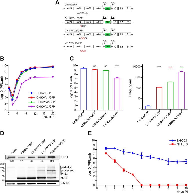
Based on the above-described data, it was reasonable to expect that similar to SINV, some mutations in VLoop of CHIKV nsP2 may abolish nuclear function(s) of the protein. Therefore, we focused our efforts on introducing mutations into the VLoop-coding sequence in the CHIKV genome and testing their effect on virus-induced RPB1 degradation, inhibition of cellular transcription, and cytopathic effect (CPE) development. The first approach was based on replacement of one or more amino acids in CHIKV-specific peptide by combinations of those found in the corresponding VLoop sequences of attenuated SINV mutants. Accordingly, the CHIKV/V1/GFP mutant encoded QTLG instead of 674ATLG677, CHIKV/V2/GFP encoded an AQQG peptide, and nsP2 of CHIKV/V3/GFP had the entire 674ATLG677 replaced by QQA (Fig. 2A). Since at least some of the new mutants were expected to be less cytopathic, their genomes and that of control CHIKV/GFP were designed to encode green fluorescent protein (GFP) under the control of the subgenomic promoter. GFP expression was used to indirectly monitor the levels of viral replication and infection spread in cultured cells.
FIG 2.
SINV-specific mutations in CHIKV-specific VLoop differentially affect viral replication rates and activation of the antiviral response. (A) Schematic presentation of recombinant CHIKV genomes encoding indicated peptides. (B) NIH 3T3 cells were infected with the indicated viruses at an MOI of 20 PFU/cell. Media were replaced at the indicated times p.i., and viral titers were determined by plaque assay on BHK-21 cells. (C) NIH 3T3 cells were infected with control and mutant viruses at an MOI of 20 PFU/cell. Viruses were harvested at 18 h p.i. Infectious titers and concentrations of the released IFN-β were determined in the same samples as described in Materials and Methods. The significances of differences in titers and IFN-β concentrations were estimated by one-way analysis of variance (ANOVA; n = 3). ****, P < 0.0001. (D) NIH 3T3 cells were infected with the indicated viruses at an MOI of 20 PFU/cell and harvested at 8 h p.i. The lysates were analyzed by Western blotting for the levels of RPB1 and for the ns polyprotein processing using RPB1-, nsP2-, and tubulin-specific Abs and corresponding infrared dye-labeled secondary Abs. Membranes were scanned on Odyssey imager (LI-COR). (E) BHK-21 and NIH 3T3 cells were infected with CHIKV/V3/GFP at an MOI of 10 PFU/cell and then incubated for 10 days. Media were changed every 24 h, and cells were split upon reaching confluency. Viral titers were determined by plaque assay on BHK-21 cells. Data are presented as means ± SDs from two independent experiments.
Within 8 h after electroporation of the equal amounts of the in vitro-synthesized RNAs, for all of the samples, similar numbers of cells were GFP positive (data not shown), indicating that the RNAs mutants were viable and did not require additional adaptive mutations for RNA replication. Titers of CHIKV/V1/GFP and CHIKV/V2/GFP in the stocks harvested at 24 h postelectroporation were the same as those of control CHIKV/GFP, but CHIKV/V3/GFP replication resulted in almost 100-fold-lower infectious titers, below 108 PFU/ml. The CHIKV/V3/GFP mutant also did not develop cytopathic effect (CPE) in BHK-21 cells at any time postelectroporation and expressed lower levels of GFP. However, it was still capable of producing small plaques in these cells under agarose cover in the presence of low levels of fetal bovine serum (FBS) (see Materials and Methods for details). NIH 3T3 cells, which in contrast to BHK-21 cells are fully competent in type I interferon (IFN) production and signaling, also produced CHIKV/V1/GFP and CHIKV/V2/GFP at the same rates as parental CHIKV/GFP (Fig. 2B). In contrast, at any times p.i., titers of the mutant with the replaced VLoop, CHIKV/V3/GFP, were 50- to 100-fold lower. In other experiments on NIH 3T3 cells, the designed mutants were additionally tested for replication and IFN-β induction (Fig. 2C). By 18 h p.i., CHIKV/GFP, CHIKV/V1/GFP, and CHIKV/V2/GFP, but not CHIKV/V3/GFP, accumulated in the media to the same titers (Fig. 2C). CHIKV/GFP, which encoded wt nsP2, induced IFN-β at the limit of detection, while replication of all three nsP2 mutants led to IFN-β accumulation at readily detectable levels (Fig. 2C). CHIKV/V3/GFP was the most efficient IFN-β inducer, and this result correlated with its inability to induce RPB1 degradation (Fig. 2D). At 8 h p.i. of NIH 3T3 cells, CHIKV/V1/GFP and CHIKV/V2/GFP caused partial degradation of RPB1, but not as efficiently as parental CHIKV/GFP, while in CHIKV/V3/GFP-infected cells, the amount of RPB1 did not change (Fig. 2D).
To compare the abilities of the designed mutants to induce CPE, NIH 3T3 and BHK-21 cells were infected with all of the generated viruses at a multiplicity of infection (MOI) of 10 PFU/cell, and viral replication was analyzed for 10 days. CHIKV/GFP, CHIKV/V1/GFP, and CHIKV/V2/GFP caused complete CPE in both cell lines within 48 h p.i. (data not shown). In contrast, CHIKV/V3/GFP infection was noncytopathic. The latter mutant was cleared from NIH 3T3 cells within 5 days p.i. by the autocrine effect of the released type I IFN (Fig. 2C and E). In BHK-21 cells, which are deficient in type I IFN response, this mutant established persistent infection.
Taken together, the data indicated that nuclear functions of nsP2 encoded by CHIKV/V1/GFP and CHIKV/V2/GFP were partially affected, but viral attenuation was incomplete. These mutants remained cytopathic and relatively inefficient IFN-β inducers. Mutations in CHIKV/V3/GFP nsP2, in turn, made the mutant dramatically less cytopathic but strongly compromised viral replication rates.
Mutations in VLoop of CHIKV SAM MTase-like domain of nsP2 can affect P12 processing.
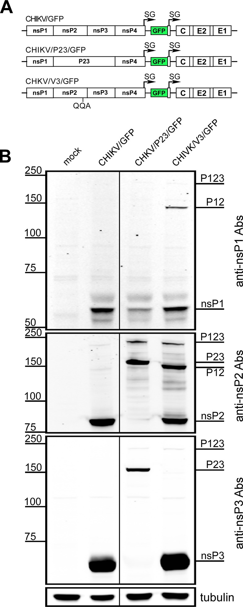
Western blot analysis of nsP2 accumulation in infected cells revealed that the introduced nsP2-specific mutations in CHIKV/V2/GFP and CHIKV/V3/GFP variants altered the processing of P123/P1234 polyproteins (Fig. 2D). In addition to nsP2, the high-molecular-weight proteins were readily detectable by anti-nsP2 monoclonal antibody (MAb). The effect of mutations in the surface-located peptide of the SAM MTase-like domain of nsP2 was unexpected and thus deserved additional investigation. Since the products of partial processing, P12 and P23, have similar sizes, it remained unclear which particular step of P123 cleavage was affected by introduced mutations. To distinguish between the possibilities, NIH 3T3 cells were infected with CHIKV/GFP, CHIKV/V3/GFP, and an additional, previously designed virus, CHIKV/P23/GFP, at an MOI of 20 PFU/cell. The CHIKV/P23/GFP mutant contained a mutation in nsP2̂3 cleavage site and was applied as a P23-producing virus. At 8 h p.i., the nonstructural proteins were analyzed by Western blot using nsP1-, nsP2-, and nsP3-specific MAbs (Fig. 3). The CHIKV/GFP-infected cells contained only individual nsPs. In CHIKV/P23/GFP-infected cells, we detected nsP1, P23, and P123 but not nsP2 or nsP3. Cells infected with CHIKV/V3/GFP, in contrast, exhibited the presence of a readily detectable fraction of P123 and P12 but not P23. Thus, processing of the entire ns polyprotein of CHIKV/V3/GFP mutant and 1̂2 cleavage site, in particular, were strongly affected. This alteration of nsP processing was likely at least partially responsible for lower viral replication rates and alteration of nsP2 nuclear functions and made this mutant a potent IFN-β inducer.
FIG 3.
The QQA mutation in CHIKV nsP2 VLoop impairs P12 processing. (A) Schematic presentation of recombinant CHIKV genomes encoding either mutated VLoop or mutated nsP2/3 cleavage site. (B) NIH 3T3 cells were infected with the indicated viruses at an MOI of 20 PFU/cell and harvested at 8 h p.i. The presence of individual nsPs and incompletely cleaved polyproteins in cell lysates was analyzed by Western blotting using antibodies specific to CHIKV nsP1, nsP2, and nsP3 proteins. Membranes were analyzed on an Odyssey imager (LI-COR).
Selection of efficiently replicating CHIKV nsP2 mutants that lack transcription inhibitory functions.
The above-described mutations did not generate CHIKV variants exhibiting wt levels of replication and high levels of type I IFN induction, which usually indicates loss of nuclear function of nsP2. The designed mutants also demonstrated alterations in ns polyprotein processing. However, these experiments showed that mutations in VLoop (674ATLG677) of CHIKV nsP2 had negative effects on the ability of the virus to interfere with the activation of cellular antiviral response. Moreover, the previously designed noncytopathic SINV, which contained the P683Q mutation in VLoop (44), did not exhibit any defects in the P123 processing. Taken together, these data suggested that the development of attenuated CHIKV mutants with a higher level of replication and wt pattern of P123 processing may be feasible. However, it could require designing and testing too many amino acid combinations in VLoop. Therefore, in order to generate and evaluate a wide collection of mutants, we applied an alternative approach.
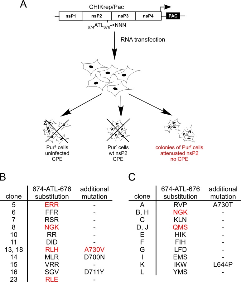
A CHIKV replicon (CHIKrep/Pac) which contained puromycin acetyltransferase (Pac) gene under the control of subgenomic promoter (Fig. 4A) was used as a basic construct. Its replication in BHK-21 cells was highly cytopathic. In repeated experiments, after transfections of the in vitro-synthesized CHIKrep/Pac RNA into BHK-21 cells followed by puromycin selection, no colonies of Purr cells were detected. This was a strong indication that the generation of a noncytopathic phenotype likely requires more than a single spontaneous adaptive point mutation. The sequential replacements of VLoop sequence by combinations of amino acids followed by analysis of their effects on cytopathogenicity of the replicon or virus could be endless. Therefore, we replaced codons encoding VLoop (674ATL676) in CHIKrep/Pac nsP2 with a randomized nucleotide sequence (Fig. 4A). The library of plasmids from ∼104 clones of Escherichia coli was used for the in vitro transcription of the replicon genomes, and the entire RNA pool was electroporated into BHK-21 cells. Following selection with puromycin resulted in ∼400 colonies of Purr cells. The untransfected cells and those containing nonviable replicons died due to translational arrest caused by puromycin. Most of the transfected cells likely received cytopathic variants of the replicon and died because of nsP2-induced transcriptional shutoff that caused cell death. The residual surviving cells contained noncytopathic CHIKrep/Pac mutants, which were probably unable to induce RPB1 degradation and transcription inhibition. We randomly selected 24 cell colonies and expanded them in the presence of a higher concentration of puromycin. After this additional step, 12 colonies were also selected for sequencing of the mutated nsP2 gene. The identified sequences of VLoop and the second-site spontaneous mutations found in some variants are presented in Fig. 4B.
FIG 4.
Defined amino acid sequences of VLoop in CHIKV nsP2 make CHIKrep/Pac replicon noncytopathic in BHK-21 cells. (A) Schematic presentations of CHIKV replicon with a cloned library of VLoops in nsP2 protein and the scheme of selection of noncytopathic replicons. Cells were electroporated with the in vitro-synthesized replicon RNA and then treated with puromycin to select colonies of Purr cells. (B) Amino acid sequences identified in nsP2 of the noncytopathic replicons in randomly selected clones of Purr cells. (C) Amino acid sequences of VLoop and detected additional mutations in CHIKV nsP2 identified in the noncytopathic replicons after 3-week-long passaging of the pool of Purr cells (see the text for details). In panels B and C, amino acid sequences used in further experiments are indicated in red.
In an additional selection, the pool of selected Purr cell clones was further passaged in the presence of puromycin for 3 weeks. This procedure was aimed at selecting the most efficiently growing cells, which likely contained more attenuated replicons. Total RNA was then isolated from the cell pool, and the nsP2 sequence was amplified by PCR and cloned into plasmids. Insertions in the plasmids of 12 randomly selected clones of E. coli were sequenced. The identified variants of the VLoop sequences and second site mutations are presented in Fig. 4C.
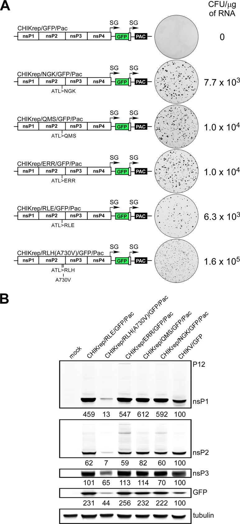
Analysis of the mutated VLoop sequences (Fig. 4B and C) revealed the following. (i) Many of them contained positively charged amino acids, arginine or lysine, in the first or third position of the peptide. (ii) A few noncytopathic replicons contained additional second site mutations in the nsP2 gene. Most of second-site mutations were mapped to α-helices 9 and 10 (35). Several mutations in this region, which strongly reduced virus replication, have been previously identified in several alphaviruses. These included A713P (SFV), Q735L (Venezuelan equine encephalitis virus [VEEV]), K733A, R740A, R751A, K752A (SINV), etc. (41, 45, 46). D711G has been also previously identified as a second-site mutation in an CHIKV replicon that already contained an attenuating P718S mutation (32). This double mutant became noncytopathic but replicated less efficiently than the wt replicon. We have previously found that presence of mutations in the same α-helix also prevented translocation of SINV nsP2 into the nucleus (44, 47). Thus, the second-site mutation likely affected RNA replication. (iii) Three amino acid sequences [RLH(A730V), NGK, and QMS] were repeatedly detected in the selected mutants. (iv) The NGK sequence was detected in the cells selected by both protocols.
The newly selected VLoop sequences affect cytopathogenicity of CHIKV replicons.
Sequences encoding some of the identified mutations (indicated in red in Fig. 4B and C) were cloned into CHIKrep/GFP/Pac to replace the original 674ATL676-encoding nsP2 fragment. The latter replicon had both GFP and Pac genes under the control of subgenomic promoters (Fig. 5A). In these experiments, GFP expression was used as a means of indirect assessment of G RNA replication and SG RNA synthesis. Equal amounts of the in vitro-synthesized RNAs of the designed replicons and CHIKrep/GFP/Pac, encoding wt nsP2, were electroporated into BHK-21 cells, followed by selection of colonies of Purr cells. As expected, no colonies were formed upon transfection of parental CHIKrep/GFP/Pac, despite the fact that all of the cells were GFP positive within 24 to 48 h posttransfection and thus contained replicating RNA. In contrast, cells transfected with mutated replicons produced large numbers of colonies of Purr, GFP-positive cells. The replicon with the additional mutation in nsP2, CHIKrep/RLH(A730V)/GFP/Pac, was the most efficient in colony formation, indicating that it had the most attenuated phenotype. However, based on relatively low GFP fluorescence in the Purr cells (data not shown) and Western blot analysis of GFP, nsP1, nsP2 and nsP3 expression (Fig. 5B), this mutant replicated less inefficiently. Importantly, all mutant replicons demonstrated either very small or no defects in P123 processing (Fig. 5B). These data demonstrated that the selected VLoop sequences attenuated cytopathogenicity of nsP2 and made CHIKV replicons capable of persistent replication at least in BHK-21 cells.
FIG 5.
The replacement of VLoop in CHIKrep/GFP/Pac by selected sequences makes replicons noncytopathic and capable of persistent replication. (A) Schematic presentation of the designed replicons containing indicated mutations in VLoop and additional mutation in nsP2 (in CHIKrep/RLH(A730V)/GFP/Pac) and their efficiencies in the formation of colonies of Purr, GFP-positive cells upon electroporation of the in vitro-synthesized RNAs. (B) BHK-21 cells were electroporated with equal amounts of the in vitro-synthesized replicon RNAs and harvested either at 8 h posttransfection (CHIKV/GFP RNA-transfected cells) or 3 to 7 days after transfection and puromycin selection of cells with mutant replicons. Equal numbers of cells were used for analysis. Levels of indicated nsPs and GFP in the cell lysates were evaluated by Western blotting using specific Abs. Membranes were scanned on an Odyssey imager (LI-COR).
The selected VLoop sequences made CHIKV incapable of inducing transcriptional shutoff.
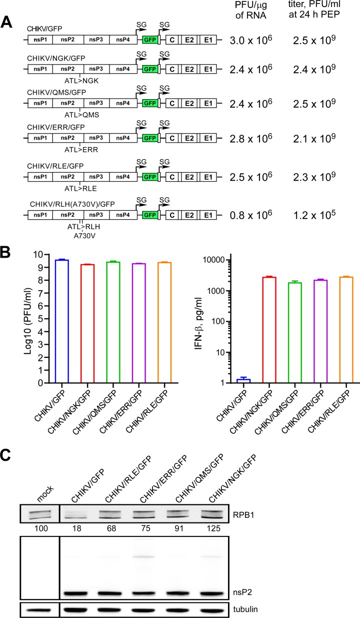
Next, we tested the effects of the above-described mutations on viral replication. The wt nsP2 gene in CHIKV/GFP genome was replaced by mutated counterparts (Fig. 6A). In the infectious center assay, the in vitro-synthesized RNAs of the mutants and parental CHIKV/GFP demonstrated the same infectivities (Fig. 6A). This suggested that the designed constructs did not require additional adaptive mutations for virus viability. Four mutants were able to replicate in electroporated BHK-21 cells to the same titers as CHIKV/GFP (Fig. 6A). However, the final titers in the electroporation-derived stocks of the double mutant CHIKV/RLH(A730V)/GFP were reproducibly very low, and in standard plaque assay, this mutant’s plaques were almost undetectable. Further analysis of the nsP2-specific A730V mutation demonstrated that it had a strong negative effect on CHIKV RNA replication (data not shown), which confirmed our hypothesis that second-site mutations affected RNA replication. Therefore, experiments with the CHIKV/RLH(A730V)/GFP mutant were discontinued. Other second-site mutants were not investigated for the same reason.
FIG 6.
CHIKV variants containing selected, mutated VLoop sequences efficiently replicate and are highly potent IFN-β inducers. (A) Schematic presentation of recombinant CHIKV genomes containing indicated substitutions in VLoop and additional mutation in nsP2 [CHIKV/RLH(A730V)/GFP], RNA infectivities in the infectious-center assay, and infectious titers in the stocks harvested at 24 h after electroporation of BHK-21 cells. (B) NIH 3T3 cells were infected with the indicated viruses at an MOI of 50 PFU/cell, and samples were harvested at 22 h p.i. Viral titers were determined by plaque assay on BHK-21 cells, and concentrations of IFN-β in the same samples were assessed by ELISA as described in Materials and Methods. (C) NIH 3T3 cells were infected with the indicated viruses at an MOI of 20 PFU/cell and harvested at 9 h p.i. The levels of degradation of RPB1 and levels of nsP2 were determined by Western blotting using specific Abs. Membranes were analyzed on an Odyssey imager (LI-COR). These experiments were reproducibly repeated more than three times, and the results of one of the representative experiments are presented.
In the next experiments, NIH 3T3 cells were infected with the designed VLoop mutants and parental CHIKV/GFP at the same MOI, and both viral titers and the levels of IFN-β release were compared at 22 h p.i. All the viruses accumulated in the media to similar titers (Fig. 6B). However, in contrast to parental CHIKV/GFP and previously developed constructs with mutated VLoop (Fig. 2C), the newly designed variants were very potent IFN-β inducers. As expected, they were inefficient in stimulation of RPB1 degradation (Fig. 6C), and this alteration of nuclear function provided a plausible explanation for detected high levels of IFN-β induction (Fig. 6B). As in the experiments with corresponding replicons, processing of mutant P123 was essentially the same as in the cells infected with the parental CHIKV/GFP (Fig. 6C), and the uncleaved products were present at very low levels, if at all.
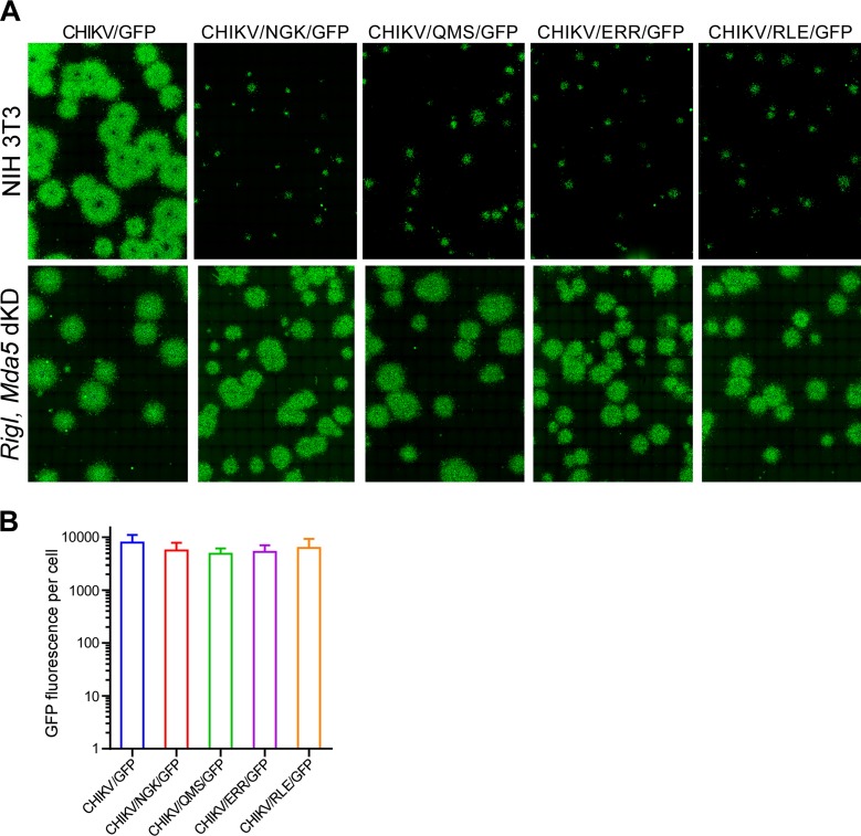
All four variants were able to rapidly spread in BHK-21 cells or Rig-I Mda5 double-knockdown (dKD) cells, both deficient in type I IFN induction (Fig. 7A and data not shown). However, in contrast to CHIKV/GFP, they could not spread in murine NIH 3T3 cells, which were fully competent in type I IFN induction and signaling (Fig. 7A). Additional comparative analysis of these images did not reveal significant differences in the intensity of GFP fluorescence per cell in NIH 3T3 cells (Fig. 7B), and thus, the inability to spread was most likely the result of IFN-β release by the already infected cells and activation of an antiviral state in the surrounding ones.
FIG 7.
Mutations in VLoop strongly affect spread of CHIKV infection in type I IFN-competent cells. (A) Monolayers of NIH 3T3 cells and their Rig-I Mda5 dKD derivatives were infected with indicated variants and then covered by agarose-containing media supplemented with 3% fetal calf serum (FCS). At 3 days p.i., cells were fixed with 4% paraformaldehyde (PFA) and imaged on a Cytation 5 cell imaging multimode reader (BioTek). GFP-positive foci are presented. (B) Average GFP intensities in infected NIH 3T3 cells were assessed using Gen5 software (BioTek).
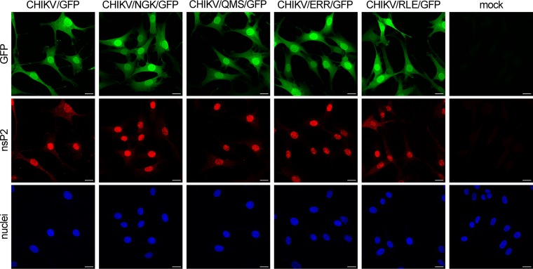
In additional experiments, we evaluated the possible effects of the introduced mutations on CHIKV nsP2 compartmentalization in virus-infected cells and on the ability of nsP2 to translocate into the nuclei. Cells were infected with CHIKV/GFP and the above-described mutants and, at 6 h p.i., were stained with anti-nsP2 MAbs. The results presented in Fig. 8 demonstrate that none of the mutations had any detectable effect on nsP2 intracellular distribution. Similar to the case with CHIKV/GFP infection, mutated nsP2 proteins very efficiently accumulated in the nuclei. However, they were no longer able to induce RPB1 degradation (Fig. 6C) and downregulate activation of type I IFN response (Fig. 6B).
FIG 8.
Mutations in VLoop do not affect nuclear translocation of CHIKV nsP2. NIH 3T3 cells were infected with indicated recombinant viruses and at 6 h p.i., fixed with 4% PFA, permeabilized, and stained with pan-anti-nsP2 MAb, anti-mouse secondary antibodies labeled with Alexa Fluor 555 (Thermo Fisher) and Hoechst nuclear staining. The image stacks of 6 optical sections were acquired on a Zeiss LSM700 confocal microscope with a 63 × 1.4 numerical aperture (NA) PlanApochromat oil objective. The multiple intensity projection images were assembled using Imaris software (Bitplane AG).
CHIKV nsP2 mutants do not interfere with transcription of cellular mRNA and rRNA.
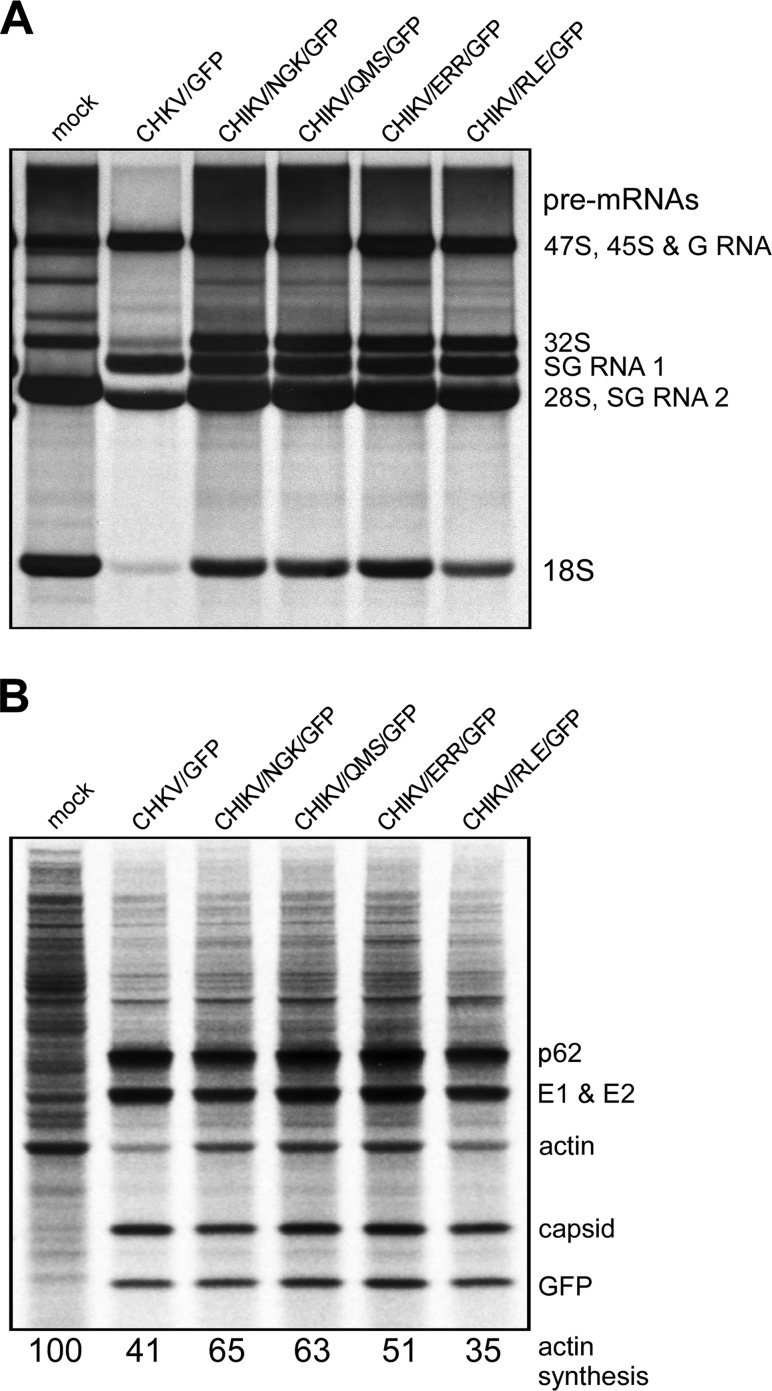
To further characterize the effects of CHIKV nsP2 mutants on cellular transcription and translation, NIH 3T3 cells were infected at the same MOI with the designed variants and parental CHIKV/GFP. Cellular and viral RNAs were metabolically labeled with [3H]uridine in the absence of actinomycin D (ActD) between 4 and 8 h p.i. RNAs were analyzed by agarose gel electrophoresis (Fig. 9A). CHIKV/GFP efficiently inhibited synthesis of both rRNAs and mRNAs, as was previously described for SINV (44). In contrast, cells infected with the mutant viruses continued to efficiently produce rRNA and mRNA. High-molecular-weight pre-mRNAs were clearly detectable above viral G RNA, and both 28S and 18S rRNA bands were present as well (Fig. 9A). The band corresponding to SG RNA1, which encodes both GFP and viral structural genes, was essentially the same in all samples. This was an additional indication that the introduced mutations had no effect on enzymatic functions of CHIKV nsP2 in viral RNA synthesis.
FIG 9.
The designed CHIKV mutants do not induce transcriptional shutoff in the infected cells and inefficiently downregulate translation of cellular mRNAs. (A) NIH 3T3 cells were infected with indicated viruses at an MOI of 20 PFU/cell. RNAs were metabolically labeled with [3H]uridine in the absence of ActD between 4 and 8 h p.i. Labeled RNAs were analyzed by agarose gel electrophoresis under denaturing conditions as described in Materials and Methods. (B) NIH 3T3 cells were infected at an MOI of 20 PFU/cell, and at 6 h p.i., proteins were metabolically labeled for 30 min with [35S]methionine and analyzed by SDS-PAGE as described in Materials and Methods.
Protein synthesis was analyzed at 6 h after infection of NIH 3T3 cells with the indicated viruses. By this time p.i., wt SINV and SFV downregulate translation of cellular mRNAs to undetectable levels (44, 48–50). However, even parental CHIKV inhibits translation of cellular templates relatively inefficiently (Fig. 9B). At this time p.i., synthesis of actin and other cellular proteins remained readily detectable, and it was only slightly lower than in the mock-infected cells. Based on our experience, the double subgenomic CHIKV variants that contain heterologous genes (GFP gene in this study) under the control of SG promoter express these proteins more efficiently than SINV (44) and probably SFV. The expression of heterologous genes by CHIKV likely does not require translational enhancers that were identified in SINV- and SFV-specific SG RNAs and shown to be essential for gene expression under the conditions of virus-induced translational shutoff (50, 51). Indeed, to date, bioinformatic approaches have not predicted the existence of the G-C rich RNA stem-loop enhancer in CHIKV SG RNA (data not shown).
The designed CHIKV nsP2 mutants develop noncytopathic infections in murine cells.
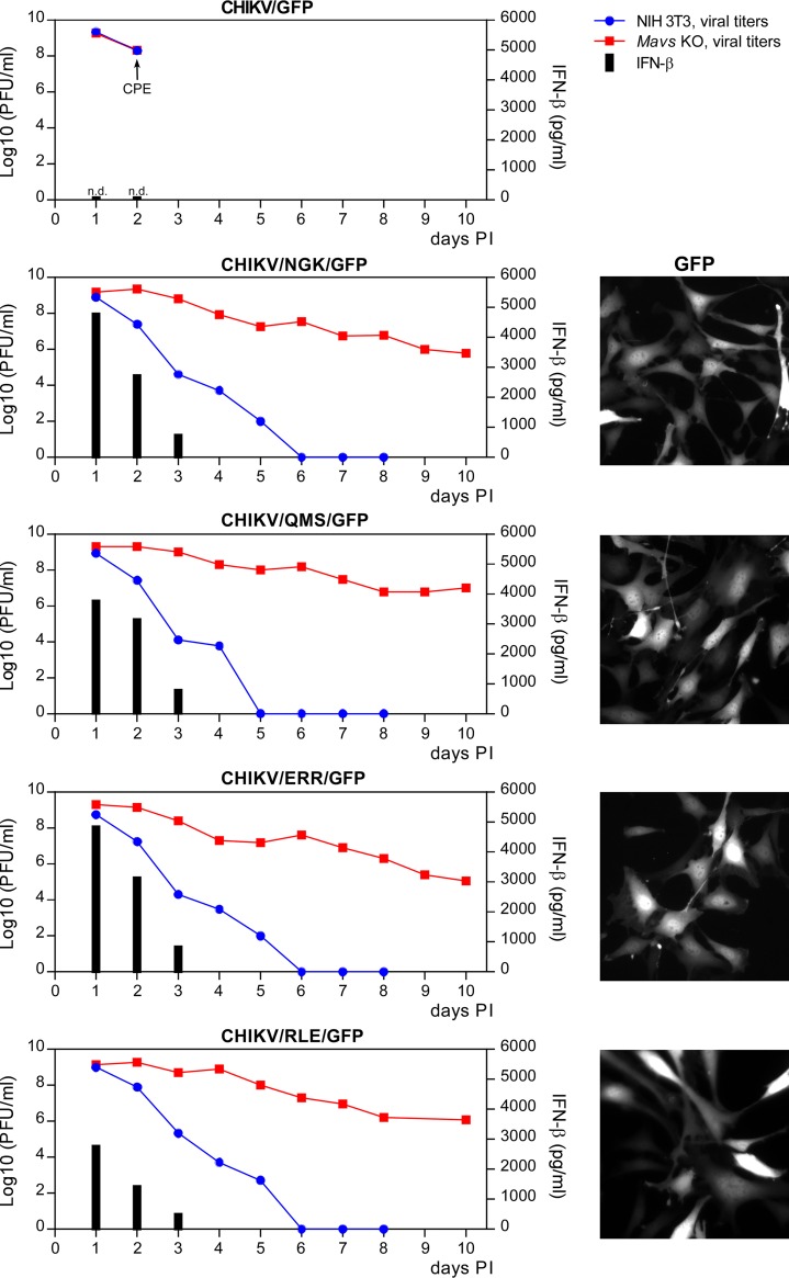
The above-described data demonstrated that in murine cells, the designed CHIKV mutants do not inhibit cellular transcription and their replication induces translational shutoff less inefficiently than that of SINV and SFV. Therefore, it was reasonable to expect that these mutants became noncytopathic. To test this possibility, NIH 3T3 cells and their Mavs knockout (KO) derivatives were infected with CHIKV/GFP and nsP2 VLoop mutants at an MOI of 20 PFU/cell. Within 8 h p.i., all of the cells became GFP positive. At 2 days p.i., CHIKV/GFP caused complete CPE in both cell lines. However, replication of the nsP2 mutants was noncytopathic, although the CHIKV/RLE/GFP noticeably affected the rates of cell growth (data not shown). In response to replication of the mutants, NIH 3T3 cells released high levels of IFN-β and cleared the infection (Fig. 10). Within 5 to 6 days p.i., cells became GFP negative, and infectious virus was no longer present in the media. In contrast, NIH 3T3 Mavs KO cells continued to express GFP (Fig. 10) and to release viral mutants for the entire duration of the 10-day-long experiment.
FIG 10.
The CHIKV nsP2 mutants are cleared from NIH 3T3 cells without cytopathic effect and persistently replicate in Mavs KO NIH 3T3 cells. NIH 3T3 and Mavs KO derivative cells were infected with the indicated viruses at an MOI of 20 PFU/cell. Media were replaced every 24 h, and cells were split upon reaching confluency. Viral titers were determined by plaque assay on BHK-21 cells, and the samples harvested from infected NIH 3T3 cells were used for assessment of IFN-β concentration (black bars) as described in Materials and Methods. The fluorescence images of Mavs KO NIH 3T3 cells were acquired at day 9 p.i. using a Nikon A1 fluorescence microscope with a 20× objective to demonstrate that they remained GFP positive and thus contained persistently replicating viruses.
SFV nsP2 with mutations in VLoop does not induce RPB1 degradation.
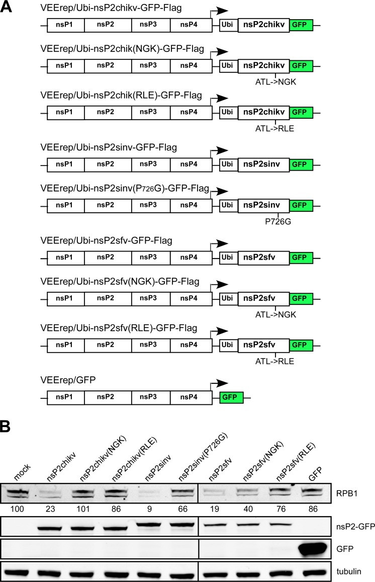
The results of this and previous studies demonstrated that VLoop plays a critical role in nuclear functions of SINV and CHIKV (44). They suggested that this highly variable peptide might similarly function in other OW alphaviruses during nsP2-mediated inhibition of cellular transcription. To test this possibility, we cloned wt SFV nsP2 and its variants with mutated VLoop into VEEV replicons as GFP fusions (Fig. 11A). Since CHIKV and SFV VLoop sequences exhibit some similarity (Fig. 1A), we argued that above-described CHIKV-specific mutations could have the same effect on the nuclear function of SFV nsP2. NIH 3T3 cells were infected at the same MOI with the replicons expressing the indicated cassettes of wt or mutant SINV, CHIKV, and SFV nsP2. At 8 h p.i., cells expressing all of the wt nsP2 proteins demonstrated the same levels of RPB1 degradation. All of the mutated versions of nsP2 became less efficient in the induction of such degradation (Fig. 11B). These results supported the hypothesis that VLoop represents an excellent target for modifications in development of attenuated OW alphaviruses that are less efficient in interfering with the antiviral response and thus are strong inducers of type I IFN.
FIG 11.
VLoop sequences in both CHIKV and SFV nsP2 proteins play critical roles in protein function in RPB1 degradation. (A) Schematic presentation of VEEV replicons encoding different forms of SINV, CHIKV, and SFV nsP2 fused with GFP. (B) NIH 3T3 cells were infected with indicated packaged replicons at an MOI of 20 infectious units/cell. At 8 h p.i., cells were harvested and cell lysates were analyzed by Western blotting using RPB1-, GFP-, and tubulin-specific Abs. Membranes were scanned on an Odyssey imager (LI-COR) and processed using the manufacturer’s software. The experiment was repeated twice, with reproducible results.
DISCUSSION
The alphavirus genome encodes a few structural and nonstructural proteins that facilitate replication of the viral genome and its packaging into released virions. The same proteins also modify multiple cellular processes. The resulting changes promote more efficient viral replication while inhibiting induction of the cellular antiviral response that can activate cell signaling and interfere with the infection spread. Alphaviruses have developed distinct means of downregulating the development of the antiviral state. The NW alphaviruses, such as Venezuelan (VEEV) and eastern (EEEV) equine encephalitis viruses, utilize their capsid protein to block the function of nuclear pores and nucleocytoplasmic traffic (31, 52–54). This, in turn, leads to rapid transcriptional shutoff, causes CPE, and makes cells incapable of both inducing cytokine expression and activating interferon-stimulated genes (ISGs). Capsid proteins of the OW alphaviruses, such as CHIKV, SINV, and SFV, do not exhibit this function (31), but instead, their nsP2 proteins mediate degradation of RPB1, the catalytic subunit of cellular DNA-dependent RNA polymerase II (29). Expression of the OW alphavirus nsP2 alone is cytotoxic and sufficient to cause transcriptional shutoff (47). Thus, transcriptional inhibition is a common characteristic of both OW and NW alphaviruses and is one of the critical contributors to their abilities to cause CPE in cultured cells. It is also an important determinant of alphavirus pathogenesis. Alterations of VEEV TC-83 capsid protein-specific nuclear function without affecting viral replication rates made such VEEV mutants (i) dramatically less cytopathic and (ii) capable of persistent replication in type I IFN signaling-deficient murine cells (55, 56). In cells competent in IFN signaling, these mutants were very efficient inducers of type I IFN, which rapidly activated ISGs in uninfected cells and eliminated replicating mutants from those already infected. The designed VEEV mutants also became less pathogenic in vivo (57). In another line of research, chimeric VEE/CHIKV or EEE/CHIKV viruses encoding the combination of the OW alphavirus-specific capsid and the NW alphavirus-specific nsP2, neither of which has transcription inhibitory function, were unable to cause CPE in vitro or any detectable disease in vivo (24, 58). Thus, alteration of viral nucleus-specific inhibitory functions can be used as an important means of alphavirus attenuation.
Development of OW alphavirus mutants, and CHIKV in particular, encoding nsP2 deficient in induction of transcriptional shutoff, is more challenging than designing modified NW alphaviruses with mutated capsid protein. The OW alphavirus nsP2 has a complicated, multidomain structure and numerous functions in viral RNA replication. Therefore, even small modifications, such as point mutations, usually affect a variety of critical processes in viral replication and alter the viability of mutants (40). Nevertheless, a few studies have identified a small set of point mutations in SINV and SFV nsP2 that made viral replicons less cytopathic and capable of persistent replication in vertebrate cell lines defective in type I IFN production/signaling (40, 41). However, the selected attenuating point mutations also strongly affected RNA replication rates. In viral context, lower levels of RNA replication, in turn, open an opportunity for viral evolution and rapid selection of more efficiently replicating true and pseudorevertants. Another complication was that the mutations selected in the context of nsP2 of one viral species, such as SINV, had a different or no effect on cytopathogenicity of other alphavirus representatives, such as CHIKV (32). In contrast to the case with SINV and SFV replicons, which required single point mutations in nsP2 for transformation to a noncytopathic phenotype, CHIKV nsP2 had to be more significantly mutated to produce a similar phenotype. The latter extensive modifications had deleterious effects on replication and transcription of CHIKV-specific RNAs (43). Therefore, most likely such nsP2 mutants of CHIKV could not be used for the development of attenuated and, at the same time, efficiently replicating viruses.
In a recent study, we identified new mutations in SINV nsP2 having strong negative effects on viral transcription inhibitory functions and cytopathogenicity without affecting viral replication rates (44). These mutations were located in a small highly variable peptide that we termed VLoop on the surface of the carboxy-terminal SAM MTase-like domain of nsP2. In the initial experiments, we introduced similar mutations into corresponding VLoop of CHIKV and explored their effects on the ability of the virus to cause CPE and inhibit cellular transcription. These modifications had detectable negative effects on CHIKV nsP2 nuclear functions. As did the mutations in the previously developed attenuated CHIKV replicon (43), VLoop mutations also caused alterations in ns polyprotein cleavage and probably affected a variety of processes in viral replication and interaction with the cells. Based on these results, the development of more attenuated and efficiently replicating viral mutants looked like an achievable goal, but on the other hand, it could require designing and testing too many additional versions of VLoop. Thus, the SINV-based data were found not to be directly applicable to CHIKV, amore pathogenic member of the genus.
The use of a library of CHIKV variants with randomized VLoop sequence opened an opportunity to evaluate a wide range of mutations for the ability to produce the noncytopathic phenotype in self-replicating CHIKV RNAs (Fig. 4). Application of this library-based approach led to the selection of hundreds of colonies of Purr cells, in which replication of CHIKV-specific RNA and expression of nsPs had no deleterious effect on cell viability. Subsequent sequencing of the persistently replicating CHIKV replicons identified a variety of VLoop sequences that rendered RNA replication noncytopathic. After replacement of the wt VLoop-encoding sequence in CHIKV replicon (CHIKrep/GFP/Pac) by those identified, the mutated constructs became capable of developing persistent noncytopathic replication in BHK-21 cells. Their efficiency to form Purr colonies depended on the particular VLoop sequence and also on the level of viral RNA replication. The least efficiently replicating CHIKrep/RLH(A730V)/GFP/Pac construct produced the highest numbers of colonies. However, other noncytopathic replicons demonstrated higher levels of replication that could be sufficient to support the replication of corresponding infectious viruses. Indeed, CHIKV variants encoding VLoop of more efficiently replicating replicon constructs were viable, did not require additional adaptive mutations, and replicated to titers very similar to those of CHIKV/GFP expressing wt nsP2. Most importantly, despite retaining efficient replication, in contrast to parental CHIKV/GFP, they all were very potent IFN-β inducers. Accordingly, they could not develop a spreading infection in cells competent in type I IFN production and signaling (Fig. 7). All of them were cleared from infected murine cells within 5 to 6 days p.i. by autocrine IFN-β signaling that likely led to ISG activation (Fig. 10) (59). Because they were noncytopathic, these viruses readily developed a persistent infection in NIH 3T3 Mavs KO cells, which were unable to mount a type I IFN response (Fig. 10). The designed viruses did not cause degradation of RPB1 and inhibition of transcription of cellular mRNA and rRNA (Fig. 9A). Compared to SINV, CHIKV replication is relatively inefficient in inhibition of cellular translation in murine cells (Fig. 9B); therefore, these mutants, in contrast to SINV, did not require additional mutations in nsP3 (44). Previously, such mutations in the nsP3 macrodomain had a negative effect on the development of SINV-induced translational shutoff and were absolutely required for selection of noncytopathic SINV. These new variants represent the first example of CHIKV mutants that demonstrate essentially wt levels of replication in cell culture while being incapable of inhibiting cellular transcription and antiviral response.
Prior studies demonstrated that presence of SAM MTase-like domain is a requirement for nsP2-specific helicase and protease activities, and mutations can make SINV, SFV, and CHIKV either very poorly replicating or nonviable (43, 47). On the other hand, this domain also plays a critical role in the nuclear function of nsP2, although the mechanism remains insufficiently understood (29). In the case of NW alphaviruses, such as VEEV and EEEV, nsP2 has no nuclear functions (54). Since the NW alphavirus nsP2 is not transported to the nucleus (54), it remains unclear whether its nuclear function is completely lost or the lack of it is a result of a change in nsP2 compartmentalization during viral replication. In any scenario, despite having a high level of identity with the OW alphavirus-specific counterparts, the NW alphavirus-specific SAM MTase-like nsP2 domain is no longer active in inhibition of the antiviral response, but it retains functions in the enzymatic activities of the protein. A lack of nuclear function appears to be a logical step in nsP2 evolution because expression of VEEV capsid protein in chimeric SINV/VEEV completely blocks translocation of SINV nsP2 into the nuclei (54).
In summary, this study resulted in the identification of a small peptide loop on the surface of the OW alphavirus CHIKV nsP2 that plays a critical role in determining the nuclear function of this nonstructural protein. The selected amino acid sequences that replaced original wt VLoop had no negative effect on CHIKV replication in tested rodent cell types. However, they had a deleterious effect on the transcription-inhibitory functions of CHIKV nsP2 and made the corresponding CHIKV variants dramatically less cytopathic. Inhibition of transcription and downregulation of the innate immune response are fundamental characteristics of CHIKV replication and, as in many other viral infections, are likely major components of viral pathogenesis. The newly designed CHIKV mutants and their replicons display a wide range of possibilities for their application in different areas of research. First, modified VLoop-encoding sequences can be further applied in development of new vaccine candidates and to additionally improve already attenuated strain CHIKV 181/25, which previously remained capable of inducing some adverse effects (60, 61). The wt level of replication of these mutants suggests that during the serial viral passage, there may be no selection pressure to further evolve to a more efficiently replicating phenotype. The replacement of three amino acids in nsP2 instead of making point mutations also makes an additional contribution to the stability of the attenuated phenotype during virus production in cells deficient in type I IFN induction and signaling. Second, similar mutants designed on the basis of wt CHIKV genome can be used to study critical aspects of CHIKV-host interactions and pathogenesis. Third, new noncytopathic CHIKV replicons demonstrate a high level of expression of heterologous genes and can be applied for expression of therapeutic proteins or antigens. Lastly, stable cell lines that contain noncytopathic CHIKV replicons may be applicable for screening of antiviral drugs without using replication-competent infectious CHIKV.
MATERIALS AND METHODS
Cell cultures.
NIH 3T3 cells were obtained from the American Type Culture Collection (Manassas, VA). BHK-21 cells were kindly provided by Paul Olivo (Washington University, St. Louis, MO). These cell lines were maintained at 37°C in alpha minimum essential medium (αMEM) supplemented with 10% fetal bovine serum (FBS) and vitamins. The Mavs KO NIH 3T3 cell line was generated using clustered regularly interspaced short palindromic repeat (CRISPR) Cas9 nuclease vector plasmid according to the manufacturer’s instructions (Invitrogen) as described elsewhere (11). Cell clones were initially analyzed in terms of target protein expression, and then KO of both alleles was confirmed by sequencing the targeted fragment in the cell genome. The Rig-I Mavs dKD NIH 3T3 cell line was described elsewhere (62).
Plasmid constructs.
The original plasmid containing the infectious cDNA of the attenuated strain CHIKV 181/25 was kindly provided by Scott Weaver (University of Texas Medical Branch, Galveston, TX). The derivative of this infectious cDNA clone that encodes GFP under the control of the subgenomic promoter, CHIKV/GFP, is described elsewhere (63). CHIKV 181/25 and parental AF15561 strains differ only by 4 aa in structural proteins and 1 aa in nsP1, of which only 2 substitutions in E2 glycoprotein determine the attenuated phenotype of 181/25 (64). Thus, CHIKV 181/25 was used as a representative model of virulent CHIKV for analysis of virus interaction with host cells, inhibition of cellular transcription, and antiviral response during CHIKV infection. The presence of tissue culture-adaptive mutations in E2 increased infectivity of the virus in vitro and prevented viral evolution. To simplify presentation, the CHIKV 181/25 strain is referred as CHIKV. pCHIKrep/Pac and pCHIKrep/GFP/Pac were designed using the standard PCR-based techniques. Mutations in the VLoop sequence of replicons were introduced by standard PCR-based approach. A library of replicons with randomized VLoop was made using a gene block with randomized corresponding nucleotide sequence (Integrated DNA Technologies). All of the introduced modifications were confirmed by sequencing. Sequences of the plasmids and details of the cloning procedures can be provided upon request.
In vitro RNA transcription and transfection.
Plasmids were purified by ultracentrifugation in CsCl gradients. Then they were linearized using NotI restriction sites located downstream of the 3ʹ-terminal poly(A) sequence in viral and replicon genomes. RNAs were synthesized by SP6 RNA polymerase in the presence of a cap analog (New England BioLabs). The quality and concentrations of the synthesized RNAs were tested by agarose gel electrophoresis, and aliquots of the transcription reactions were used for electroporation without additional RNA purification. Electroporations of BHK-21 cells by in vitro-synthesized virus-specific RNAs were performed under previously described conditions (65, 66). Briefly, 5 × 10⁶ BHK-21 cells in 450 μl of phosphate-buffered saline (PBS) were electroporated with replicon or viral RNAs in 2-mm cuvettes by 2 pulses at 1,500 V/25 μF using Gene Pulser II (Bio-Rad). Viruses were harvested at 20 to 24 h postelectroporation, and titers were determined by plaque assay on BHK-21 cells (67).
Cells electroporated with replicons were seeded into 100-mm dishes, and after ∼24 h of incubation at 37°C, media were supplemented with puromycin (10 μg/ml). Incubation continued until the development of colonies of Purr cells. The cells were either stained by crystal violet or collected for further analysis of persistently replicating replicons.
Infectious center assay (ICA).
To compare infectivities of the in vitro-synthesized RNAs, equal amounts were electroporated into BHK-21 cells. Ten-fold dilutions of electroporated cells were seeded in 6-well Costar plates containing subconfluent monolayers of naive BHK-21 cells. After 2-h-long incubation at 37°C, cells were overlaid with agarose supplemented with MEM and 3% FBS. Plaques were stained with crystal violet after 3 days of incubation at 37°C, and RNA infectivity was determined as PFU per microgram of electroporated RNA.
Analysis of viral replication.
In standard experiments, 5 × 10⁵ cells in 6-well Costar plates were infected with recombinant CHIKV at MOIs indicated in the figure legends in 200 μl of phosphate-buffered saline (PBS) supplemented with 1% FBS. After 1-h-long virus adsorption, the inoculum was replaced by complete media, and cells were incubated at 37°C. At the time points indicated in the figures, media were replaced, and viral titers in the harvested samples were determined by plaque assay on BHK-21 cells (67). In the analyses of viral clearance or persistent replication, cells were trypsinized and replated upon reaching confluency.
IFN-β measurement.
Cells were infected with recombinant viruses as described in the figure legends. Media were harvested at the time points indicated in the figure legends. In these samples, the pH was stabilized by adding HEPES buffer (pH 7.5) to 0.01 M. Concentrations of IFN-β were measured with the VeriKine IFN-β enzyme-linked immunosorbent assay (ELISA) kit (PBL InterferonSource) according to the manufacturer’s recommendations.
Western blotting.
Equal amounts of protein lysates were separated in 4% to 12% gradient NuPAGE gels (Invitrogen). After protein transfer, the membranes were incubated in blocking buffer and then with primary antibodies, followed by incubation with corresponding, infrared dye-labeled secondary antibodies. For imaging and quantitative analysis, membranes were scanned on an Odyssey imager (LI-COR). The following primary antibodies were used: anti-tubulin rat MAb (custom-made), pan-anti-alphavirus nsP2 MAb (custom-made), anti-CHIKV nsP1 MAb (custom-made), anti-CHIKV nsP3 MAb (custom-made), and anti-RPB1 MAb (clone F12; Santa Cruz Biotechnology). All custom MAbs were developed in UAB Epitope Recognition & Immunoreagent Core (11, 29).
Analysis of RNA synthesis.
NIH 3T3 cells were infected with recombinant viruses at an MOI of 20 PFU/cell. Viral and cellular RNAs were metabolically labeled between 4 and 8 h p.i. in 0.8 ml of complete medium supplemented with [3H]uridine (20 μCi/ml). RNAs were isolated from the cells by TRIzol reagent as recommended by the manufacturer (Invitrogen) and then denatured with glyoxal in dimethyl sulfoxide as described elsewhere (39). RNAs were analyzed by agarose gel electrophoresis in 0.01 M sodium phosphate buffer (pH 7.0). Gels were impregnated with 2,5-diphenyloxazol (PPO) and used for autofluorography (39).
Analysis of protein synthesis.
A total of 5 × 10⁵ NIH 3T3 cells were seeded into six-well Costar plates and infected with recombinant CHIKV variants at an MOI of 20 PFU/cell for 1 h. After 6 h of incubation at 37°C, cells were washed with PBS and then incubated for 30 min at 37°C in Dulbecco’s modified Eagle’s medium lacking methionine, supplemented with 0.1% FBS and 20 μCi of [35S]methionine/ml. Cells were dissolved in standard loading buffer for protein electrophoresis, and equal amounts of lysates were analyzed by electrophoresis in 10% NuPAGE gels, followed by autoradiography.
ACKNOWLEDGMENTS
We thank Scott Weaver for providing infectious cDNA clone of CHIKV 181/25 and Mary Accavitti-Loper for providing anti-CHIKV nsP1 and anti-CHIKV nsP3 MAbs and for assistance in production of anti-nsP2 MAbs.
This study was supported by Public Health Service grants AI073301, AI118867, and AI133159 to E.I.F. and AI119627 to I.F.
REFERENCES
- 1.Strauss JH, Strauss EG. 1994. The alphaviruses: gene expression, replication, evolution. Microbiol Rev 58:491–562. [DOI] [PMC free article] [PubMed] [Google Scholar]
- 2.Lanciotti RS, Valadere AM. 2014. Transcontinental movement of Asian genotype chikungunya virus. Emerg Infect Dis 20:1400–1402. doi: 10.3201/eid2008.140268. [DOI] [PMC free article] [PubMed] [Google Scholar]
- 3.Nappe TM, Chuhran CM, Johnson SA. 2016. The Chikungunya virus: an emerging US pathogen. World J Emerg Med 7:65–67. doi: 10.5847/wjem.j.1920-8642.2016.01.012. [DOI] [PMC free article] [PubMed] [Google Scholar]
- 4.Weaver SC, Frolov I. 2005. Togaviruses, p 1010–1024. In Mahy BWJ, Meulen VT (ed), Virology, vol 2, Hodder Arnold, London, UK. [Google Scholar]
- 5.Weaver SC, Forrester NL. 2015. Chikungunya: evolutionary history and recent epidemic spread. Antiviral Res 120:32–39. doi: 10.1016/j.antiviral.2015.04.016. [DOI] [PubMed] [Google Scholar]
- 6.Weaver SC, Lecuit M. 2015. Chikungunya virus infections. N Engl J Med 373:94–95. doi: 10.1056/NEJMc1505501. [DOI] [PubMed] [Google Scholar]
- 7.Weaver SC, Lecuit M. 2015. Chikungunya virus and the global spread of a mosquito-borne disease. N Engl J Med 372:1231–1239. doi: 10.1056/NEJMra1406035. [DOI] [PubMed] [Google Scholar]
- 8.McSweegan E, Weaver SC, Lecuit M, Frieman M, Morrison TE, Hrynkow S. 2015. The global virus network: challenging chikungunya. Antiviral Res 120:147–152. doi: 10.1016/j.antiviral.2015.06.003. [DOI] [PMC free article] [PubMed] [Google Scholar]
- 9.Tsetsarkin KA, Vanlandingham DL, McGee CE, Higgs S. 2007. A single mutation in chikungunya virus affects vector specificity and epidemic potential. PLoS Pathog 3:e201. doi: 10.1371/journal.ppat.0030201. [DOI] [PMC free article] [PubMed] [Google Scholar]
- 10.Meshram CD, Agback P, Shiliaev N, Urakova N, Mobley JA, Agback T, Frolova EI, Frolov I. 2018. Multiple host factors interact with hypervariable domain of chikungunya virus nsP3 and determine viral replication in cell-specific mode. J Virol 92:e00838-18. doi: 10.1128/JVI.00838-18. [DOI] [PMC free article] [PubMed] [Google Scholar]
- 11.Kim DY, Reynaud JM, Rasalouskaya A, Akhrymuk I, Mobley JA, Frolov I, Frolova EI. 2016. New World and Old World alphaviruses have evolved to exploit different components of stress granules, FXR and G3BP proteins, for assembly of viral replication complexes. PLoS Pathog 12:e1005810. doi: 10.1371/journal.ppat.1005810. [DOI] [PMC free article] [PubMed] [Google Scholar]
- 12.Hardy WR, Strauss JH. 1989. Processing the nonstructural proteins of Sindbis virus: nonstructural proteinase is in the C-terminal half of nsP2 and functions both in cis and trans. J Virol 63:4653–4664. [DOI] [PMC free article] [PubMed] [Google Scholar]
- 13.Lemm JA, Rice CM. 1993. Roles of nonstructural polyproteins and cleavage products in regulating Sindbis virus RNA replication and transcription. J Virol 67:1916–1926. [DOI] [PMC free article] [PubMed] [Google Scholar]
- 14.Shirako Y, Strauss JH. 1990. Cleavage between nsP1 and nsP2 initiates the processing pathway of Sindbis virus nonstructural polyprotein P123. Virology 177:54–64. doi: 10.1016/0042-6822(90)90459-5. [DOI] [PubMed] [Google Scholar]
- 15.Shirako Y, Strauss JH. 1994. Regulation of Sindbis virus RNA replication: uncleaved P123 and nsP4 function in minus-strand RNA synthesis, whereas cleaved products from P123 are required for efficient plus-strand RNA synthesis. J Virol 68:1874–1885. [DOI] [PMC free article] [PubMed] [Google Scholar]
- 16.Strauss EG, Rice CM, Strauss JH. 1984. Complete nucleotide sequence of the genomic RNA of Sindbis virus. Virology 133:92–110. doi: 10.1016/0042-6822(84)90428-8. [DOI] [PubMed] [Google Scholar]
- 17.Rikkonen M, Peranen J, Kaariainen L. 1994. ATPase and GTPase activities associated with Semliki Forest virus nonstructural protein nsP2. J Virol 68:5804–5810. [DOI] [PMC free article] [PubMed] [Google Scholar]
- 18.Karpe YA, Aher PP, Lole KS. 2011. NTPase and 5ʹ-RNA triphosphatase activities of Chikungunya virus nsP2 protein. PLoS One 6:e22336. doi: 10.1371/journal.pone.0022336. [DOI] [PMC free article] [PubMed] [Google Scholar]
- 19.Das PK, Merits A, Lulla A. 2014. Functional cross-talk between distant domains of chikungunya virus non-structural protein 2 is decisive for its RNA-modulating activity. J Biol Chem 289:5635–5653. doi: 10.1074/jbc.M113.503433. [DOI] [PMC free article] [PubMed] [Google Scholar]
- 20.Rausalu K, Utt A, Quirin T, Varghese FS, Zusinaite E, Das PK, Ahola T, Merits A. 2016. Chikungunya virus infectivity, RNA replication and non-structural polyprotein processing depend on the nsP2 protease’s active site cysteine residue. Sci Rep 6:37124. doi: 10.1038/srep37124. [DOI] [PMC free article] [PubMed] [Google Scholar]
- 21.Vasiljeva L, Merits A, Auvinen P, Kaariainen L. 2000. Identification of a novel function of the alphavirus capping apparatus. RNA 5ʹ-triphosphatase activity of Nsp2. J Biol Chem 275:17281–17287. doi: 10.1074/jbc.M910340199. [DOI] [PubMed] [Google Scholar]
- 22.Michel G, Petrakova O, Atasheva S, Frolov I. 2007. Adaptation of Venezuelan equine encephalitis virus lacking 51-nt conserved sequence element to replication in mammalian and mosquito cells. Virology 362:475–487. doi: 10.1016/j.virol.2007.01.009. [DOI] [PMC free article] [PubMed] [Google Scholar]
- 23.Volkova E, Frolova E, Darwin JR, Forrester NL, Weaver SC, Frolov I. 2008. IRES-dependent replication of Venezuelan equine encephalitis virus makes it highly attenuated and incapable of replicating in mosquito cells. Virology 377:160–169. doi: 10.1016/j.virol.2008.04.020. [DOI] [PMC free article] [PubMed] [Google Scholar]
- 24.Kim DY, Atasheva S, Foy NJ, Wang E, Frolova EI, Weaver S, Frolov I. 2011. Design of chimeric alphaviruses with a programmed, attenuated, cell type-restricted phenotype. J Virol 85:4363–4376. doi: 10.1128/JVI.00065-11. [DOI] [PMC free article] [PubMed] [Google Scholar]
- 25.Fayzulin R, Frolov I. 2004. Changes of the secondary structure of the 5ʹ end of the Sindbis virus genome inhibit virus growth in mosquito cells and lead to accumulation of adaptive mutations. J Virol 78:4953–4964. doi: 10.1128/JVI.78.10.4953-4964.2004. [DOI] [PMC free article] [PubMed] [Google Scholar]
- 26.Frolova EI, Fayzulin RZ, Cook SH, Griffin DE, Rice CM, Frolov I. 2002. Roles of nonstructural protein nsP2 and alpha/beta interferons in determining the outcome of Sindbis virus infection. J Virol 76:11254–11264. doi: 10.1128/JVI.76.22.11254-11264.2002. [DOI] [PMC free article] [PubMed] [Google Scholar]
- 27.Rikkonen M, Peranen J, Kaariainen L. 1992. Nuclear and nucleolar targeting signals of Semliki Forest virus nonstructural protein nsP2. Virology 189:462–473. doi: 10.1016/0042-6822(92)90570-F. [DOI] [PubMed] [Google Scholar]
- 28.Peranen J, Rikkonen M, Liljestrom P, Kaariainen L. 1990. Nuclear localization of Semliki Forest virus-specific nonstructural protein nsP2. J Virol 64:1888–1896. [DOI] [PMC free article] [PubMed] [Google Scholar]
- 29.Akhrymuk I, Kulemzin SV, Frolova EI. 2012. Evasion of the innate immune response: the Old World alphavirus nsP2 protein induces rapid degradation of Rpb1, a catalytic subunit of RNA polymerase II. J Virol 86:7180–7191. doi: 10.1128/JVI.00541-12. [DOI] [PMC free article] [PubMed] [Google Scholar]
- 30.Garmashova N, Gorchakov R, Frolova E, Frolov I. 2006. Sindbis virus nonstructural protein nsP2 is cytotoxic and inhibits cellular transcription. J Virol 80:5686–5696. doi: 10.1128/JVI.02739-05. [DOI] [PMC free article] [PubMed] [Google Scholar]
- 31.Garmashova N, Gorchakov R, Volkova E, Paessler S, Frolova E, Frolov I. 2007. The Old World and New World alphaviruses use different virus-specific proteins for induction of transcriptional shutoff. J Virol 81:2472–2484. doi: 10.1128/JVI.02073-06. [DOI] [PMC free article] [PubMed] [Google Scholar]
- 32.Fros JJ, van der Maten E, Vlak JM, Pijlman GP. 2013. The C-terminal domain of chikungunya virus nsP2 independently governs viral RNA replication, cytopathicity, and inhibition of interferon signaling. J Virol 87:10394–10400. doi: 10.1128/JVI.00884-13. [DOI] [PMC free article] [PubMed] [Google Scholar]
- 33.Fros JJ, Liu WJ, Prow NA, Geertsema C, Ligtenberg M, Vanlandingham DL, Schnettler E, Vlak JM, Suhrbier A, Khromykh AA, Pijlman GP. 2010. Chikungunya virus nonstructural protein 2 inhibits type I/II interferon-stimulated JAK-STAT signaling. J Virol 84:10877–10887. doi: 10.1128/JVI.00949-10. [DOI] [PMC free article] [PubMed] [Google Scholar]
- 34.Frolov I, Akhrymuk M, Akhrymuk I, Atasheva S, Frolova EI. 2012. Early events in alphavirus replication determine the outcome of infection. J Virol 86:5055–5066. doi: 10.1128/JVI.07223-11. [DOI] [PMC free article] [PubMed] [Google Scholar]
- 35.Russo AT, White MA, Watowich SJ. 2006. The crystal structure of the Venezuelan equine encephalitis alphavirus nsP2 protease. Structure 14:1449–1458. doi: 10.1016/j.str.2006.07.010. [DOI] [PubMed] [Google Scholar]
- 36.Strauss EG, De Groot RJ, Levinson R, Strauss JH. 1992. Identification of the active site residues in the nsP2 proteinase of Sindbis virus. Virology 191:932–940. doi: 10.1016/0042-6822(92)90268-T. [DOI] [PMC free article] [PubMed] [Google Scholar]
- 37.Shin G, Yost SA, Miller MT, Elrod EJ, Grakoui A, Marcotrigiano J. 2012. Structural and functional insights into alphavirus polyprotein processing and pathogenesis. Proc Natl Acad Sci U S A 109:16534–16539. doi: 10.1073/pnas.1210418109. [DOI] [PMC free article] [PubMed] [Google Scholar]
- 38.Gorchakov R, Frolova E, Frolov I. 2005. Inhibition of transcription and translation in Sindbis virus-infected cells. J Virol 79:9397–9409. doi: 10.1128/JVI.79.15.9397-9409.2005. [DOI] [PMC free article] [PubMed] [Google Scholar]
- 39.Gorchakov R, Frolova E, Sawicki S, Atasheva S, Sawicki D, Frolov I. 2008. A new role for ns polyprotein cleavage in Sindbis virus replication. J Virol 82:6218–6231. doi: 10.1128/JVI.02624-07. [DOI] [PMC free article] [PubMed] [Google Scholar]
- 40.Frolov I, Agapov E, Hoffman TA Jr, Prágai BM, Lippa M, Schlesinger S, Rice CM. 1999. Selection of RNA replicons capable of persistent noncytopathic replication in mammalian cells. J Virol 73:3854–3865. [DOI] [PMC free article] [PubMed] [Google Scholar]
- 41.Perri S, Driver DA, Gardner JP, Sherrill S, Belli BA, Dubensky TW Jr, Polo JM. 2000. Replicon vectors derived from Sindbis virus and Semliki forest virus that establish persistent replication in host cells. J Virol 74:9802–9807. doi: 10.1128/JVI.74.20.9802-9807.2000. [DOI] [PMC free article] [PubMed] [Google Scholar]
- 42.Casales E, Rodriguez-Madoz JR, Ruiz-Guillen M, Razquin N, Cuevas Y, Prieto J, Smerdou C. 2008. Development of a new noncytopathic Semliki Forest virus vector providing high expression levels and stability. Virology 376:242–251. doi: 10.1016/j.virol.2008.03.016. [DOI] [PubMed] [Google Scholar]
- 43.Utt A, Das PK, Varjak M, Lulla V, Lulla A, Merits A. 2015. Mutations conferring a noncytotoxic phenotype on chikungunya virus replicons compromise enzymatic properties of nonstructural protein 2. J Virol 89:3145–3162. doi: 10.1128/JVI.03213-14. [DOI] [PMC free article] [PubMed] [Google Scholar]
- 44.Akhrymuk I, Frolov I, Frolova EI. 2018. Sindbis virus infection causes cell death by nsP2-induced transcriptional shutoff or by nsp3-dependent translational shutoff. J Virol 92:e01388-18. doi: 10.1128/JVI.01388-18. [DOI] [PMC free article] [PubMed] [Google Scholar]
- 45.Petrakova O, Volkova E, Gorchakov R, Paessler S, Kinney RM, Frolov I. 2005. Noncytopathic replication of Venezuelan equine encephalitis virus and eastern equine encephalitis virus replicons in mammalian cells. J Virol 79:7597–7608. doi: 10.1128/JVI.79.12.7597-7608.2005. [DOI] [PMC free article] [PubMed] [Google Scholar]
- 46.Mayuri, Geders TW, Smith JL, Kuhn RJ. 2008. Role for conserved residues of Sindbis virus nonstructural protein 2 methyltransferase-like domain in regulation of minus-strand synthesis and development of cytopathic infection. J Virol 82:7284–7297. doi: 10.1128/JVI.00224-08. [DOI] [PMC free article] [PubMed] [Google Scholar]
- 47.Frolov I, Garmashova N, Atasheva S, Frolova EI. 2009. Random insertion mutagenesis of sindbis virus nonstructural protein 2 and selection of variants incapable of downregulating cellular transcription. J Virol 83:9031–9044. doi: 10.1128/JVI.00850-09. [DOI] [PMC free article] [PubMed] [Google Scholar]
- 48.Gorchakov R, Frolova E, Williams BR, Rice CM, Frolov I. 2004. PKR-dependent and -independent mechanisms are involved in translational shutoff during Sindbis virus infection. J Virol 78:8455–8467. doi: 10.1128/JVI.78.16.8455-8467.2004. [DOI] [PMC free article] [PubMed] [Google Scholar]
- 49.Barry G, Breakwell L, Fragkoudis R, Attarzadeh-Yazdi G, Rodriguez-Andres J, Kohl A, Fazakerley JK. 2009. PKR acts early in infection to suppress Semliki Forest virus production and strongly enhances the type I interferon response. J Gen Virol 90:1382–1391. doi: 10.1099/vir.0.007336-0. [DOI] [PMC free article] [PubMed] [Google Scholar]
- 50.Frolov I, Schlesinger S. 1994. Translation of Sindbis virus mRNA: effects of sequences downstream of the initiating codon. J Virol 68:8111–8117. [DOI] [PMC free article] [PubMed] [Google Scholar]
- 51.Kutchko KM, Madden EA, Morrison C, Plante KS, Sanders W, Vincent HA, Cruz Cisneros MC, Long KM, Moorman NJ, Heise MT, Laederach A. 2018. Structural divergence creates new functional features in alphavirus genomes. Nucleic Acids Res 46:3657–3670. doi: 10.1093/nar/gky012. [DOI] [PMC free article] [PubMed] [Google Scholar]
- 52.Atasheva S, Fish A, Fornerod M, Frolova EI. 2010. Venezuelan equine encephalitis virus capsid protein forms a tetrameric complex with CRM1 and importin alpha/beta that obstructs nuclear pore complex function. J Virol 84:4158–4171. doi: 10.1128/JVI.02554-09. [DOI] [PMC free article] [PubMed] [Google Scholar]
- 53.Garmashova N, Atasheva S, Kang W, Weaver SC, Frolova E, Frolov I. 2007. Analysis of Venezuelan equine encephalitis virus capsid protein function in the inhibition of cellular transcription. J Virol 81:13552–13565. doi: 10.1128/JVI.01576-07. [DOI] [PMC free article] [PubMed] [Google Scholar]
- 54.Atasheva S, Garmashova N, Frolov I, Frolova E. 2008. Venezuelan equine encephalitis virus capsid protein inhibits nuclear import in mammalian but not in mosquito cells. J Virol 82:4028–4041. doi: 10.1128/JVI.02330-07. [DOI] [PMC free article] [PubMed] [Google Scholar]
- 55.Atasheva S, Krendelchtchikova V, Liopo A, Frolova E, Frolov I. 2010. Interplay of acute and persistent infections caused by Venezuelan equine encephalitis virus encoding mutated capsid protein. J Virol 84:10004–10015. doi: 10.1128/JVI.01151-10. [DOI] [PMC free article] [PubMed] [Google Scholar]
- 56.Atasheva S, Frolova EI, Frolov I. 2014. Interferon-stimulated poly(ADP-ribose) polymerases are potent inhibitors of cellular translation and virus replication. J Virol 88:2116–2130. doi: 10.1128/JVI.03443-13. [DOI] [PMC free article] [PubMed] [Google Scholar]
- 57.Atasheva S, Kim DY, Frolova EI, Frolov I. 2015. Venezuelan equine encephalitis virus variants lacking transcription inhibitory functions demonstrate highly attenuated phenotype. J Virol 89:71–82. doi: 10.1128/JVI.02252-14. [DOI] [PMC free article] [PubMed] [Google Scholar]
- 58.Wang E, Kim DY, Weaver SC, Frolov I. 2011. Chimeric Chikungunya viruses are nonpathogenic in highly sensitive mouse models but efficiently induce a protective immune response. J Virol 85:9249–9252. doi: 10.1128/JVI.00844-11. [DOI] [PMC free article] [PubMed] [Google Scholar]
- 59.Atasheva S, Akhrymuk M, Frolova EI, Frolov I. 2012. New PARP gene with an anti-alphavirus function. J Virol 86:8147–8160. doi: 10.1128/JVI.00733-12. [DOI] [PMC free article] [PubMed] [Google Scholar]
- 60.Levitt NH, Ramsburg HH, Hasty SE, Repik PM, Cole FE Jr, Lupton HW. 1986. Development of an attenuated strain of chikungunya virus for use in vaccine production. Vaccine 4:157–162. doi: 10.1016/0264-410X(86)90003-4. [DOI] [PubMed] [Google Scholar]
- 61.Edelman R, Tacket CO, Wasserman SS, Bodison SA, Perry JG, Mangiafico JA. 2000. Phase II safety and immunogenicity study of live chikungunya virus vaccine TSI-GSD-218. Am J Trop Med Hyg 62:681–685. doi: 10.4269/ajtmh.2000.62.681. [DOI] [PubMed] [Google Scholar]
- 62.Akhrymuk I, Frolov I, Frolova EI. 2016. Both RIG-I and MDA5 detect alphavirus replication in concentration-dependent mode. Virology 487:230–241. doi: 10.1016/j.virol.2015.09.023. [DOI] [PMC free article] [PubMed] [Google Scholar]
- 63.Reynaud JM, Kim DY, Atasheva S, Rasalouskaya A, White JP, Diamond MS, Weaver SC, Frolova EI, Frolov I. 2015. IFIT1 differentially interferes with translation and replication of alphavirus genomes and promotes induction of type I interferon. PLoS Pathog 11:e1004863. doi: 10.1371/journal.ppat.1004863. [DOI] [PMC free article] [PubMed] [Google Scholar]
- 64.Gorchakov R, Wang E, Leal G, Forrester NL, Plante K, Rossi SL, Partidos CD, Adams AP, Seymour RL, Weger J, Borland EM, Sherman MB, Powers AM, Osorio JE, Weaver SC. 2012. Attenuation of Chikungunya virus vaccine strain 181/clone 25 is determined by two amino acid substitutions in the E2 envelope glycoprotein. J Virol 86:6084–6096. doi: 10.1128/JVI.06449-11. [DOI] [PMC free article] [PubMed] [Google Scholar]
- 65.Liljeström P, Lusa S, Huylebroeck D, Garoff H. 1991. In vitro mutagenesis of a full-length cDNA clone of Semliki Forest virus: the small 6,000-molecular-weight membrane protein modulates virus release. J Virol 65:4107–4113. [DOI] [PMC free article] [PubMed] [Google Scholar]
- 66.Gorchakov R, Hardy R, Rice CM, Frolov I. 2004. Selection of functional 5ʹ cis-acting elements promoting efficient sindbis virus genome replication. J Virol 78:61–75. doi: 10.1128/JVI.78.1.61-75.2004. [DOI] [PMC free article] [PubMed] [Google Scholar]
- 67.Lemm JA, Durbin RK, Stollar V, Rice CM. 1990. Mutations which alter the level or structure of nsP4 can affect the efficiency of Sindbis virus replication in a host-dependent manner. J Virol 64:3001–3011. [DOI] [PMC free article] [PubMed] [Google Scholar]


