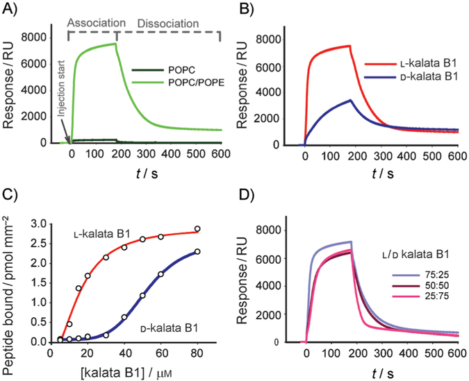
Figure 5.
Membrane binding of native and D-kalata B1 studied by surface plasmon resonance. Kalata B1 and its d isomer were injected for 180 s (association phase) over lipid surfaces deposited on an L1 chip, and dissociation was monitored after the injection had stopped (dissociation phase). A) Sensorgrams obtained with 50 μM native kalata B1 over POPC and POPC/POPE (4:1) lipid surfaces. B) Comparison of 50 μM L- and D-kalata B1 injected over POPC/POPE (4:1). C) Amount of L- and D-kalata B1 bound to the POPC/POPE (4:1) lipid surface at the end of the association phase as a function of different peptide concentrations. D) Sensorgrams obtained with mixtures of L and D isomers of kalata B1 in ratios as indicated on the figure).

