Abstract
Introduction
The aim of the study was the application and evaluation of real-time PCRs based on the fluorescence of SYBR Green I intercalating dye for the detection of three Bacillus anthracis genes in contaminated liver and blood samples. The goals for detection were rpoB gene as a chromosomal marker, pag gene located on plasmid pXO1, and capC gene located on plasmid pXO2.
Material and Methods
Five B. anthracis strains were used for the experiments. Additionally, single strains of other species of the genus Bacillus, i.e. B. cereus, B. brevis, B. subtilis, and B. megaterium, and strains of six other species were used for evaluation of the specificity of the tests. Three SYBR Green I real-time PCRs were conducted allowing confirmation of B. anthracis in the biological samples.
Results
The observation of amplification curves in real-time PCRs enabled the detection of the chromosomally encoded rpoB gene, pag gene, and capC gene of B. anthracis. The specificity of the tests was confirmed by estimation of the melting temperature of the PCR products. The sensitivity and linearity of the reactions were determined using regression coefficients. Strains of other microbial species did not reveal real-time PCR products.
Conclusion
All real-time PCRs for the detection of B. anthracis in biological samples demonstrated a significant sensitivity and high specificity.
Keywords: Bacillus anthracis, real-time PCR, SYBR Green I
Introduction
Anthrax is an infectious disease caused by Bacillus anthracis which usually takes the form of septicaemia. The characteristic lesions include an enlarged spleen and poorly clotted blood. The disease is most common in herbivorous animals, and less frequent in omnivorous and carnivorous animals and humans (3, 28).
In cattle, the course of the disease is often acute and leads to death after one or two days. In several cases seemingly healthy animals infected with anthrax suddenly fall and convulse on the pasture. Anthrax also demonstrates an acute or peracute course in sheep and goats. These animals show nervous system symptoms during this short-term illness. Horses infected with anthrax present symptoms of colic resulting from the disease processes developing in the gastrointestinal tract. In pigs, as in the animal species mentioned above, the disease follows the course of septicaemia or is limited to lesions in the larynx and trachea. B. anthracis is also pathogenic for carnivores and causes inflammation of the stomach, intestines, and throat, and gum ulceration. Anthrax has also been reported in domestic fowl although these birds are far less sensitive to it than herbivorous mammals (3, 27).
In humans anthrax may demonstrate one of three forms depending on the route of infection. The cutaneous form is associated with skin damage and infection including a blister with a serosanguineous exudate that dries into a black gangrenous scab. The pulmonary form develops as a result of inhaling anthrax spores and proceeds in the form of bronchopneumonia usually leading to death. Contaminated food or water may result in the gastrointestinal form with the symptoms of bloody diarrhoea, vomiting, cyanosis, vascular system disorders, and death (6, 27, 28).
The basis for B. anthracis identification is bacterial culture. However, the entailed laboratory diagnostics, based on the assessment of colony morphology, physiological and biochemical properties, are time-consuming and require specialised tests. The identification of B. anthracis is complicated because the pathogen is closely related to several species of environmental bacilli including B. cereus, B. mycoides, B. pseudomycoides, B. thuringiensis, and B. weihenstephanesis (4, 15, 23). B. cereus species show a similar cellular structure and physiology, but they differ from B. anthracis in pathogenicity. The high genetic similarity of these species may pose identification problems when conventional diagnostic methods are used (10, 12, 24). Molecular biological methods are developed on the elaboration of the specific and sensitive PCR and real-time PCRs that are used to identify B. anthracis.
The aim of the study was the application and evaluation of a real-time PCR based on the fluorescence of SYBR Green I dye for detection of the rpoB gene as a chromosomal marker and genes located on the pXO1 (pag gene) and pXO2 (capC gene) plasmids of B. anthracis strains in contaminated biological samples (5, 7, 25).
Material and Methods
Bacterial strains. The study involved five B. anthracis strains (B.a. 1/47, B.a. 7/51, B.a. 13/93, B.a. 15/93, and B.a. 16/96). Strains of other species of the genus Bacillus, i.e. single strains of B. cereus ATCC 11778, B. brevis 1114, B. subtilis ATCC 6633, and B. megaterium 1534 were also included in the experiment. To assess the specificity of the tests, the experiments involved strains of other species, including Staphylococcus aureus ATCC 6538, Listeria monocytogenes ATCC 7644, Pasteurella multocida M1404, Escherichia coli ATCC 25922, Salmonella Typhimurium ATCC 14028, and Klebsiella pneumoniae ATCC 13883. The strain of P. multocida M1404 originated from National Animal Disease Centre, Ames, USA and other strains from the NVRI, Department of Microbiology’s own collection.
Isolation of DNA. Each strain was streaked on TSB medium (bioMérieux, France) and incubated for 18 h at 37ºC. Bacterial cultures of the examined strains were used to contaminate bovine liver and blood. The samples were preliminarily incubated with mutanolysin or lysozyme, and then kits from A&A Biotechnology (Poland) and Qiagen (USA) were used, respectively. Isolation of DNA from liver samples was performed using a DNA Genomic Mini (A&A Biotechnology, Poland) and a DNeasy Blood and Tissue Kit (Qiagen, USA). In the case of blood samples, DNA was isolated with the use of a Genomic Blood Mini and a DNeasy Blood and Tissue Kit.
The sensitivity and linearity of real-time PCRs for detection of B. anthracis were determined by the 10-fold dilution technique. The initial concentration of strain B.a. 1/47 was about 3.5 × 10⁴ CFU/reaction.
SYBR Green I real-time PCR. A QuantiTect SYBR Green PCR Kit (Qiagen, USA) was used. The reaction was performed in a 25 μL mixture containing the appropriate concentration of each primer and 5 μL of DNA. Sequences of primers for amplification were based on the literature data in accordance with Ellerbrok et al. (7). The primers were synthesised in the Laboratory of DNA Sequencing and Oligonucleotide Synthesis at the Institute of Biochemistry and Biophysics, Polish Academy of Sciences (IBB PAN), in Poland. The characteristics of the real-time PCR primers are shown in Table 1. The effect of primer concentrations (0.1 mM, 0.25 mM, 0.5 mM, 1.0 mM, 1.5 mM, and 2.0 mM) was evaluated to optimise the reaction. Reactions were run on a Rotor-Gene Q cycler (Qiagen, USA) and Rotor-Gene Q Series software version 2.0.2 with the following programme: 10 min at 95°C and 40 cycles of 30 s at 95°C, 30 s at 55°C, and 90 s at 72°C. To confirm amplification specificity, the melting temperature of PCR products was determined by gradually raising the reaction mixture temperature from 55°C to 95°C and continuously measuring fluorescence.
Table 1.
Characteristics of the primers for real-time PCRs
| Primers | Sequence (5’–3’) | Concentration |
|---|---|---|
| rpoB gene | rpoB-F CCACCAACGTAGAAAATGCC |
1.0 μM |
| rpoB-R AAATTTCACCAGTTTCTGGAT CT | ||
| pag gene | PA-F CGGATCAAGTATATGGGAAT ATAGCAA |
0.1 μM |
| PA-R CCGGTTTAGTCGTTTCTAATG GAT | ||
| capC gene | Cap-F ACGTATGGTGTTTCAAGATTC ATG |
1.5 μM |
| Cap-R ATTTTCGTCTCATTCTACCTC ACC |
Results
The first stage of experiment focused on the influence of different primer concentrations. It was found that in the case of real-time PCR for detecting rpoB gene with the rpoB-F and rpoB-R primers, their optimal concentration was 1.0 μM. For detection of pag gene with the PA-F and PA-R primers, the optimal concentration was 0.1 μM, whereas for detecting capC gene with the Cap-F and Cap-R primers it was 1.5 μM.
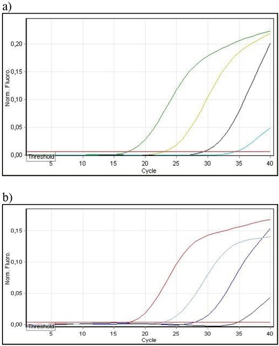
In the next stage, a SYBR Green I real-time PCR was performed for detecting the rpoB gene with template DNA obtained from the liver samples contaminated experimentally with five B. anthracis strains. Ct values ranged from 25.59 to 30.97 for the Genomic Mini (A&A Biotechnology, Poland) and from 14.56 to 25.34 for the DNeasy Blood and Tissue Kit (Qiagen, USA). When the Genomic Blood Mini and DNeasy Blood and Tissue Kit isolated DNA from contaminated blood samples, Ct values from 21.29 to 26.89 and from 11.65 to 25.62 were obtained, respectively. In all five examined strains the rpoB gene was detected. The specificity of amplification was confirmed by determination of the PCR product melting temperature, which was found to be about 78°C. The sensitivity of the real-time PCR was also determined. Isolating DNA from contaminated liver samples using the Genomic Mini, the sensitivity was 3.5 × 100 CFU/reaction, and using the DNeasy Blood and Tissue Kit it was 3.5 × 101 CFU/reaction. The sensitivity was established at 3.5 × 101 CFU/reaction for isolation of DNA from contaminated blood samples, with the use of the kits from both manufacturers (Fig. 1). In the case of template DNA obtained from experimentally contaminated biological samples, with the use of the kits from A&A Biotechnology (Poland) and Qiagen (USA), a high regression coefficient 0.99 was demonstrated.
Fig. 1.

Determination of the sensitivity of SYBR Green I real-time PCR with rpoB-F/R primers for the detection of B. anthracis strain 1/47 in liver (a) and blood (b) samples using a DNeasy Blood and Tissue Kit for DNA isolation
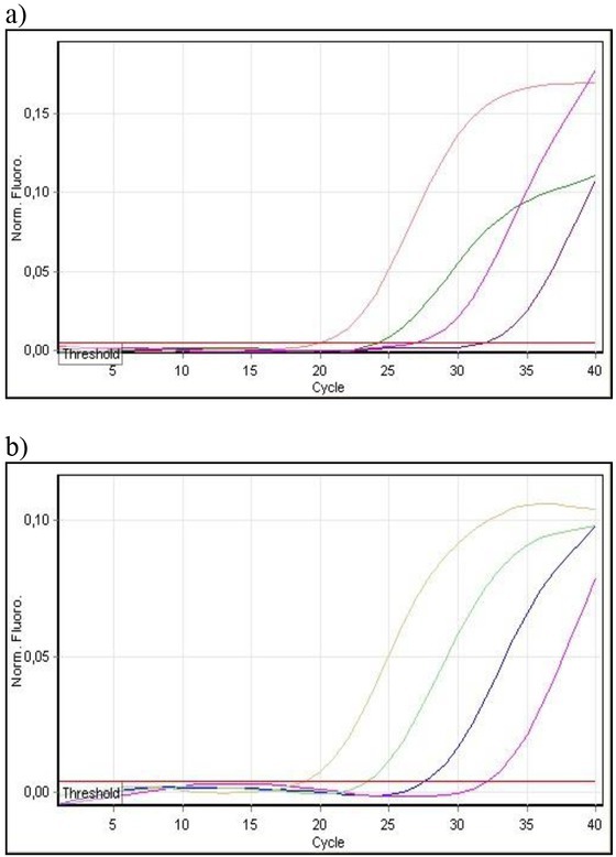
Next, real-time PCR detection of the pag gene located on plasmid pXO1 was carried out. For template DNA isolated from contaminated liver samples, Ct values fell within of 17.72 to 22.90 using the Genomic Mini and 18.24 to 22.83 for the DNeasy Blood and Tissue Kit. Ct values ranged from 15.12 to 20.69 and from 15.85 to 20.78 for DNA isolated from contaminated blood samples with the use of the Genomic Blood Mini and DNeasy Blood and Tissue Kit, respectively. One of the five examined B. anthracis strains (B.a. 7/51) did not show any real-time PCR product, which indicates the absence of the pag gene. The melting temperature of the reaction products was found to be about 79°C. The sensitivity of real-time PCR with the use of PA-F and PA-R primers was 3.5 × 101 CFU/reaction for DNA obtained from contaminated liver samples using both kits. Regarding DNA isolated from contaminated blood samples using the Genomic Blood Mini the sensitivity was 3.5 × 101 CFU/reaction and with the use of the DNeasy Blood and Tissue Kit was 3.5 × 100 CFU/reaction (Fig. 2). A high regression coefficient 0.99 was also shown.
Fig. 2.

Determination of the sensitivity of SYBR Green I real-time PCR with PA-F/R primers for the detection of B. anthracis strain 1/47 in liver (a) and blood (b) samples using Genomic Mini or Genomic Blood Mini Kits for DNA isolation
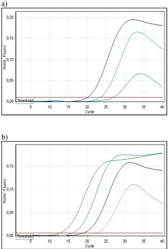
Subsequently real-time PCR detection of the capC gene located on plasmid pXO2 was performed. For DNA isolated from contaminated liver samples with the use of the Genomic Mini and the DNeasy Blood and Tissue Kit, Ct values ranged from 23.60 to 27.51 and from 16.72 to 18.43, respectively. Regarding DNA isolated from contaminated blood samples using the Genomic Blood Mini, Ct values were from 14.46 to 19.11 and using the DNeasy Blood and Tissue Kit from 9.56 to 14.95. Again one of the five examined B. anthracis strains (B.a. 13/93) did not show any reaction product, which indicates absence of the capsule. Specific amplification products dissociated at about 76°C. For DNA isolated from contaminated liver samples using the Genomic Mini the sensitivity was 3.5 × 10³ CFU/reaction and using the DNeasy Blood and Tissue Kit 3.5 × 10² CFU/reaction. For DNA isolated from contaminated blood samples with the use of kits from the both manufacturers, the sensitivities were 3.5 × 10² CFU/reaction and 3.5 × 101 CFU/reaction, respectively (Fig. 3). For template DNA obtained from experimentally contaminated biological samples using the A&A Biotechnology and Qiagen kits, a high regression coefficient above 0.99 was demonstrated.
Fig. 3.

Determination of the sensitivity of SYBR Green I real-time PCR with capC-F/R primers for the detection of B. anthracis strain 1/47 in liver (a) and blood (b) samples using a DNeasy Blood and Tissue Kit for DNA isolation
Examination of a range of bacterial DNA was undertaken to demonstrate the specificity of all SYBR Green I real-time PCRs. The template DNA was obtained from liver and blood samples experimentally contaminated with five strains of B. anthracis, single strains of other species of the genus Bacillus, i.e. B. cereus, B. brevis, B. subtilis, and B. megaterium, and other microbial species including Staphylococcus aureus, Listeria monocytogenes, Pasteurella multocida, Escherichia coli, Salmonella Typhimurium, and Klebsiella pneumoniae. All sets of primers were highly specific. Products of amplification specific for B. anthracis were obtained in all real-time PCRs, while isolates of other species failed to yield real-time PCR products.
Discussion
Identification of bacteria using traditional phenotypic methods is limited in comparison to genotypic methods. Tests based on the analysis of nucleic acids allow the direct detection of the microorganism in bacterial cultures or clinical samples. The PCR and real-time PCR techniques for identifying microorganisms at the strain, species, or genus level save time and increase sensitivity, and these methods have been used for fast and sensitive detection of Bacillus anthracis. Specific primers detect B. anthracis genes located on the DNA of plasmid pXO1, including the pag, lef, and cya genes (1, 16, 18, 21, 22). The second plasmid, pXO2, contains the sequences of the cap gene (A, B, and C) responsible for the production of the capsule (8, 19, 30). The confirmation of the presence of the two plasmids in B. anthracis indicates the virulence of the strain tested and has diagnostic value (7, 11, 21). Specific chromosomal markers for B. anthracis including Ba813 and rpoB are also available (7, 17, 25, 26). The conventional PCR and real-time PCR tests used in laboratories are valuable diagnostic tools. The use of real-time PCR may resolve most problems connected with the classical PCR method. This technique is less time consuming, reduces the risk of contamination of the sample due to the lack of post-amplification treatment, and may increase the sensitivity and efficiency of the method (9, 13, 20, 29). The optimisation of reaction conditions is crucial for the effectiveness of real-time PCR. The optimal concentration of the pair of primers was determined by testing different concentrations to achieve the lowest Ct value with the lowest primer concentration.
Khandia et al. (14) obtained a detection limit of 2 × 10⁻⁶ pg DNA, which corresponded to 3.47 copies for a pure culture SYBR Green I real-time PCR using a pag target. Ellerbrok et al. (7) achieved a detection limit of 10 plasmid copies per reaction for pure cultures in a TaqMan real-time PCR for the detection of the pag, capC, and rpoB genes. In our research, the results of SYBR Green I real-time PCRs for detection of the chromosomally encoded rpoB and the pag genes for DNA isolated from experimentally contaminated liver and blood samples using kits from A&A Biotechnology and Qiagen were comparable. Only in the case of real-time PCR detecting the capC gene slightly lower sensitivity was achieved. The reaction sensitivity for liver samples using Cap-S and Cap-R primers ranged from 3.5 × 10² to 3.5 × 10³ CFU/reaction and for blood samples from 3.5 × 101 to 3.5 × 10² CFU/reaction.
Khandia et al. (14) obtained a regression coefficient higher than 0.989 for pure cultures in SYBR Green I real-time PCR using the pag gene. For the detection of the cap gene, Bentahir et al. (2) achieved a regression coefficient higher than 0.995 for pure cultures. In our studies, the high regression coefficient attained in SYBR Green I real-time PCRs with the use of the rpoB, pag, and capC genes indicates a high degree of reaction linearity.
In our examinations, evaluation of real-time PCRs showed the high specificity of the tests. Products specific to B. anthracis rpoB gene as a chromosomal marker, pag gene, and capC gene were obtained. Isolates of various species of the genus Bacillus and strains of six other microorganisms gave no real-time PCR products to be found. Similarly, Bode et al. (4) and Qi et al. (25) examining B. anthracis strains and other species of the genus Bacillus showed 100% specificity of the reaction. In contrast, Ellerbrok et al. (7) demonstrated the presence of the rpoB gene in four of the five B. cereus strains tested. Although the researchers used a comparable number of bacteria for the reaction, the Ct values obtained for B. cereus were found 10 cycles later than in the case of B. anthracis. It may be a result of the close relationship of these species (4, 7).
Summing up, the real-time PCR method based on the fluorescence of SYBR Green I light-emitting dye in intercalation with double-stranded DNA successfully detected genes located on the pXO1 and pXO2 plasmids and the rpoB gene as a chromosomal marker. It was shown that real-time PCRs revealed considerable sensitivity and specificity. The specificity of the tests was confirmed by the melting points of the PCR products, and the sensitivity and linearity of the reaction were determined using the regression coefficient (29).
In order to fully utilise the advantages of the real-time PCR method, TaqMan probes will be used for further identification of B. anthracis from experimentally contaminated biological samples. The use of fluorescently labelled probes may additionally increase the sensitivity and specificity of the method (9, 29).
Footnotes
Conflict of Interests Statement: The authors declare that there is no conflict of interests regarding the publication of this article.
Financial Disclosure Statement: This work was financed with the funds of the National Veterinary Research Institute in Pulawy, Poland.
Animal Rights Statement: None required.
References
- 1.Adone R., Pasquali P., La Rosa G., Marianelli C., Muscillo M., Fasanella A., Francia M., Ciuchini F.. Sequence analysis of the genes encoding for the major virulence factors of Bacillus anthracis vaccine strain Carbosap. J Appl Microbiol. 2002;93:117–121. doi: 10.1046/j.1365-2672.2002.01660.x. [DOI] [PubMed] [Google Scholar]
- 2.Bentahir M., Ambroise J., Delcorps C., Pilo P., Gala J.L.. Sensitive and specific recombinase polymerase amplification for fast screening, detection, and identification of Bacillus anthracis in a field setting. Appl Environ Microbiol. 2018;84:1–13. doi: 10.1128/AEM.00506-18. [DOI] [PMC free article] [PubMed] [Google Scholar]
- 3.Beyer W., Turnbull P.C.B.. Anthrax in animals. Mol Aspects Med. 2009;30:481–489. doi: 10.1016/j.mam.2009.08.004. [DOI] [PubMed] [Google Scholar]
- 4.Bode E., Hurtle W., Norwood D.. Real-time PCR assay for a unique chromosomal sequence of Bacillus anthracis. J Clin Microbiol. 2004;42:5825–5831. doi: 10.1128/JCM.42.12.5825-5831.2004. [DOI] [PMC free article] [PubMed] [Google Scholar]
- 5.Budniak S., Kędrak-Jabłonska A., Reksa M., Szczawińska A., Borkowska-Opacka B.. Application of the real-time PCR method with the intercalating dye SYBR Green I for the detection of genes located on plasmids pXO1 and pXO2 as well as the specific chromosomal rpoB sequence of Bacillus anthracis strains. Med Weter. 2010;66:630–634. [Google Scholar]
- 6.D’Amelio E., Gentile B., Lista F., D’Amelio R.. Historical evolution of human anthrax from occupational disease to potentially global threat as bioweapon. Environ Int. 2015;85:133–146. doi: 10.1016/j.envint.2015.09.009. [DOI] [PubMed] [Google Scholar]
- 7.Ellerbrok H., Nettermann H., Özel M., Beutin L., Appel B., Pauli G.. Rapid and sensitive identification of pathogenic and apathogenic Bacillus anthracis by real-time PCR. FEMS Microbiol Lett. 2002;214:51–59. doi: 10.1111/j.1574-6968.2002.tb11324.x. [DOI] [PubMed] [Google Scholar]
- 8.Fouet A.. The surface of Bacillus anthracis. Mol Aspects Med. 2009;30:374–385. doi: 10.1016/j.mam.2009.07.001. [DOI] [PubMed] [Google Scholar]
- 9.Heid C.A., Stevens J., Livak K.J., Williams P.M.. Real time quantitative PCR. Genome Res. 1996;6:986–994. doi: 10.1101/gr.6.10.986. [DOI] [PubMed] [Google Scholar]
- 10.Helgason E., Okstad O.A., Caugant D.A., Johansen H.A., Fouet A., Mock M., Hegna I., Kolstro A.B.. Bacillus anthracis Bacillus cereus and Bacillus thuringiensis – one species on the basis of genetic evidence. Appl Environ Microbiol. 2000;66:2627–2630. doi: 10.1128/aem.66.6.2627-2630.2000. [DOI] [PMC free article] [PubMed] [Google Scholar]
- 11.Kaspar R.L., Robertson D.L.. Purification and physical analysis of Bacillus anthracis plasmids pXO1 and pXO2. Biochem Biophys Res Commun. 1987;149:362–368. doi: 10.1016/0006-291x(87)90375-5. [DOI] [PubMed] [Google Scholar]
- 12.Keim P., Klevystka A.M., Price L.B., Schupp J.M., Zinser G., Smith K.L., Hugh-Jones M.E., Okinaka R., Hill H.K., Jackson P.J.. Molecular diversity in Bacillus anthracis. J Appl Microbiol. 1999;87:215–217. doi: 10.1046/j.1365-2672.1999.00873.x. [DOI] [PubMed] [Google Scholar]
- 13.Kędrak-Jabłońska A., Budniak S., Krupa M., Szczawińska A., Reksa M., Szulowski K., Iwaniak W.. Detection of Listeria spp. and Listeria monocytogenes in biological samples by SYBR Green I and TaqMan Probe-based real-time PCRs. J Vet Res. 2017;61:427–432. doi: 10.1515/jvetres-2017-0069. [DOI] [PMC free article] [PubMed] [Google Scholar]
- 14.Khandia R., Pattnaik B., Rajukumar K., Pateriya A.K., Murugkar H.V., Bhatia S., Prakash A., Pradhan H.K.. Evaluation of a protective antigen gene based SYBR Green I real time PCR for detection of Bacillus anthracis in field samples. Ind J Anim Sci. 2013;83:118–123. [Google Scholar]
- 15.Koehler T.M.. Bacillus anthracis physiology and genetics. Mol Aspects Med. 2009;30:386–396. doi: 10.1016/j.mam.2009.07.004. [DOI] [PMC free article] [PubMed] [Google Scholar]
- 16.Liu S., Moayeri M., Leppla S.H.. Anthrax lethal and oedema toxins in anthrax pathogenesis. Trends Microbiol. 2014;6:317–325. doi: 10.1016/j.tim.2014.02.012. [DOI] [PMC free article] [PubMed] [Google Scholar]
- 17.Jackson P.J., Hugh-Jones M.E., Adair D.M., Green G., Hill K.K., Kuske C.R., Grinberg L.M., Abramova F.A., Keim P.. RCR analysis of tissue samples from the 1978 Sverdlovsk anthrax victims: the presence of multiple Bacillus anthracis strains in different victims. Proc Natl Acad Sci 1998. 95:1224–1229. doi: 10.1073/pnas.95.3.1224. [DOI] [PMC free article] [PubMed] [Google Scholar]
- 18.Makino S., Cheun H.I., Watarai M., Uchida I., Takeshi K.. Detection of anthrax spores from air by real-time PCR. Lett Appl Microbiol. 2001;33:237–240. doi: 10.1046/j.1472-765x.2001.00989.x. [DOI] [PubMed] [Google Scholar]
- 19.Makino S., Uchida I., Terakado N., Sasakawa C., Yoshikawa M.. Molecular characterization and protein analysis of the cap region, which is essential for encapsulation in Bacillus anthracis. J Bacteriol. 1989;171:722–730. doi: 10.1128/jb.171.2.722-730.1989. [DOI] [PMC free article] [PubMed] [Google Scholar]
- 20.Morrison T.B., Weis J.J., Wittwer C.T.. Quantification of low copy transcripts by continuous SYBR Green I monitoring during amplification. Biotechniques. 1998;24:954–962. [PubMed] [Google Scholar]
- 21.Okinaka R., Cloud K., Hampton O., Hoffmaster A.R., Hill K.K., Keim P., Koehler T.M., Lamke G., Kumano S., Manter D., Martinez Y., Ricke D., Svensson R., Jackson P.. Sequence assembly and analysis of pXO1 and pXO2. J Appl Microbiol. 1999;87:261–262. doi: 10.1046/j.1365-2672.1999.00883.x. [DOI] [PubMed] [Google Scholar]
- 22.Okinaka R., Cloud K., Hampton O., Hoffmaster A.R., Hill K.K., Keim P., Koehler T.M., Lamke G., Kumano S., Mahillon J., Manter D., Martinez Y., Ricke D., Svensson R., Jackson P.J.. Sequence and organization of pXO1, the large Bacillus anthracis plasmid harboring the anthrax toxin genes. J Bacteriol. 1999;181:6509–6515. doi: 10.1128/jb.181.20.6509-6515.1999. [DOI] [PMC free article] [PubMed] [Google Scholar]
- 23.Pilo P., Frey J.. Pathogenicity, population genetics, and dissemination of Bacillus anthracis. Infect Genet Evol. 2018;64:115–125. doi: 10.1016/j.meegid.2018.06.024. [DOI] [PubMed] [Google Scholar]
- 24.Prince L.B., Hugh-Jones M., Jackson P.J., Keim P.. Genetic diversity in the protective antigen gene of Bacillus anthracis. J Bacteriol. 1999;181:2358–2362. doi: 10.1128/jb.181.8.2358-2362.1999. [DOI] [PMC free article] [PubMed] [Google Scholar]
- 25.Qi Y., Patra G., Liang X., Williams L.E., Rose S., Redkar R.J., DelVecchio V.G.. Utlization of the rpoB gene as a specific chromosomal marker for real-time PCR detection of Bacillus anthracis. Appl Environ Microbiol. 2001;67:3720–3727. doi: 10.1128/AEM.67.8.3720-3727.2001. [DOI] [PMC free article] [PubMed] [Google Scholar]
- 26.Ramisse V., Patra G., Garrigue H., Guesdon J.L., Mock M.. Identification and characterization of Bacillus anthracis by multiplex PCR analysis of sequences on plasmids pXO1 and pXO2 and chromosomal DNA. FEMS Microbiol Lett. 1996;145:9–16. doi: 10.1111/j.1574-6968.1996.tb08548.x. [DOI] [PubMed] [Google Scholar]
- 27.Schwartz M.. Dr. Jekyll and Mr. Hyde: a short history of anthrax. Mol Aspects Med. 2009;30:347–355. doi: 10.1016/j.mam.2009.06.004. [DOI] [PubMed] [Google Scholar]
- 28.Spencer R.C.. Bacillus anthracis. J Clin Pathol. 2003;56:182–187. doi: 10.1136/jcp.56.3.182. [DOI] [PMC free article] [PubMed] [Google Scholar]
- 29.Stadejek T.. Recent developments in polymerase chain reaction – real-time PCR. Med Weter. 2006;62:390–394. [Google Scholar]
- 30.Turnbull P.C., Hutson R.A., Ward M.J., Jones M.N., Quinn C.P., Finnie N.J., Duggleby C.J., Kramer J.M., Melling J.. Bacillus anthracis but not always anthrax. J Appl Bacteriol. 1992;72:21–28. doi: 10.1111/j.1365-2672.1992.tb04876.x. [DOI] [PubMed] [Google Scholar]


