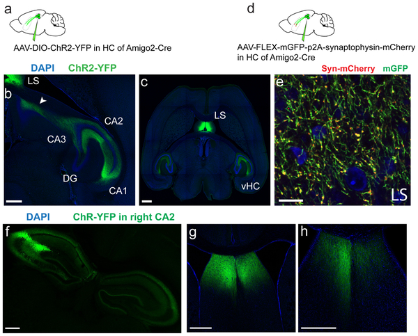
Extended Data Figure 1: CA2 projections to LS in horizontal sections.
a-c. Horizontal sections from an Amigo2-Cre mouse brain injected with rAAV5-EF1a-DIO-hChR2(E123T/T159C)-eYFP into CA2. The arrow on b shows the CA2 axons extending up to the dLS. Drawing was inspired from ref.49 c. is more ventral and shows the CA2 projection to the lateral septum as well as to the ventral hippocampus. d-e. Enlarged view of a coronal dLS section of an Amigo2-Cre mouse injected in CA2 with rAAV5-mGFP-p2A-Synatophysin-mCherry labelling CA2 projections (green) and presynaptic terminals (red). f-h. Coronal sections of an Amigo2-Cre mouse brain injected unilaterally with rAAV5-EF1a-DIO-hChR2(E123T/T159C)-eYFP into the right CA2 area. f. Hippocampal section. g-h. LS sections. 3 mice injected per experiment. All mice presented similar staining pattern. All scale bars 500 μm except e (20 μm).

