Abstract
AIM:
To investigate the efficacy of plastic surgery in the treatment of giant congenital melanocytic nevus (GCMN).
METHODS:
We enrolled 20 patients with 44 lesions and performed one of the following procedures: serial excision, skin grafting, tissue expansion, primary skin closure, distant flap, and adjacent flap. We assessed the outcome at 10 days and 6 months after surgery.
RESULTS:
Of 44 surgical sites, the most commonly used reconstruction surgeries were serial excision (16), skin grafting (16), and tissue expansion (6). Other types were rarely used. All patients with serial excision had good outcome. A total of 81% and 19% of the patients with skin grafting had good and fair outcome, respectively. Around 83% and 17% of the patients with tissue expansion had good and fair outcome. No cases had bad outcome.
CONCLUSION:
In conclusion plastic surgery is effective in the treatment of GCMN. There are different techniques but serial excision, skin grafts, and tissue expansion are most commonly used.
Keywords: Melanocytic nevus, Giant congenital melanocytic nevus, Plastic surgery
Introduction
Giant congenital melanocytic nevus (GCMN) is a rare restricted dysplasia, with embryonic origin and associated with high risk of malignant melanoma (6.3%). In addition, they also cause aesthetic lacking, especially while appears in open areas, such as face and neck [1], [2]. Treatment of GCMN is mainly surgical and it depends on the position, size and malignant tendency of the nevus. Many surgical techniques are avaible and they could be very difficult, mostly with GCMN of excessive size, with very large injured area [3], [4].
We aimed to investigate the efficacy of plastic surgery in the treatment of giant congenital melanocytic nevus (GCMN)
Methods
We enrolled at National Hospital of Dermatology and Venereology and Saint Paul Hospital 20 patients who had been diagnosed with GCMN and operated through 44 surgeries, then monitored and evaluated from 2006 to 2010.
Results
We evaluated the results of each main type of reconstruction surgeries to propose the suitable method for every specific case: serial excision, skin grafting and tissue expansion. Other types of surgery are rarely used, so we cannot evaluate the results. There were no cases with bad result as shown in Table 1 and Table 2.
Table 1.
Distribution of skin reconstructing methods after GCMN removal
| Plastic surgery | n | % |
|---|---|---|
| Serial excision | 16 | 36 % |
| Skin grafting | 16 | 36 % |
| Tissue expansion | 6 | 14 % |
| Primary skin closure | 3 | 7 % |
| Adjacent flap | 2 | 5 % |
| Distant flap | 1 | 2 % |
| Total | 44 | 100 % |
Table 2.
Surgical results
| Surgery methods | Good | Fair | Bad | Total |
|---|---|---|---|---|
| Serial excision | 16 | 0 | 0 | 16 |
| 100% | 0% | 0% | 100% | |
| Skin grafting | 13 | 3 | 0 | 16 |
| 81% | 19% | 0% | 100% | |
| Tissue expansion | 5 | 1 | 0 | 6 |
| 83% | 17% | 0% | 100% | |
| Total | 34 | 4 | 0 | 38 |
Serial excision was used 16/44 sites, the result is 100% good. It is a method that takes advantage of natural skin dilation with many advantages: fast surgery, low cost, high aesthetic, no additional damage to the adjacent skin but it cannot be applied to the malignant de-generalized nevus.
Arneja [3] recommended carrying out this method when the nevus can be completely removed after only 3 or 4 surgeries, otherwise the tissue expansion should be suggested. We found out that, the serial excision can be used simply to treat GCMN, especially the GCMN in the patient’s back or abdomen.
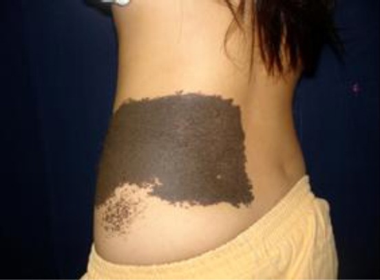
Figure 1.

Clinical presentation of GCMN before surgery, baseline
Tissue expansion can create a sufficient volume of tissue to cover a large cell shortage. It is a complex technique with high cost, prolonged duration, capability of many complications. Tissue expansion was used 6/44 sites, the results were 83% good, 17% fair.
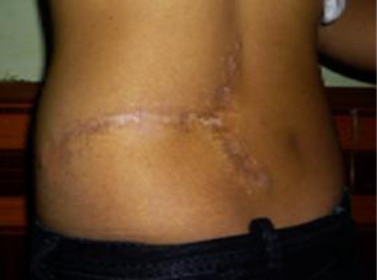
Figure 2.

Clinical presentation of GCMN after three serial excision. The aesthetic outcome was satisfactory for the patient
None patients had infections and there was only one case of partial necrosis was not completely removed from the skin, however, it did not significantly affect the results of the surgery, so we assessed it fair, accounting for 17%. The remaining 5 cases, having no necrosis and scarring immediately and well, are rated as good, accounting for 83%. Arneja [3] recommends the use of tissue expansion for GCMN that cannot be operated by serial excision after the fourth surgery, available on children aged over 3 months [5].
Discussion
The flap is used as an indigenous or distant flap or as material for a skin graft. This is also the best indication for the scalp [5], [6].
Skin grafting can cover a very large area especially when it is associated with tissue expansion. It is an easy and low cost method. In our study skin grafting was used 16/44 sites, the results reached 81% good, 19% fair. There were 4 cases of thick skin grafts that were all slightly infected due to fluid gathered under grafts, get partial necrosis, which affected the outcome of the surgery. These 4 cases are evaluated as fair, accounting for 19%. The remaining cases were good, accounting for 81%. Skin graft should be used only if neither serial excision nor tissue expansion can be performed [7].
In conclusion, to reconstructing the skin after GCMN excision, the serial excision and tissue expansion surgeries are preferentially selected; skin grafting is an alternative.
Footnotes
Funding: This research did not receive any financial support
Competing Interests: The authors have declared that no competing interests exist
References
- 1.Etchevers HC, Rose C, Kahle B, Vorbringer H, Fina F, Heux P, Berger I, Schwarz B, Zaffran S, Macagno N, Krengel S. Giant congenital melanocytic nevus with vascular malformation and epidermal cysts associated with a somatic activating mutation in BRAF. Pigment cell & melanoma research. 2018;31(3):437–41. doi: 10.1111/pcmr.12685. https://doi.org/10.1111/pcmr.12685 PMid:29316280. [DOI] [PubMed] [Google Scholar]
- 2.Graham B Colver. Primary malignant melanoma. Skin cancer:a practical guide to management. 2002:64. [Google Scholar]
- 3.Arneja JS, Gosain AK. Giant congenital melanocytic nevi of the trunk and an algorithm for treatment. Journal of Craniofacial Surgery. 2005;16(5):886–93. doi: 10.1097/01.scs.0000183356.41637.f5. https://doi.org/10.1097/01.scs.0000183356.41637.f5. [DOI] [PubMed] [Google Scholar]
- 4.Lehrer M, Zieve D. Giant congenital nevus. Mediline Plus Medical Encyclopedia. 2009 [Google Scholar]
- 5.Bhatnagar V, Mukherjee MK, Bhargava P. A case of giant hairy pigmented nevus of face. Medical Journal Armed Forces India. 2005;61(2):200–2. doi: 10.1016/S0377-1237(05)80029-2. https://doi.org/10.1016/S0377-1237(05)80029-2. [DOI] [PMC free article] [PubMed] [Google Scholar]
- 6.Ravi KV, Ramaswamy CN. Congenital giant hairy nevus treated by serial excision using tissue expanders. Journal of Indian Association of Pediatric Surgeons. 2004;9(2):92–94. [Google Scholar]
- 7.Jabaiti SK. Use of lower abdominal full-thickness skin grafts for coverage of large skin defects. European Journal of Scientific Research. 2010;39(1):134–142. [Google Scholar]
- 8.Tchernev G, Lozev I, Pidakev I, et al. Giant Congenital Melanocytic Nevus (GCMN) - A New Hope for Targeted Therapy? Open Access Maced J Med Sci. 2017;5(4):549–550. doi: 10.3889/oamjms.2017.121. https://doi.org/10.3889/oamjms.2017.121 PMid:28785360 PMCid:PMC5535685. [DOI] [PMC free article] [PubMed] [Google Scholar]
- 9.Chokoeva AA, Fioranelli M, Roccia MG, Lotti T, Wollina U, Tchernev G. Giant congenital melanocytic nevus in a bulgarian newborn. J Biol Regul Homeost Agents. 2016;30(2 Suppl 2):57–60. PMid:27373137. [PubMed] [Google Scholar]


