



Abstract
Self-hardening calcium phosphate cements present ideal bone tissue substitutes from the standpoints of bioactivity and biocompatibility, yet they suffer from (a) weak mechanical properties, (b) negligible bone growth gene effects without the use of exogenous growth factors, and (c) a lack of intrinsic antibacterial activity. Here we attempt to improve on these deficiencies by studying the properties of self-setting Fe-doped bone-integrative cements containing two different concentrations of the dopant: 0.49 and 1.09 wt.% Fe. The hardening process, which involved the transformation of Fe-doped β-tricalcium phosphate (Fe-TCP) to nanocrystalline brushite, was investigated in situ by continuously monitoring the cements using the Energy Dispersive X-Ray diffraction technique. The setting time was 20 minutes and the hardening time 2 h, but it took 50 h for the cement to completely stabilize compositionally and mechanically. Still, compared to other similar systems, the phase transformation during hardening was relatively fast and it also followed a relatively simple reaction path, virtually free of complex intermediates and noisy background. Mössbauer spectrometry demonstrated that 57Fe atoms in Fe-TCP were located in two non-equivalent crystallographic sites and distributed over positions with a strong crystal distortion. The pronounced presence of ultrafine crystals in the final, brushite phase contributed to the reduction of the porosity and thereby to the enhancement of the mechanical properties. The compressive strength of the hardened TCP cements increased by more than twofold when Fe was added as a dopant, i.e., from 11.5 ± 0.5 to 24.5 ± 2.0 MPa. The amount of iron released from the cements in physiological media steadied after 10 days and was by an order of magnitude lower than the clinical threshold that triggers the toxic response. The cements exhibited osteoinductive activity, as observed from the elevated levels of expression of genes encoding for osteocalcin and Runx2 in both undifferentiated and differentiated MC3T3-E1 cells challenged with the cements. The osteoinductive effect was inversely proportional to the content of Fe ions in the cements, indicating that an excessive amount of iron can have a detrimental effect on the induction of bone growth by osteoblasts in contact with the cement. In contrast, the antibacterial activity of the cement in the agar assay increased against all four bacterial species analysed (E. coli, S. enteritidis, P. aeruginosa, S. aureus) in direct proportion with the concentration of Fe ions in it, indicating their key effect on the promotion of the antibacterial effect in this material. This effect was less pronounced in broth assays. Experiments involving co-incubation of cements with cells in an alternate magnetic radiofrequency field for 30 min demonstrated a good potential for the use of these magnetic cements in hyperthermia cancer therapies. Specifically, the population of the glioblastoma cells decreased six-fold at the 24 h time point following the end of the magnetic field treatment, while the population of the bone cancer cells dropped approximately twofold. The analysis of the MC3T3-E1 cell/cement interaction reiterated the effects of iron in the cement on the bone growth marker expression by showing signs of adverse effects on the cell morphology and proliferation only for the cement containing the higher concentration of Fe ions (1.09 wt.%). Biological testing concluded that the effects of iron are beneficial from the perspective of a magnetic hyperthermia therapy and antibacterial prophylaxis, but its concentration in the material must be carefully optimized to avoid the adverse effects induced above a certain level of iron concentrations.
Keywords: iron-doped tricalcium phosphate, bone cement, hardening behaviour, osteoblastic MC3T3-E1, Escherichia coli, Salmonella enteritidis, Pseudomonas aeruginosa, Staphylococcus aureus
1. Introduction
Due to their chemical similarity with the inorganic component of bone and teeth, calcium phosphate materials have been widely used for grafts, substitutions, void fillers and implant coatings in these hard tissues. Such materials almost invariably elicit a favourable biological response and can be employed in orthopaedics and dentistry in a wide range of forms, e.g. as granules, solid blocks, injectable pastes, etc. [1,2,3,4] Although hydroxyapatite is the calcium phosphate most similar to the mineral component of natural bone, having decent mechanical properties too [5], it is poorly resorbed in the body and different calcium phosphates have been preferred to it, more resorbable and capable of inducing a more optimal bone regeneration process. Among these resorbable calcium phosphates, monetite and brushite are the currently favourite candidates [6,7]. However, although calcium phosphates present ideal bone tissue substitutes from the standpoints of bioactivity and biocompatibility, their state-of-the-art, self-setting formulations suffer from (a) weak mechanical properties, (b) negligible bone growth gene effects without the use of exogenous growth factors, and (c) a lack of intrinsic antibacterial activity. In this study, we aim to improve on these deficiencies by investigating iron ions as dopants in a self-hardening brushite-forming beta-tricalcium phosphate (TCP) cement. Although the capacity of hydroxyapatite to accommodate foreign ions is unrivalled among calcium phosphates due to its exceptional crystal lattice versatility and flexibility, TCPs are also shown to be able to incorporate a number of cationic substitutions without significant lattice distortions [8,9].
Iron is a ubiquitous micromineral in the human body. Almost two-thirds of it are employed for the synthesis of metalloproteins (e.g., hemoproteins, mainly hemoglobin and myoglobin) constituting the oxygen carriers between the lung and other tissues [10]. Iron also forms essential complexes with enzymes (e.g., heme enzymes, such as peroxidase, catalase and cytochromes C/a/a3/b) involved in electron transfer mechanisms, oxidative metabolism and energy production in mitochondria [11,12]. Finally, iron is present in non-heme enzymes too (e.g., iron sulfur complexing flavoproteins, oxidase and hydroxylase) [10,11,13] and in compounds with storage function (e.g., ferritin, hemosiderin) or iron-transferring function (e.g., transferrin) [10,11,14,15]. Considering the pervasive presence of the abovementioned enzymes (heme and non-heme) in the body, the deficiency of this micronutrient or its excess can cause a variety of pathologies and squilibria [10,16,17], including most evidently the metabolic ones (e.g., the formation of thyroid hormones is iron-sensitive [18]) and the immunocompetence one (as iron alters proportion and function of various T lymphocyte subsets)[19,20,21,22]; furthermore, iron is involved in the delicate role of formation of the connective tissue of several neurotransmitters in the brain [23,24] and in free-radical controlled process for microbe killing [25,26].
Even if iron has no primary, direct effect in the processes of bone mineralization and vascularization, another issue should be taken into account: in addition to a positive effect on cell physiological functions, including osteoblastic proliferation and remineralization of the inserted material, an ideal bone substitute should also possess an intrinsic antibacterial activity in order to avoid serious hospital-acquired infections originating from opportunistic pathogens adsorbed on the implant surface [27]. Given the peculiar nature of bone, the only mineralized tissue in the body, entailing limited perfusion of nutrients and consequently scarce penetration of systemically administered drugs, one way of preventing infections is to bind antibiotics to the calcium phosphate matrix of the implant and allow it to be released in loco [28]. The use of antibiotics is, however, costly and linked with the problem of resistance-rising in pathogens [29], The use of antibacterial cations doping the structure of one of the precursors of the cements (usually TCP) has been proposed as an alternative way to solve this issue [30]. A certain number of such ions (e.g., Ag+, Cu2+, and Zn2+) doping calcium phosphate cements have been extensively studied for their physiological and antibacterial effects on tissue cultures and whole organisms [31,32,33,34], being a part of the larger research topic (extended also to other mono-, bi-, tri- and tetravalent ions, e.g. Na+, K+, Li+, Mg2+, Sr2+, Co2+, Mn2+/3+, Cr3+, Si4+) recently explored [35,36,37,38,39,40] in the field of calcium phosphate cements in biomedical engineering. Mechanical features, particularly compressive strength, the rate of ion release and the dissolution rate of the cement should also be evaluated for every composition.
The present study provides the description and characterization of the steps to obtain and use an Fe-doped bone integrative cement, starting from a wet route for the preparation of Fe-doped TCP powder, followed by the biological analyses investigating the response of bone tissues to the different forms of this material, including osteoconductivity in contact with bone cells, gene-based osteoinductive activity, antibacterial properties and magnetic-field-induced heating properties for anticancer hyperthermia therapies. Finally, the study investigates the modification of the behaviour of the cement obtained with a selected concentration of Fe-doped TCP with respect to the pure brushite one, in relation to mechanical properties (compressive strength) and solubility, while also showing the results of the real-time monitoring of the doped-TCP → doped-brushite transformation (i.e., chemical composition of the evolving system and hardening kinetics) by means of Energy Dispersive X-Ray Diffraction (EDXRD).
2. Materials and Methods
2.1. Synthesis and physicochemical characterization
Analytical grade Ca(NO3)2, Fe(NO3)3, (NH4)2HPO4 substances were selected as precursors for the iron-tricalcium phosphate (Fe-TCP) powder synthesis. To synthesize Fe-TCP powders containing 1.8 wt% and 0.9 wt% Fe, the wet method was chosen. This method was previously described in [41] and proceeds according to the following scheme:
| Eq.1 |
where x= 0.1 and 0.05.
Synthesis procedure was as follows: a 0.5 mol/L aqueous solution of Ca(NO3)2 was mixed with a calculated amount of Fe(NO3)3 solution conforming to the intended stoichiometry; then, (NH4)2HPO4 solution was added dropwise at the rate of 20 mL/min (1). The pH level was kept at 6.5-6.9 by the dropwise addition of ammonia solution. After 30 min, the precipitate was filtered, washed with distilled water and dried at 80 °C. The precipitates were heat treated at 900 °C for 1 h for crystallization.
The elemental analysis of the final product was performed with atomic emission spectrometry with inductively coupled plasma “Ultima-2” (Perkin-Elmer, USA).
XRD investigation of powders was performed using Energy Dispersive X-ray Diffraction (EDXRD) run on a non-commercial diffractometer [42]. It consists of white X-ray radiation that is produced by a commercial W-anode X-ray tube (up to 50 keV) and a solid-state EG&G high purity Germanium photodiode detector, with energy resolution of about 1.5–2.0 % in the 20–50 keV range. The detector is connected to a PC via the ADC AM hardware and the signal is processed by a Maestro software, performing the necessary analogue to digital conversions. The reciprocal space scan necessary to collect the diffraction pattern is carried out electronically. The diffraction pattern represents the diffracted intensity, i.e. the number of incident X-ray photons, as a function of the scattering parameter q, while maintaining that q = aEsinΘ, where q is the normalized momentum transfer magnitude, a is a constant, E is the energy of the incident X-ray beam and 2Θ is the scattering angle. The electronic scanning permits the experimental geometry to be kept fixed during pattern acquisition, allowing a faster recording of the Bragg peaks, since in the ED mode, the whole diffraction pattern is obtained in parallel at any q value. The corresponding 2Θ values are easily obtained by means of the previous relation. Qualitative analysis of the diffractogram was performed using the ICDD database PDF-2 [43].
μ-Raman spectroscopy was used to further check the product, by means of an in-house made instrument. TCP, 0.1Fe-TCP and 0.05Fe-TCP powders were analysed by a 632 nm laser. Exposure conditions were 20 accumulations, each 3 s long-lasting, at a 10mW power. Qualitative analysis of the spectra was performed using the RRUFF Project database [44].
The powder phase of the brushite-based cement consisted in 40.8 wt.% monocalcium phosphate monohydrate (MCPM; Sigma-Aldrich, St. Louis, MO, USA) and 54.3 wt.% Fe-doped beta-tricalcium phosphate obtained as above. The as-received MCPM powder was first sieved and only particles with sizes below 75 μm were used. The two powders were mixed with 4.9 wt.% carbonated hydroxyapatite ceramic granules. The liquid phase consisted of a 7.5 wt.% (0.4 M) citric acid solution (Sigma-Aldrich, St. Louis, MO, USA). The powder-to-liquid ratio was 4.4 g/mL. The manual mixing of the reagents requires repeated pressure applications by means of a putty knife. The product of the reaction is dicalcium phosphate dihydrate (CaHPO4 2H2O, DCPD, brushite), according to the following equation, while taking into account that some of the calcium ions will be substituted by iron, thus yielding iron-doped brushite:
| (Eq.2) |
The cement samples for the compressive strength measurements were prepared as follows: 0.55g of 0.05Fe-TCP, 0.42g of MCPM, 0.05g of carbonated hydroxyapatite and 0.3 g of ammonium citrate powders were mixed to prepare a cement powder. It was subsequently mixed with 0.5g of the liquid phase for 2-3 min to form the cement paste. Then, the paste was formed in a Teflon mould (diameter 8 mm, height 16 mm) and allowed 24 h to set under ambient conditions. The compressive strength measurements were carried out after 1 day of hardening using Instron 5581 (“TTS”, Great Britain) [33]. To obtain statistically reliable results, 4 cements samples were measured.
Iron-ion release from the 0.05Fe-TCP cement samples was studied at 37 °C in TRIS-HC1 buffer solution at pH 7.4 at a constant liquid phase volume [31]. The buffer solution was adjusted to pH = 7.4 by adding 13.25 g of TRIS (Cat. No.: 77-86-1, Sigma-Aldrich, Dorset, UK) and 125 mL of HC1 (Cat. No.: 7647-01-0, Aldrich-Aldrich, Dorset, UK). The iron-ion release measurements were performed for crushed cements with the solid-to-liquid ratio of 1g/50 mL. Three samples were used for each time point (1,3, 16 and 20 days). The iron ion concentration in the liquid phase was measured using Atomic Emission Spectrometer Ultima 2 (Jobin-Yvon, Longjumeau, France).
The EDXRD method was used also for time-resolved in situ investigation of the structural evolution of the cements [42]. A freshly made cement sample is quickly transferred (within thirty seconds) to the optical centre of the diffractometer, on a low scattering glass sample holder. Operating settings of the diffractometer were: no monochromator filters applied to the radiogen tube, W anticathode, 50kV voltage, 30 mA current intensity, apertures 200 μm wide and θ°= 5°.
SEM micrographs were obtained on a FEI Quanta 400 Scanning Electron Microscope. The images were obtained at 20 kV acceleration voltage. The cement samples were analyzed without any pre-treatment. For the image analysis, ImageJ open source processing program (NIH, Bethesda, MD) [45] was used to measure the grain size and estimate pore diameters.
The 57Fe Mössbauer spectra were recorded at room temperature (RT) using a conventional constant-acceleration spectrometer. The radiation source 57Co(Rh) was kept at room temperature (RT). All isomer shifts refer to the α-Fe at RT. The spectra were processed and analysed using the SpectrRelax program [46] by considering a suitable number of quadrupole doublets with Lorentzian lines. The samples consisted of thin layers of powder with about 15 mg of Fe/sm2. The proportions of each iron form were estimated from the relative absorption area of their respective components, assuming the same recoilless Lamb- Mössbauer factor values [47].
2.2. Biological assaying
Cell culture assays were conducted on MC3T3-E1 subclone 4 pre-osteoblastic murine calvarial cells (ATCC), K7M2 murine osteosarcoma cells (ATCC) and E297 human glioblastoma cells (gift of Herbert Engelhard, Department of Neurosurgery, ETC). All cells were cultured at 37 °C and 5 % CO2 in either DMEM (Gibco, MC3T3-E1, E297) or MEM-α (Gibco, K7M2) media with 10 % fetal bovine serum (FBS) and 1 % antibiotic-antimycotic (Life Technologies, Carlsbad, CA) to prevent bacterial and fungal contamination. For individual assays, cells were seeded at 5 × 10⁴ cells/well in 24-well plates in 1 mL of the DMEM/MEM-α cell culture medium. Upon reaching the confluency, freshly prepared cements were added to the culture and the incubation continued under the same cell growth conditions, specifically 37 °C and 5 % CO2. To mimic the clinical scenario, cements were added to the cells 30 minutes after the formation, set, but not hardened. Normalized to the amount of the cell culture medium in the wells, the cement concentrations in different assays ranged between 5 and 20 mg/ml, which was equivalent to the 2.5 – 10 mg/cm2 range when normalized to the surface area occupied by the cells in the wells. The following formulations were used in the assays: 0.1Fe-TCP and 0.05Fe-TCP. The cell response was compared against negative, cement-untreated controls and positive controls either treated with appropriate assay-dependent cell-obliterating agents or containing cell-free culture medium. All assays were performed on undifferentiated K7M2 cells unless otherwise noted. Osteoblastic differentiation was performed by adding 50 μg/mL L-ascorbic acid and 10 mM β-glycerophosphate to the cell culture medium.
Cell nuclei, f-actin microfilaments and calcium phosphate particles were fixed and stained 72 hours after the onset of the treatment of the cells with the cements at the concentration of 5 mg/mL. Cells were fixed for 5 minutes in 4 % paraformaldehyde (PFA) and washed 3 × 10 min in PBS, then blocked at room temperature for 1 h in the blocking solution (2 % bovine serum albumin, 0.5 % Triton-X in PBS), washed 3 × 10 min with PBS again and stained with Alexa Fluor 568 phalloidin (1:400), OsteoImage reagent (1:100) and NucBlue® Fixed Cell ReadyProbes™ reagent (Molecular Probes, Life Technologies) for 1 hour at room temperature. After the incubation, cells were washed 3 × 5 min with OsteoImage wash buffer and mounted using Prolong Diamond mounting media (Life Technologies). Images were acquired on a Nikon T1-S/L100 inverted epifluorescent confocal microscope. All the samples were analysed in triplicates.
Total RNA was extracted from MC3T3-E1 cells using the RNeasy kit (Qiagen) and cDNA synthesized using the High-Capacity cDNA reverse transcription kit (Applied Biosystems) from 100 ng of total RNA. cDNA was quantified using custom TaqMan probes for osteocalcin (BGLAP) and Runx2 on a StepOne Real Time PCR System (Applied Biosystems). The real-time PCR results were analysed using the ΔΔCt method [48] and all the data were normalized to the expression levels of β-actin (ACTB) as the housekeeping gene. All the samples were analysed in three experimental triplicates and three analytical replicates (n= 3×3= 9). In the liquid inoculation test of the cements, a single colony of Staphylococcus aureus cultured on a blood agar plate over a period of 24 h was stabbed with a pipette tip and placed in 5 mL of brain heart infusion broth (Sigma Life Sciences) and incubated overnight at 37 °C and 170 rpm. The same procedure was repeated for Escherichia coli, Salmonella entiritidis and Pseudomonas aeruginosa, all of which were cultured on nutrient agar plates and inoculated in the nutrient broth (Sigma Life Sciences). Cements in the concentration of 20 mg/mL were added to the infected broths. After the overnight incubation, 200 μL of the suspension had 1M HC1 added to it to dissolve the cement. Optical density was then measured at 600 nm (BMG LABTECH FLUOstar Omega) and converted to the number of colony-forming units. In the agar diffusion antibacterial assay, 10 μL of the cement were placed onto a bacterium-infused nutrient agar plate with the spot radius of 1 cm. The agar plates were then allowed to incubate for 24 h at 37 °C. The radius of the zone of inhibition was then used to gauge the antibacterial activity of the cements. All the samples for both antibacterial assays were analyzed in triplicates.
To assess the anticancer activity of the cements in a magnetic hyperthermia setting, E297 and K7M2 cells were seeded in rectangular, μ-Slide 8-well plates (Ibidi) and grown to confluence. The heating experiments were performed under ambient conditions and the wells containing the cells were held inside an insulating box to prevent the heat from dissipating. Cements were then added to the cells at the concentration of 10 mg/cm2 in 100 μL of the DMEM cell culture medium and incubated for 30 min, before plates were placed inside a multi-turn helical induction coil of an induction heating system (Ultraflex SH2/350) and kept in the alternate magnetic radiofrequency field (1.16 μT, 350 kHz, 1 kW) for another 30 min, after which the particles were gently washed off from the cells and the cells were allowed to recover for 24 h. Temperature readings were made during the hyperthermia measurement by periodically pulling the wells out of the coils and immersing the tip of a pocket-size thermometer with water-resistant case (Fisher Scientific Traceable Total-Range Thermometer) into the medium. After 24 of the recovery time, an MTT assay was run to test for viability. For that purpose, a 12 mM 3-[4, 5-dimethylthiazol-2-yl]-2,5-diphenyl tetrazolium bromide (MTT) stock solution was prepared by adding 1 mL of sterile phosphate buffered saline (PBS) to a 5 mg vial of MTT and vortex-mixing to ensure complete dissolution. Cells were washed with PBS and 275 μL of 1:10 MTT/media v/v were added into each well. After 4 hours of incubation at 37 °C, 200 μL of the solution were removed and 125 μL of DMSO added to each well. Plates were placed in a 37 °C incubator shaker at 120 rpm for 30 minutes before measuring the absorbance at 570 nm using the BMG LABTECH FLUOstar Omega microplate reader. To account for the effect of cements per se on the absorbance in the lysate, the absorbance of the control sample containing cell-free medium and the cement was subtracted from that of the cement samples.
3. Results and Discussion
3.1. Characterization of the precursor powders
The Fe content in the Fe-TCP powder was determined to be 1.09 and 0.49 wt.% for x= 0.1 and 0.05 in Eq.1, respectively, according to the results of the elemental analysis. The discrepancy between these values and the values calculated from the stoichiometry of the precursors (1.8 and 0.9 wt.%) can be explained by the fact that 50–60 % of Fe2+ ions in the solution substituted Ca2+ ions in the TCP structure, whereas the remaining Fe2+ions formed water-soluble complexes with NH4+ ions from the reaction mixture and were, as such, washed away in the filtrate.
The X-ray diffraction patterns of the two powders confirm the formation of a mixed calcium-iron phosphate (Ca9Fe(PO4)7 PDF #450338 and Fe2P2O7·2H2O PDF #501798) and a minor presence of β-TCP (Ca3(PO4)2 PDF #090169). Hematite (α-Fe2O3 PDF #330664) is also present, as expected according to [41]. Even though calcium phosphates delay this phase transformation, mixed valence iron oxides undergo complete oxidation at circa 300 °C, at which point they transition first to maghemite, γ-Fe2O3, and then to hematite [49], α-Fe2O3 at 350–600 °C depending on the concentration of calcium phosphates in the solid mixture [50]. Oxidation of divalent ferrous ions into trivalent ferric ions limits the amount of iron that divalent calcium sites in the TCP lattice can accommodate for charge balance reasons and the excess of iron, expectedly, separates as the hematite phase. Namely, a single calcium ion vacancy is expected to form with every two Ca2+ → Fe3+ substitutions, imposing strain on the lattice due to defect accumulation and reducing the crystallinity of the material. The product of the synthesis is thus very poorly crystalline for the major part, as evident from the broadening of the diffraction peaks. An earlier Rietveld analysis has shown that Fe3+ ions preferentially occupy the six-fold coordinated Ca2+ (5) lattice site, one of five distinct calcium ion sites in the TCP lattice [51]. This was explained by the shortest mean Ca2+(5)---O bond distance among the five different calcium sites, which attracts to it the smaller Fe3+ cation, whose size for the six-fold coordination equals 0.645 Å compared to 1.000 for Ca2+, and causes lattice contraction. In Fig. 1 the EDXRD pattern is shown for 0.05Fe-TCP.
Fig. 1.

EDXRD of the 0.05Fe-TCP powder.
Raman spectra were collected on 0.05Fe-TCP and 0.1Fe-TCP powders as well as on hematite secondary phase, appearing as red grains under the microscope and being more abundant in the 0.1Fe-TCP than in the 0.05Fe-TCP (Figure 2). The patterns are predominantly attributable to the structure of β-TCP, as the main phosphate triad 951-972-1048 cm−1 is present, involving the symmetric stretch, ν1, doublet and the asymmetric stretch, ν3, singlet, respectively. The following major features are present only in the spectra of the doped-TCPs, while they are absent in the control spectrum collected on pure TCP, suggesting their origination from Fe-O vibrations: a peak at 397cm−1, originating from an Fe-O stretch in Fe-TCP, a peak at 840 cm−1, originating possibly from the Fe-OH bend involving surface hydroxyls, a couple of peaks at 897-912 cm−1, originating possibly from the Fe-OH stretch, a peak at 967 cm−1 and a shoulder at 985 cm−1, originating from P-O-Fe symmetric stretch. Other minor features are: 408, 430, 592, 716, 725, 762, 1068, 1133, 1156 cm−1. Some of these bands, such as 408 and 430 cm−1, originate from the P-0 vibration of TCP [52] and some, such as 716, 1133 and 1156 indicate the presence of Ca9Fe(PO4)7 as a secondary phase [53,54]. The signature band of TCP at 408 cm−1 (ν2 in-plane bending oscillations) is indicative of the presence of TCP [52] and its shift to a higher wavenumber (420 cm−1) in 0.1Fe-TCP compared to 0.05Fe-TCP is caused by the substitution of lighter Ca2+ ions with heavier Fe2+/3+ ions, an effect that increases the frequency of bending vibration modes. This shift makes also the peak at 397 cm−1 distinguishable in the 0.1Fe-TCP powder, whereas in the 0.05Fe-TCP it is convoluted. The shoulder at 725 cm−1 and the slight shift of the peak at 1048 cm−1 towards 1043 cm−1, characteristic of the pyrophosphate group, indicated the presence of this phase in the doped powder. The major bands in the spectrum of the red grains, appearing as the impurity of the synthesis procedure according to [41], coincide with the major bands in the hematite standard. These bands include the A1g mode at 229 and 500 cm−1, the Eg mode at 249, 295, 414 and 615 cm−1, and the broad longitudinal optical Eu mode at 660 cm−1.
Fig. 2.

Raman spectra collected on 0.05Fe-TCP, 0.1Fe-TCP and red grains.
The aim of Mössbauer study was to provide local information about the valence state and local surrounding of iron atoms located in the present samples.
Figure 3 represents 57Fe Mössbauer spectra of 0.05Fe-TCP and 0.1Fe-TCP powders recorded at T= 298 K. The rather low statistics of the spectra was due to the low iron content in the samples (it took 5-7 days for recording using a powerful radioactive source). The spectra can be represented as a superposition of two quadrupole doublets Fe(i) with significantly different isomer shifts (δi) and quadrupole splittings (Δi), thus underlying that 57Fe atoms are located in two non-equivalent crystallographic sites. The best-fit hyperfine parameters (δi, Δi) and relative intensities (Ii) of the partial spectra are given in Table 1. The absence in the spectra of the Zeeman magnetic hyperfine structure indicates that the both Fe(1) and Fe(2) subspectra correspond to iron ions in the paramagnetic or superparamagnetic states. The isomer shift (δ1) and quadrupole splitting (Δ1) values of the most intense (I1 >> I2) quadrupole doublet Fe(1) well correspond to the high-spin ions Fe3+(d5, 6S0) in an octahedral oxygen coordination [47]. The isomer shift (δ2) for the second doublet Fe(2) is very close to the quadrupole doublet observed earlier [55] and attributed to the two-fold coordinated Fe3+ ions. However, without any additional structural information, we are not ready to attribute the Fe(2) subspectrum to a particular iron state in the 0.05Fe-TCP and 0.1Fe-TCP structure. For the 0.05Fe-TCP powder, there was an increase in the relative population of the Fe(2) sites (I2/I1, see Table 1). Very large values of the quadrupole splittings for the both doublets suggest that the iron ions are distributed over positions with a strong crystal distortion. Both subspectra have very broadened components Wi (Table 1) in comparison with the reference α-Fe2O3 (W = 0.24(1) mm/s), which is indicative of the inhomogeneous, non-uniform crystalline order surrounding the iron atoms in Fe-TCP.
Fig. 3.

57Fe Mössbauer spectra (experimental hollow dots) of (A) 0.1Fe-TCP powder and (B) 0.05Fe-TCP powder recorded at T = 298 K
Table 1.
Hyperfine parameters of the 57Fe Mössbauer subspectra at RT. The symbols δ and Δ denote isomer shift and quadrupole splitting, respectively, I denote relative contribution of the particular subspectrum, while the symbol W stands for the absorber line width.
| Samples | Subspectrum | δ, mm/s | Δ, mm/s | W, mm/s | I, % |
|---|---|---|---|---|---|
| 0.1Fe-TCP Powder | Fe(1) | 0.17(2) | 0.62(2) | 0.61(2) | 37(6) |
| Fe(2) | 0.39(2) | 1.08(2) | 0.61* | 63(6) | |
| 0.05Fe-TCP powder | Fe(1) | 0.01(1) | 1.02(1) | 0.44(2) | 29(2) |
| Fe(2) | 0.26(1) | 1.52(1) | 0.44* | 71(3) |
These parameters were taken to be equal to each other.
3.2. Characterization of cements and their self-hardening kinetics
In addition to the characterization of the TCP precursors, the self-hardening reaction of a selected Fe-doped cement was also investigated using EDXRD. Continuous acquisitions lasted for 100 h and the acquisition time setup was as follows: 60 seconds for the first 10 spectra (until the 10th minute from the mixing), 120 seconds for the successive 10 spectra (30th minute reached), 5 minutes for the successive 6 spectra (1st hour reached), 10 minutes for the successive 12 spectra (3rd hour reached), and 15 minutes for the successive 388 spectra (until the 100th hour is reached). In Figure 4, the initial (60 s) and the final (100 h) spectra of the Fe-doped cement are shown. The pattern obtained at the 100th hour shows the presence of two phases: predominantly brushite, the phase whose presence is expected from Eq.1, with a very small contribution of hematite (α-Fe2O3) and calcium iron phosphate (Ca9Fe(PO4)7) that could result from an incomplete reaction. The detailed report of the peaks in the final diffractogram of the reaction is as follows:
Fig. 4.

Initial (60 s) and final (100 h) EDXRD spectra of the Fe-doped cement. CaFePh refers to calcium iron phosphate; DCPD refers to brushite; Fe2O3 refers to hematite.
-
i)
Brushite: (041) at q=2.081 Å−1; (241)&(022) at q=2.609 Å−1; (260)&(223) at q=3.389 Å−1; (062)&(241) at q=3.553 Å−1.
-
ii)
Calcium iron phosphate: (110) at q=1.207 Å−1; (024) at q=1.559 Å−1; (4 0 10) at q=3.278 Å−1.
-
iii)
Coupled brushite and calcium iron phosphate peaks: brushite (171) & calcium iron phosphate (048) at q=3.119 Å−1.
-
iv)
Hematite: (104) at q=2.328 Å−1; (110) at q=2.495 Å−1; (113) at q=2.848 Å−1.
-
v)
Coupled hematite and calcium iron phosphate peaks: hematite (116) & calcium iron phosphate (2 0 20) at q=3.694 Å−1.
The detailed report of the peaks in the initial diffractogram of the reaction is as follows:
-
i)
Brushite: (041) at q=2.081 Å−1 (shoulder); (221) at q=2.134 Å−1; (241)&(022) at q=2.562 Å−1; (062)&(241) at q=3.571 Å−1.
-
ii)
Calcium iron phosphate: (110) at q=1.207 Å−1; (220) at q=2.433 Å−1; (4 0 10) at q=3.278 Å−1.
-
iii)
Hematite: (104) at q=2.328 Å−1.
-
iv)
Coupled hematite and calcium iron phosphate: hematite (116) & calcium iron phosphate (2 0 20) at q=3.694 Å−1.
The narrow and sharp (041) diffraction peak at 2.081 Å1 demonstrates that brushite is comparatively crystalline by the end of the hardening reaction. Originating from an expected phase in the final product, i.e. brushite, and also being the most intense peak for the given phase in the spectral range considered, it has been the key to noticing the consistence of the discrepancies between the observed and the expected q values for brushite. These shifts, which are induced by the substitution of calcium ions by iron ions, are listed in Table 2, where an asterisk marks the discrepancies significant with respect to the EDXRD resolution. For the coupled peaks, the observed value refers to the resulting sum peak found in the spectrum, while the expected value refers to the expected one nearest to the observed value. These shifts are not all concordant in terms of their sign nor do they oscillate either, as verifiable from Figure 5, confirming that this is the evidence of real phenomena, independent of a systematic instrumental error.
Table 2.
Comparison between the observed and the expected values for the EDXRD peak positions of brushite.
| Peak | q (Å−1) expected | q (Å−1) observed |
|---|---|---|
| (041) | 2.061 | 2.081* |
| (241)&(022) | 2.596 | 2.609 |
| (171) | 3.141 | 3.119* |
| (260)&(223) | 3.383 | 3.389 |
| (062)&(241) | 3.456 | 3.553* |
Fig. 5.

3D diffraction map collected during the setting-hardening of the cement: (a) perspective view and (b) frontal view.
The transformation of Fe-TCP into brushite, recognizable even in the first spectrum, acquired after 60 seconds only, suggests a very quick progress of the reaction. The q values for peaks in the 60 s spectrum are the same as the corresponding ones in the 100 h spectrum, indicating that during the setting/hardening of this specific doped cement, iron ions participate in the overall process and the result is a homogeneous composition of brushite. Differently from other dynamic systems [33], characterized by the diffusion effect of the amorphous component (corresponding mostly to the liquid phase with the contribution of the yet-to-be-crystallized products of the reaction), the pattern at 60 s shows a scarce background and prominent peaks indicating the early beginning of the crystallization process. At the very first sight, this may seem to be in disagreement with the macroscopic evidence that the paste was hard to form and that the hardening solution barely wetted the entire volume of the powder reagents: the consequent, hypothesized fast supersaturation-induced precipitation of brushite, coupled with the consumption of the solvent, would hinder the mobility of the atomic growth units, resulting in an incomplete reaction or a very fine nucleation. As a result, the crystal dimensions would be penalized and the low content of free water would have likely given a less defined, noisy pattern. Considering the profile at 95 h, the early crystallites are supposed to be thermodynamically very reactive and continue to grow in spite of the very quick variation in the stiffness of the paste.
Figure 5 shows the overall evolution of the system from the soft state to the hard one by means of a 3D representation of the corresponding diffraction patterns over time. It can be noticed that:
-
i)
diffraction peaks at q=1.55 and 3.69 Å−1 increase in intensity owing to the crystallization of a small amount of unreacted calcium iron phosphate;
-
ii)
a shift-inducing process is evident at q=2.13 and 2.369 Å−1: starting from the 60 s spectrum, one can follow the formation of a shoulder on the left side of both peaks, progressively becoming more and more intense. As for the q=2.13 Å−1 peak, the new (041) peak overlaps with the (221) one from the point of origin of the process; as for the q=2.369 Å−1 peak, the result is its enlargement. As stated before, these processes shift the q-positions with respect to the theoretical values for brushite;
-
iii)
the third series of shifted peaks at about q=2.56 Å−1: in this case, the process of increase/decrease of the intensity of the peak is coupled with the continuously increasing intensity of a shoulder at q=2.608 Å−1 of (241)/(022) brushite;
-
iv)
two transient peaks are present at q=1.29 and 4.06 Å−1: the former extinguishes within the fifth minute; the latter extinguishes within the fifteenth minute.
Comparing this 3D map with the maps we obtained earlier for other dynamic systems [33], the absence of peculiar intermediate phases and of any discontinuities in the evolving patterns could be noticed, indicating a relatively simple reaction path.
The behaviour of the cement was quantitatively described by means of the kinetic curve of the (041) peak, the most prominent peak of brushite recognized in the final spectrum: the trend followed by the diffracted intensity, i.e. the integrated area as a function of time, is shown in Figure 6. The time constant for the Boltzmann sigmoid fit is 0.31±0.03 h, corresponding to the setting time of the cement. The growth of the intensity of the (041) peak gets slower after 2 h and until this time the hardening paste should be considered to have been in a critical state. The total stabilization of the system is reached in 50 hours for both the qualitative composition and the intensity variations of the peak. The average grain size estimated by Scherrer’s equation is 13±6 nm. This value is obtained considering that the (041) peak is the only peak not overlapped by other brushite peaks among the peaks recognized in the spectrum profile, not requiring the approximate deconvolution procedures prior to spectral analysis.
Fig. 6.

Kinetic curve obtained on the peak (041) of brushite, fitted by the Boltzmann fit curve.
SEM analysis was carried out to distinguish the microstructural features of the hardening reaction and SEM micrographs shown in Figure 7 demonstrate the presence of five main microstructural components in the hardened cements:
-
i
medium-to-small sized plate-shaped crystals (light blue dotted rectangles in Figure 7a), extensively present and constituting the main part of the hardened cement, being attributed to brushite. There is no particular spatial arrangement of the crystals. The longest dimension is within the 0.6÷13.0 μm range, whereas the interval with an asymmetrical distribution was typified by the following parameters estimated from the image analysis of over 700 units: median=3.5 μm, Q1=2.7 μm, Q3=4.6 μm. The thickness was estimated at 0.22±0.05 μm;
-
ii
larger plate-shaped crystals (red dotted rectangles in Figure 7a), constituting the second main component of the cement matrix, also attributed to brushite. These crystals are homogeneous in terms of their dimensions (19±4 μm long and 0.60±0.15 μm thick, as estimated from the analysis of 50 units) and arranged in cauliflower-like structures, similarly to those reported by other authors [40], with dimensions also comparable to the earlier reports;
-
iii
very fine-grained or amorphous phase, attributed to brushite, present in the spaces between the crystals described above (Figure 7b). Their dimensions are asymmetrically distributed with the median=0.7 μm, Q1=0.6 μm, Q3=1.0 μm (as estimated from the analysis of 950 units).
-
iv
very fine-grained or amorphous phase, present as isolated spots (Figure 7c), likely attributable to residuals of unreacted calcium iron phosphate;
-
v
impurities of hematite aggregates, 200 μm wide, whose presence was detected in the EDXRD and Raman analyses of the precursor Fe-TCP powders (not shown in the Figure 7).
Fig. 7.

SEM micrographs of the cement showing the medium-to-small sized plate-shaped crystals (light blue dotted rectangles), larger plate-shaped crystals (red dotted rectangles) and the cauliflower-like structures (magnification 1,200 ×) (a); medium sized and fine sized crystals (magnification 6,000 ×) (b); unreacted residuals of doped-TCP (magnification 3,000 ×) (c).
Compositional and microstructural features defining the crystallization of brushite are in agreement with the aforementioned quick variation in the stiffness of the paste, which could not allow a consistent increase of crystallite dimensions across the sample volume. This also explains the discrepancy between the small value of the crystallite size estimated by Scherrer’s equation and the markedly larger grain size values obtained from SEM micrographs.
Porosity of the hardened cements was indirectly estimated from the image analysis of Feret’s diameters, i.e. the longest distances between any two points along the selection boundary. The distribution of the pore size, as estimated from the analysis of circa 3700 units in four different micrographs, ranged within the 0.04÷M.25μm interval, as shown in Figure 8. The descriptors are median=0.52 μm, Q1=0.26 μm, Q3=0.91 μm and modal value=0.20 μm. A log-normal fit curve gives further parameters to model the asymmetry of the distribution.
Fig. 8.
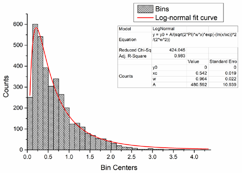
Histogram of the frequencies of Feret’s diameter values of the pores obtained from the SEM micrograph analysis. Log-normal fit curve was used to analytically fit the count distribution.
Iron release measurements were performed on 0.05Fe-TCP-derived cement samples. The sample mass for volume unit of the isotonic solution was 20 mg/mL. The plateau value for the concentration considered, corresponding to the equilibrium, was estimated by the fit to be 0.313±0.003 mg/mL and it is reached after about 10 days, as shown in Figure 9a. Clinical studies refer to an Fe “threshold” of about 4.188 mg/mL in the blood (total iron binding capacity) [56]. The amount released from the samples of the studied composition is an order of magnitude below the threshold cited above, meaning that toxic effects in vivo are not expected and a large amount of cement can be used for implantation.
Fig. 9.

(a) Cumulative iron release curve obtained on 0.05FeTCP-derived cement. Relative uncertainty for all the measures is 1 %. Reduced chi squared for the estimated fit is . (b) Comparison between compressive strength of hardened doped and non-doped TCP-derived cements. *** = p<0.001
The effect of the doping concentration of Fe on the compressive strength (σMAX) of the cements was investigated by comparing two series of samples: non-doped cements as controls and 0.05Fe-TCP derived cements. Repeated measurements gave the average σMAX, non-doped=( 11.5±0.5) MPa and σMAX, doped=(24.5±2.0) MPa (Fig.9b). The values are significantly different at a confidence level <0.001 (t=12.717; df=3.5), indicating a clear enhancement of the mechanical features due to the presence of iron. It is clear that the smaller-sized crystals of brushite contributed to the reduction of the porosity, which is more pronounced in systems formed by larger and well-formed crystals. However, these ultrafine crystals comprising the final, brushite phase are likely to have given a positive contribution to the enhancement of the mechanical properties demonstrated by the results shown in Fig.9b. The role of the hematite component in the enhancement of the compressive strength by inducing dislocation slide impediment at grain boundaries could be also hypothesized, considering that the amount of hematite in the doped-TCP powder was estimated at up to 3 wt.% [41], but additional studies with increasing wt.% of hematite crystals should be performed to investigate these effects.
3.3. Characterization of the biological response
The osteoinductive effect of the cements derived from Fe-TCPs was assessed by measuring the mRNA expression relative to the expression of the housekeeping gene, β-actin using real-time PCR. The expression of osteocalcin, an osteogenic protein playing the role of the mineralization promoter, was expectedly significantly higher in MC3T3-E1 cells differentiated using the standard chemical agents, including ascorbic acid and β-glycerophosphate. However, the expression of osteocalcin in MC3T3-E1 cells challenged with the cements greatly exceeded that in both undifferentiated and differentiated cells, suggesting the pronouncedly osteoinductive activity of the cements (Figure 10a). When measured in terms of the expression the transcription factor, Runx2, this effect was equally significant compared to the undifferentiated cells. The expression of Runx2 in the cements with the lower content of Fe was in the same range as that in the differentiated cells, but it was markedly lower in the cements with the higher content of Fe compared to the differentiated cells (Figure 10b). The lesser effect observed for Runx2 than for osteocalcin may be the consequence of the fact that the peak in the expression of Runx2 precedes that of osteocalcin in the process of differentiation and activation of the osteogenic phenotype by the cells. The osteoinductive effect was consistently inversely proportional to the content of Fe in the cements, indicating that an excessive amount of iron in the cements can have a detrimental effect on the induction of bone growth in cells.
Fig. 10.

mRNA expression of osteocalcin (BGLAP) (a) and the transcription factor Runx2 (b) in MC3T3-E1 cells incubated for 7 days with 5 mg/ml of 0.49 wt.% or 1.09 wt.% Fe-TCP cement. mRNA expression was detected by quantitative RT-polymerase chain reaction and normalized to the expression of the housekeeping gene β-actin (ACTB). C− and C+ represent the expressions in undifferentiated and differentiated (50 μg/ml ascorbic acid and 10 mM β-glycerophosphate) negative, untreated control cells, respectively. Data normalized to the expression of ACTB are shown as averages with error bars representing standard deviation (n = 3 × 3). Statistically significant difference between sample groups is represented with * (p < 0.05), ** (p < 0.005), and *** (p < 0.0001).
To prove the feasibility of the use of iron-doped TCP cements in hyperthermia anticancer therapies, we measured the ability of the material to increase the temperature of the aqueous medium. As seen from Figure 11a, after the initial lag time lasting for 5 minutes, the temperature of the liquid medium increased linearly from the 5th to the 30th and the final minute of exposure to the alternating magnetic field. The total temperature increase in this timespan was 21.7 °C and it took place at the rate of 0.87 °C/min. Given that 41-42 °C is considered the threshold above which cells start to die out, it will take approximately 12 minutes under these conditions for necrotic effects on the cancer cells to be induced. We also tested the ability of the Fe-doped cements to affect the viability of the population of two different types of cancer cells, human E297 glioblastoma and mouse K7M2 osteosarcoma after co-incubating the cement and the cells for 30 minutes in an alternating magnetic field, then washing off the solid phase and allowing the cells to recover for 24 h under regular conditions. E297 cell line was chosen for this assay in addition to K7M2 osteosarcoma cells so as to increase the number of sample groups and thus the confidence in the response of the cells to the treatment with the cements. In addition, E297 cells are a patient-derived cell line proven to possess an exceptional level of resistance to any therapy [57], so any positive therapeutic effect observed on it is likely to apply to the majority of other neoplastic cell types. In addition to this, our previous work [58] demonstrated successful use of calcium phosphates in targeting glioblastoma and we were curious to see if an equally intense hyperthermia effect as the one observed for hydroxyapatite combined with iron oxide nanoparticles would apply to iron-doped TCP/brushite cements. As a result of the temperature increase in the alternate magnetic field, the population of the glioblastoma cells dropped six-fold at the 24 h time point following the end of the magnetic field treatment, while the population of the bone cancer cells dropped approximately twofold (Figure 11b). At this concentration of the cement, it had no negative effect on any of the two cell types in the absence of the magnetic field. Based on Eq.2, there is no release of protons or hydroxyls accompanying the hardening reaction, which allows the cements to be brought into contact with the cells even during this reaction, without concern that this would cause a negative impact on the cell viability. At the tested concentration of the cement, 10 mg/cm2, the effect on the cell viability is rather fully controllable by turning the magnetic field on/off.
Fig. 11.

Temperature increase of the aqueous medium (a) and the viability of human E297 glioblastoma and mouse K7M2 osteosarcoma cells (b) challenged with 10 mg/cm2 of 1.09 wt.% Fe-TCP cement for 30 minutes in an alternate magnetic field (300 kHz, 1.16 μT) compared to the negative, untreated control. All the cells were allowed 24 h to recover after the 30 min treatment in the magnetic field. Treatments of the cells in the magnetic field (Fe-TCP 1.09 wt.% + H) were compared against the no-cement controls and against the treatments with the cements in the absence of the magnetic field (Fe-TCP 1.09 wt.%). Data points are shown as averages with error bars representing the standard deviation (n = 2). Statistically significant difference between the sample groups is represented with either * (p < 0.05) or ** (p < 0.005).
Tests performed to assess the antibacterial activity of Fe-TCP cements and their precursor components showed that the activity of the cements differs against different bacterial species. The activities also greatly differed depending on whether the cements were tested in the diffusive, broth conditions or in the semisolid, agar conditions. Thus, in the broth assay, the activity of the cements was pronounced against the Gram-negative E. coli, present to a minor, but significant extent against Gram-negative S. enteritidis and nonexistent against Gram-negative P. aeruginosa and Gram-positive S. aureus (Figure 12). Previous research has shown that E. coli is, in general, most susceptible to calcium phosphates out of the most common opportunistic pathogens [59]. The higher content of Fe in the cements did not lead to more pronounced bacteriostatic effects in the broth tests, suggesting that the effect of iron is not critical in ensuring the antibacterial activity of the cements under these conditions. In contrast, the antibacterial efficacy of one particular component of the starting formulation, carbonated hydroxyapatite (HAp), was very high against all species but S. enteritidis. Earlier research has shown that the presence of HAp component in alpha-TCP cements improves their cytocompatibility [60], indicating the possibility that the leftovers of this precursor component could exert a key selective influence, such that it would be lethal for the bacteria while reinforcing the growth of primary cells.
Fig. 12.

Optical densities of liquid broths, directly indicative of the number of colony forming units, following a 24 h inoculation with Escherichia coli, Salmonella enteritidis, Pseudomonas aeruginosa or Staphylococcus aureus without (control) or with either 20 mg/ml of a precursor HAp component of the cement formulation (carbonated HAp) or Fe-TCP cements with different concentrations of Fe (low Fe = 0.49 wt.%, high Fe = 1.09 wt.%). Data are shown as averages with error bars representing standard deviation. Data points significantly lower than the untreated control (p < 0.05) are topped with an asterisk.
Whereas antibacterial assays in broths test for the ability of the material to inhibit the growth of suspended pathogens diffusing freely through a liquid medium, the agar assays test for the ability to inhibit the growth of pathogens migrating along a solid, albeit viscous, surface. Such tests are usually more relevant for anti-infective bone replacement materials, given that the migration of pathogens onto the bone surface rarely proceeds through a broth-mimicking, very dilute liquid environment. In contrast, the agar conditions more veritably mimic the extracellular space that bacteria cross en route to the bone surface. Agar assays demonstrated that the increase of the iron content in the cements invariably increased their bacterial inhibitory potential. Distinct inhibition zones were observed around all the cement compositions, including their carbonated HAp precursor component, and were in all cases statistically significant compared to the control (Figure 13). The carbonated HAp precursor, which presumably owed its superior antibacterial activity in broths to its powder form and higher dispersability than that of the cements, displayed a diminished activity under the agar conditions, the reason being its immobilization on the agar surface. Its activity was still consistently higher than the activity of the cement containing the lower concentration of Fe ions (0.49 wt.%). However, its activity was lower than the activity of the cement containing the higher concentration of Fe ions (1.09 wt.%) against all the bacterial species except P. aeruginosa. At the same time, the activity of the cement increased against all the species in direct proportion with the concentration of Fe ions in it, indicating their key effect on the promotion of the antibacterial effect in this material. The antibacterial effect of ferrous ions is usually mechanistically associated with the Fenton reaction, during which OH radicals as powerful reactive oxygen species form from H2O2, capable of damaging biomolecules including DNA, proteins and lipids [61].
Fig. 13.
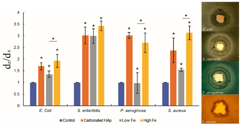
(a) The ratio of the diameter of the inhibition zone (dz) to the diameter of the spherical cement sample deposit (ds) on an agar plate following a 24 h inoculation with Escherichia coli, Salmonella enteritidis, Pseudomonas aeruginosa or Staphylococcus aureus without (control) or with a precursor HAp component of the cement formulation (carbonated HAp) or Fe-TCP cements with different concentrations of Fe (low Fe = 0.49 wt.%, high Fe = 1.09 wt.%). Data are shown as averages with error bars representing standard deviation. Data points significantly different from the untreated control (p < 0.05) are topped with an asterisk. (b) Visual appearance of either homogeneous (E. coli, S. aureus) or concentrically circled (S. enteritidis, P. aeruginosa) inhibition zones around Fe-TCP cements for different bacterial cultures.
The analysis of the cell/particle interaction in an osteoblastic MC3T3-E1 culture reiterated the effects of iron delivered using the TCP cement on the bone growth marker expression, namely their downregulation due to an increase in iron concentration in the cement. Correspondingly, whereas the control calcium phosphate sample, nanoparticulate HAp, exhibited no negative effects at the concentration of 5 mg/mL after 72 h co-incubation nor did the TCP cement containing the lower concentration of Fe (0.49 wt.%), the adverse effects on the cell morphology and proliferation were evident upon incubation with the cement containing the higher concentration of Fe (1.09 wt.%). Thus, actin microfilaments ceased to exhibited a healthy, striated pattern and adopted the clumped and round, apoptotic morphology, together with the nucleus (Figure 14). It is conceivable that the cells can mitigate the ROS production in the Fenton process only to a certain level, above which, i.e. at higher concentrations of iron, lethal effects on cells start to be exhibited. Therefore, the effects of iron are beneficial from the perspective of a magnetic hyperthermia therapy and antibacterial prophylaxis or bactericidal effects, but its concentration in the material must be carefully optimized, lest the adverse effects take over above a certain limit in the amount of iron.
Fig. 14.

Single-plane confocal optical micrographs of fluorescently stained osteoblastic MC3T3-E1 cells (cytoskeletal f-actin - red; nucleus – blue; TCP cement - green) in interaction with 5 mg/ml of either (a) the control HAp or (b, c) Fe-TCP cements with different concentrations of Fe (low Fe = 0.49 wt.%, high Fe = 1.09 wt.%) following a 72 h incubation with the particles.
4. Conclusions
The present study provided insights into the physicochemical and biological properties of self-setting Fe-doped bone-integrative cements. The in situ, continuous monitoring of the cement hardening using EDXRD evidenced the presence of iron-doped brushite as the final product, the setting time point at 0.31±0.03 h, the hardening time point at 2 h, and complete stabilization after 50 h. Compared to other similar systems, the phase transformation from TCP to brushite during hardening was relatively fast and it also followed a relatively simple reaction path, virtually free of complex intermediates and noisy background. SEM studies revealed that in addition to the large plateshaped crystals with circa 20 μm in length and the medium-sized plate-shaped crystals ranging between 3 and 5 μm in size, brushite was also composed of amorphous and/or very fine crystals ranging between 0.6 and 1.0 μm in size. The small size of these crystals contributed to the reduction of the porosity and thereby to the enhancement of the mechanical properties. Specifically, the compressive strength of the hardened product doubled due to iron doping. The amount of iron released from the cements in physiological media steadied after 10 days and was within the range of clinically acceptable iron blood levels. The cements containing two different concentrations of the dopant were studied (0.49 and 1.09 wt.% Fe) in interaction with MC3T3-E1 osteoblasts, yet only the former cement, containing the lower amount of iron, was fully biocompatible with the cells. The osteoinductive effect was pronounced in the osteoblastic cells challenged with the cement containing the lower amount of iron, but inversely proportional to the content of iron in the cements. Antibacterial properties were tested for the two concentrations of the dopant both in broth and on agar, resulting in different activities. Specifically, in the broth assay, the iron concentration did not exert a critical effect and the cements were active against E. coli and S. enteritidis strains, but not against P. aeruginosa and S. aureus. On the contrary, in the agar assays, the iron concentration increase in the cements had a positive effect on the antibacterial properties against all the species except P. aeruginosa. Magnetic-field-induced heating was attempted on two different cultures of cancer cells, demonstrating an effective reduction of the cell population by the treatment and the prospect of the cements for future use in hyperthermia therapies.
Overall, these findings indicate that iron-doped cements undergoing TCP → brushite transformation during hardening are promising in view of the positive effect they have on the mechanical properties, on the antibacterial activity, and on osteoinduction at the gene expression level. As such, they may provide a solution to some of the key factors limiting the clinical application of these cements for the replacement of regions of bone that are moderately load-bearing in nature, that are highly prone to infection or that have low remineralization potentials. In spite of these promising insights, the concentration of the dopant must be carefully optimized to avoid adverse clinical effects.
Highlights.
-
-
Self-setting Fe-doped bone cements with two dopant concentrations are prepared.
-
-
Cements exhibite osteoinductivity inversely proportional to the Fe ions content.
-
-
Cements exhibite antibacterial activity against E. coli, S. enteritidis, P. aeruginosa, S. aureus.
-
-
Glioblastoma and bone cancer cells decreased after end of magnetic field treatment.
-
-
Effects of Fe are beneficial from the perspective of a magnetic hyperthermia therapy.
Acknowledgments
Authors are grateful to I. Glazkova for performing the Mössbauer spectroscopy measurements. NIH R00-DE021416 grant is acknowledged for support. The work was partially supported by the Russian Federation state assignment N° 007-00129-18-0.
Footnotes
Publisher's Disclaimer: This is a PDF file of an unedited manuscript that has been accepted for publication. As a service to our customers we are providing this early version of the manuscript. The manuscript will undergo copyediting, typesetting, and review of the resulting proof before it is published in its final citable form. Please note that during the production process errors may be discovered which could affect the content, and all legal disclaimers that apply to the journal pertain.
The authors declare no competing interests.
References
- [1].Ambard AJ, Mueninghoff L, Calcium phosphate cement: review of mechanical and biological properties, J Prosthodont. 15 (2006) 321–328. [DOI] [PubMed] [Google Scholar]
- [2].Zhang J, Liu W, Schnitzler V, Tancret F, Bouler JM, Calcium phosphate cements for bone substitution: chemistry, handling and mechanical properties, Acta Biomater. 10 (2014) 1035–49. [DOI] [PubMed] [Google Scholar]
- [3].Luo J, Ajaxon I, Ginebra MP, Engqvist H, Persson C, Compressive, diametral tensile and biaxial flexural strength of cutting-edge calcium phosphate cements, Journal of the mechanical behavior of biomedical materials 60 (2016) 617–627. [DOI] [PubMed] [Google Scholar]
- [4].Dorozhkin SV, Calcium orthophosphates (CaPO4): occurrence and properties, Progress in Biomaterials 5 (2016) 9–70. [DOI] [PMC free article] [PubMed] [Google Scholar]
- [5].Charriére E, Terrazzoni S, Pittet C, Mordasini Ph., Dutoit M, Lemaître J, Zysset Ph., Mechanical characterization of brushite and hydroxyapatite cements, Biomaterials 22 (2001) 2937–2945. [DOI] [PubMed] [Google Scholar]
- [6].Cama G, Gharibi B, Knowles JC, Romeed S, Di Silvio L, Deb S, Structural changes and biological responsiveness of an injectable and mouldable monetite bone graft generated by a facile synthetic method, J. R. Soc. Interface 11(2014) 20140727. [DOI] [PMC free article] [PubMed] [Google Scholar]
- [7].Cama G, Gharibi B, Saif Sait M, Knowles JC, Lagazzo A, Romeed S, Di Silvio L, Deb S, A novel method of forming micro- and macroporous monetite cements, J. Mater. Chem. B 1 (2013) 958–969. [DOI] [PubMed] [Google Scholar]
- [8].Quillard S, Paris M, Deniard P, Gildenhaar R, Berger G, Obadia L, Bouler J-M, Structural and spectroscopic characterization of a series of potassium and/or sodium substituted β-tricalcium phosphate, Acta Biomater. 7 (2011) 1844–1852. [DOI] [PubMed] [Google Scholar]
- [9].Bose S, Tarafder S, Baneijee SS, Davies NM, Bandyopadhyay A, Understanding in vivo response and mechanical property variation in MgO, SrO and SiO2 doped β-TCP, Bone 48 (2011) 1282–1290. [DOI] [PMC free article] [PubMed] [Google Scholar]
- [10].Abbaspour N, Hurrell R, Kelishadi R, Review on iron and its importance for human health. Journal of Research in Medical Sciences: The Official Journal of Isfahan University of Medical Sciences 19 (2014) 164–174. [PMC free article] [PubMed] [Google Scholar]
- [11].Cammack R, Wrigglesworth JM, Baum H, Iron-Dependent Enzymes In Mammalian Systems, in: Ponka P, Schulman HM, Woodworth RC, Richter GW (Eds.), Iron transport and storage, CRC Press, 1990, pp. 17–39. [Google Scholar]
- [12].Dlouhy ACA.C., Outten CE, The Iron Metallome in Eukaryotic Organisms, Metal ions in life sciences 12 (2013) 241–278. [DOI] [PMC free article] [PubMed] [Google Scholar]
- [13].Ruzicka FJ, Beinert H, A new iron-sulfur flavoprotein of the respiratory chain. A component of the fatty acid beta oxidation pathway, J Biol Chem. 252 (1977) 8440–5. [PubMed] [Google Scholar]
- [14].Hurrell RF, Bioavailability of iron, Eur J Clin Nutr. 51 (1997) S4–8. [PubMed] [Google Scholar]
- [15].McDowell LR, Minerals in Animal And Human Nutrition, Elsevier Science (2003) 660. [Google Scholar]
- [16].Jacobs A, Tissue Changes in Iron Deficiency, Brit. J. Haematol 16 (1969) 1–4. [DOI] [PubMed] [Google Scholar]
- [17].Basta SS, Soekirman S, Karyadi D, Scrimshaw NS, Iron Deficiency Anemia and the Productivity of Adult Males in Indonesia, Am. J. Clin. Nutr. 32 (1979) 916–925. [DOI] [PubMed] [Google Scholar]
- [18].Zimmermann MB, Köhrle J, The impact of iron and selenium deficiencies on iodine and thyroid metabolism: biochemistry and relevance to public health., Thyroid 12 (2002) 867–78. [DOI] [PubMed] [Google Scholar]
- [19].Chandra RK, Iron and immunocompetence, Nutr. Rev 34 (1976) 129–132. [DOI] [PubMed] [Google Scholar]
- [20].Chandra RK, Iron status, immune response and susceptibility to infection, in Ciba Foundation Symposium 5, Iron metabolism, Elsevier; (1977) 249–262. [DOI] [PubMed] [Google Scholar]
- [21].Chandra RK, Impaired Immunocompetence Associated with Iron Deficiency, J Pediat 86 (1975) 899–902. [DOI] [PubMed] [Google Scholar]
- [22].Bagchi K, Mohanram M, Reddy V, Humoral Immune Response in Children with Iron-Deficiency Anaemia, British Medical Journal 2 (1980) 1251–1253. [DOI] [PMC free article] [PubMed] [Google Scholar]
- [23].Beard J, Iron Deficiency Alters Brain Development and Functioning, J. Nutr. 133 (2003) 1468S–1472S. [DOI] [PubMed] [Google Scholar]
- [24].Zheng W, Monnot AD, Regulation of Brain Iron and Copper Homeostasis by Brain Barrier Systems: Implication in Neurodegenerative Diseases, Pharmacol. Ther 133 (2012) 177–188. [DOI] [PMC free article] [PubMed] [Google Scholar]
- [25].MacDougall LG, Anderson R, McNab GM, Katz J, Immune Response in Iron-deficient Children: Impaired Cellular Defense Mechanisms with Altered Humoral Components, J Pediat 86 (1975) 833–843. [DOI] [PubMed] [Google Scholar]
- [26].Chandra RK, Reduced Bactericidal Capacity of Polymorphs in iron Deficiency, Arch. Dis. Child 48 (1973) 863–866. [DOI] [PMC free article] [PubMed] [Google Scholar]
- [27].Furustrand Tafin U, Betrisey B, Bohner M, Ilchmann T, Trampuz A, Clauss M, Staphylococcal biofilm formation on the surface of three different calcium phosphate bone grafts: a qualitative and quantitative in vivo analysis, J. Mater. Sci. Mater. Med. 26 (2015) 130. [DOI] [PMC free article] [PubMed] [Google Scholar]
- [28].Uskoković V, Desai TA, Phase Composition Control of Calcium Phosphate Nanoparticles for Tunable Drug Delivery Kinetics and Treatment of Osteomyelitis. I. Preparation and Drug Release, J. Biomed. Mater. Res. Part A 101 (2013) 1416–1426. [DOI] [PMC free article] [PubMed] [Google Scholar]
- [29].Theuretzbacher U, Accelerating resistance, inadequate antibacterial drug pipelines and international responses, Int. J. Antimicrob. Agents 39 (2012) 295–9. [DOI] [PubMed] [Google Scholar]
- [30].Scalny AV, Rudakov IA, Bioelements in medicine, Moscow: Publishing House Onyx 21, 2004, pp. 272. [Google Scholar]
- [31].Rau JV, Fosca M, Graziani V, Egorov AA, Zobkov V.Yu, Fedotov A.Yu., Ortenzi M, Caminiti R, Baranchikov AE, Komlev VS, Silver-doped calcium phosphate bone cements with antibacterial properties, J. Funct. Biomater 7 (2016) 10. [DOI] [PMC free article] [PubMed] [Google Scholar]
- [32].Graziani V, Fosca M, Egorov AA, Zobkov V.Yu, Fedotov A.Yu., Baranchikov AE, Ortenzi M, Caminiti R, Komlev VS, Rau JV, Zinc-releasing calcium phosphate cements for bone substitute materials, Ceram. Int 42 (2016) 17310–17316. [Google Scholar]
- [33].Rau JV, Wu VM, Graziani V, Fadeeva IV, Fomin AS, Fosca M, Uskoković V, The Bone Building Blues: Self-hardening copper-doped calcium phosphate cement and its in vitro assessment against mammalian cells and bacteria, Materials Science and Engineering C 79 (2017) 270–279. [DOI] [PMC free article] [PubMed] [Google Scholar]
- [34].Pina S, Vieira ST, Torres PMC, Goetz-Neunhoeffer F, Neubauer J, da Cruz e Silva OAB, da Cruz e Silva EF, Ferreira JMF, In Vitro performance assessment of new brushite-forming Zn- and ZnSr-substituted b-TCP bone cements, J. Biomed. Mater. Res. B Appl. Biomater 94 (2010) 414–20. [DOI] [PubMed] [Google Scholar]
- [35].Bose S, Fielding G, Tarafder S, Bandyopadhyay A, Trace element doping in calcium phosphate ceramics to Understand osteogenesis and angiogenesis, Trends in Biotechnology 31 (2013) 594–605. [DOI] [PMC free article] [PubMed] [Google Scholar]
- [36].Pina S, Ferreira JMF, Brushite-Forming Mg-, Zn- and Sr-Substituted Bone Cements for Clinical Applications, Materials 3 (2010) 519–535. [Google Scholar]
- [37].Bernhardt A, Schamel M, Gbureck U, Gelinsky M, Osteoclastic differentiation and resorption is modulated by bioactive metal ions Co2+, Cu2+ and Cr3+ incorporated into calcium phosphate bone cements, PLoS ONE 12 (2017) e0182109. [DOI] [PMC free article] [PubMed] [Google Scholar]
- [38].Torres PMC, Marote A, Cerqueira AR, Calado AJ, Abrantes JCC, Olhero S, da Cruz e Silva OAB, Vieira SI, Ferreira JMF, Injectable MnSr-doped brushite bone cements with improved biological performance, J. Mater. Chem. B 5 (2017) 2775–2787. [DOI] [PubMed] [Google Scholar]
- [39].Saleh AT, Ling LS, Hussain R, Injectable magnesium-doped brushite cement for controlled drug release application, J. Mater. Sci 51 (2016) 7427–7439. [Google Scholar]
- [40].El-dek SI, Mansour SF, Ahmed MA, Ahmed MK, Microstructural features of flower like Fe brushite, Materials International 27 (2017) 520–526. [Google Scholar]
- [41].Fadeeva IV, Selezneva II, Davydova GA, Fomin AS, Antonova OS, Filippov Ya.Yu., Barinov SM, Iron-substituted tricalcium phosphate ceramics, in Doklady Akademii Nauk 468, 2 (2016)171–174. [Google Scholar]
- [42].Caminiti R et al. , Italian Patent No. 01126484-23 (1993).
- [43].International Centre for Diffraction Data, Database JCPDS, (2001).
- [44].Lafuente B, Downs RT, Yang H, Stone N, The power of databases: the RRUFF project, in Highlights in Mineralogical Crystallography, Armbruster T and Danisi RM, eds. Berlin, Germany, W. De Gruyter, 2015, pp. 1–30. [Google Scholar]
- [45].Abrámoff MD, Magalhães PJ, Ram SJ, Image Processing with ImageJ, Biophotonics Intern. 11 (2004) 36–42. [Google Scholar]
- [46].Matsnev ME and Rusakov VS, AIP Conf. Proc. 1489, 178 (2012); [Google Scholar]; Matsnev ME and Rusakov VS, AIP Conference Proceedings 1622 (2014) 40. [Google Scholar]
- [47].Menil F, J. Phys. Chem. Solids 46 (1985) 763. [Google Scholar]
- [48].Pfaffl MW, A new mathematical model for relative quantification in real-time RT-PCR, Nucleic Acids Res. 29 (2001) 45e–45. [DOI] [PMC free article] [PubMed] [Google Scholar]
- [49].Zhang X, Niu Y, Meng X, Li Y, Zhao J, Structural Evolution and Characteristics of the Phase Transformations Between α-Fe2O3, Fe3O4 and γ-Fe2O3 Nanoparticles Under Reducing and Oxidizing Atmospheres, Cryst. Eng. Comm 15 (2013) 8166–8172. [Google Scholar]
- [50].Zhao K, Liu X, Jin C, Yu F, Rykov A, Wang J, Zhang T, Influence of Hydroxyapatite on Maghemite-to-Hematite Phase Transfer of FeOx-Hydroxyapatite Composite, Hyperfine Interact. 218(2012) 1–7. [Google Scholar]
- [51].Kumar PN, Mishra SK, Kannan S, Structural elucidation and iron oxidation states in situ formed β-Ca3(PO4)2/α-Fe2O3 composites, J. Am. Ceram. Soc 100 (2017) 3746–3756. [Google Scholar]
- [52].Berzina-Cimdina L, Borodajenko N, Research of Calcium Phosphates using Fourier Transform Infrared Spectroscopy, in Infrared Spectroscopy, Materials Science, Engineering and Technology, Theophile Theophanides, ed. InTech, Rijeka: 2012. [Google Scholar]
- [53].Belik AA, Izumi F, Stefanovich S.Yu, Lazoryak BI, Oikawa K, Chemical and Structural Properties of a Whitlockite-like Phosphate, Ca9FeD(PO4)7, Chem. Mater 14 (2002) 3937–3945. [Google Scholar]
- [54].Lazoryak BI, Morozov VA, Belik AA, Khasanov SS, Shekhtman VS, Crystal Structures and Characterization of Ca9Fe(PO4)7 and Ca9FeH0.9(PO4)7, J. Solid State Chem 122 (1996) 15–21. [Google Scholar]
- [55].Gomes S, Kaur A, Grenéche J-M, Nedelec J-M, Renaudin G. Atomic scale modeling of iron-doped biphasic calcium phosphate bioceramics, Acta Biomaterialia 50 (2017) 78–88. [DOI] [PubMed] [Google Scholar]
- [56].Erber WN, Red blood cell disorders, in Bennett PN, Brown MJ, Sharma P, Clinical Pharmacology, eds Churchill Livingstone Elsevier, 2012. [Google Scholar]
- [57].Engelhard HH, Duncan HA, Kim S, Criswell PS, Van Eldik L, Therapeutic Effects of Sodium Butyrate on Glioma Cells in Vitro and in the Rat C6 Glioma Model, Neurosurgery 48(3) (2001)616–625. [DOI] [PubMed] [Google Scholar]
- [58].Pernal SP, Wu VM, Uskoković V, Hydroxyapatite as a Vehicle for the Selective Effect of Superparamagnetic Iron Oxide Nanoparticles against Human Glioblastoma Cells, ACS Appl. Mater. Interfaces 9(45) (2017) 39283–39302. [DOI] [PMC free article] [PubMed] [Google Scholar]
- [59].Ghosh S, Wu VM, Pernal S, Uskoković V, Self-Setting Calcium Phosphate Cements with Tunable Antibiotic Release Rates for Advanced Bone Graft Applications, ACS Applied Materials and Interfaces 8 (2016) 7691–7708. [DOI] [PMC free article] [PubMed] [Google Scholar]
- [60].Zima A, Czechowska J, Siek D, Olkowski R, Noga M, Lewandowska-Szumiel M, Slosarczyk A, How calcite and modified hydroxyapatite influence physicochemical properties and cytocompatibility of alpha-TCP based bone cements, J. Mater. Sci.: Mater. Med 28 (2017) 117. [DOI] [PMC free article] [PubMed] [Google Scholar]
- [61].Park SC, Kim NH, Yang W, Nah JW, Jang MK, Lee D, Polymeric micellar nanoplatforms for Fenton reaction as a new class of antibacterial agents, J. Controlled Release 221 (2016) 37–47. [DOI] [PubMed] [Google Scholar]