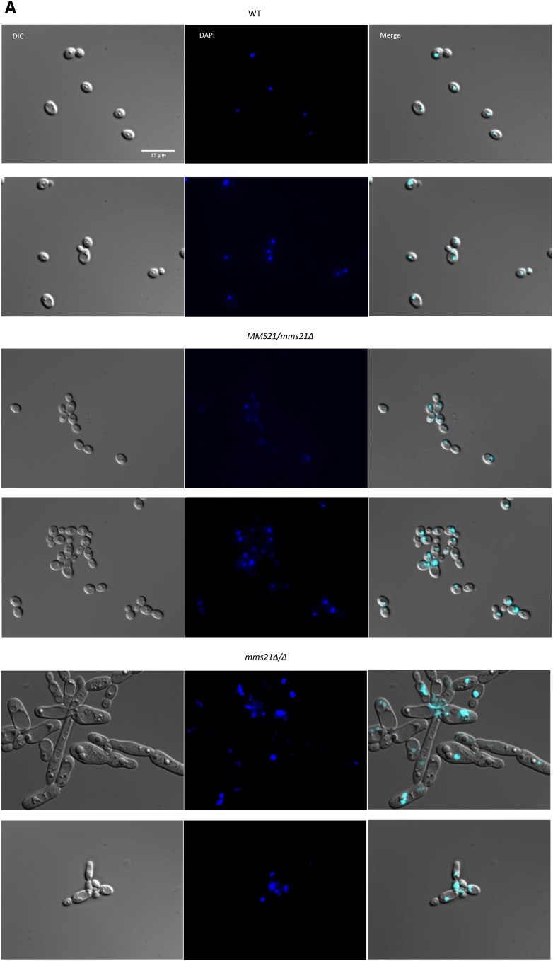
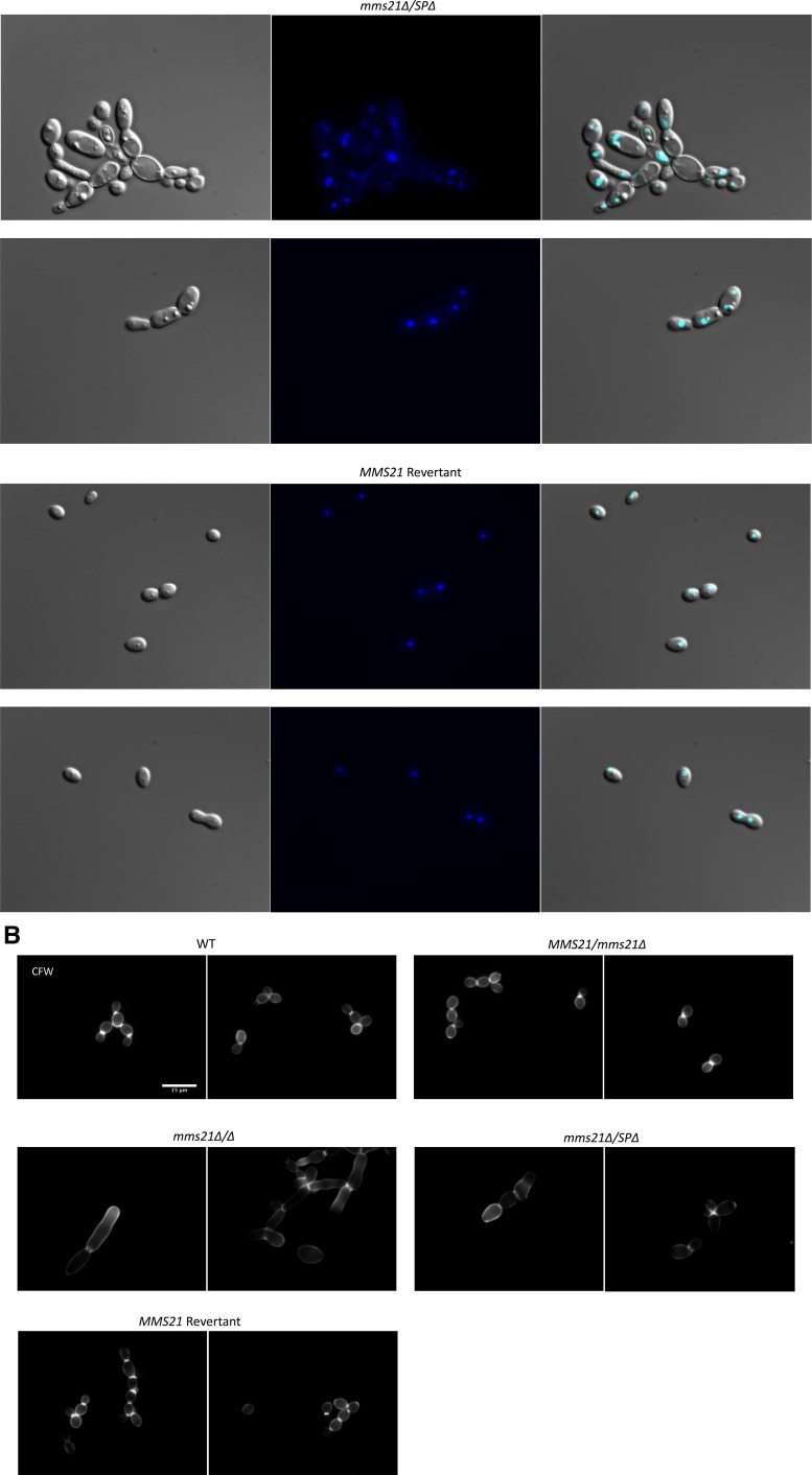
Figure 5.
Staining of the MMS21 mutants with DAPI and calcoflour. Cultures were allowed to grow for 3.5 hr for WT and revertant strains, and 6 hr for mutants under yeast growth conditions, and then stained with 2 μg/ml DAPI or 2 μg/ml calcoflour. Individual cells were examined under ×100 magnification using a Leica DM 6000 microscope, Bar, 15 μm. Only (more or less) yeast-looking cells of mutants were considered for better comparison to the WT. (A) WT cells and strains with one WT allele of MMS21 showed normal nuclear segregation. The mms21Δ/Δ and mms21Δ/SPΔ mutants displayed considerable variation in the number of nuclei present in the individual cells. (B) Calcoflour-stained cells where WT cells display even chitin distributions and prominent septa, while both mms21 mutants have abnormal chitin composition and inconspicuous septa. Reinsertion of Mms21 into the null mutant resulted in highly noticeable septa. WT, wild-type.


