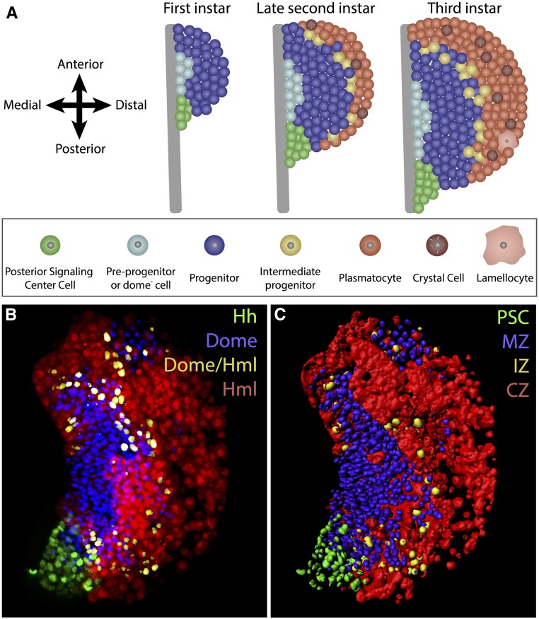
Figure 4.
LG zones and cell types in the primary lobe. (A) Schematic diagrams of LG primary lobes from first (left), late second (middle), and third (right) instar larvae. By convention, we designate the region close to the dorsal vessel as medial and the opposite edge as distal. Anterior is up in all LG images. The PSC (green) acts as a niche for LG progenitors and is present throughout development in all three instars. The majority of cells in the first-instar primary lobe are undifferentiated Domeless+ progenitors that belong to the MZ (dark blue). A small number of domeless− and Notch+ preprohemocytes (light blue) lie on the medial edge of the lobe adjacent to the dorsal vessel (gray), and are capable of contributing to the MZ population during the first 5–20 hr of first instar development. The second-instar LG develops mature hemocytes that make up the CZ (red). This zone mostly includes plasmatocytes and a small number of crystal cells. Additionally, an IZ (yellow) at the interface of the MZ and the CZ contains cells that express both progenitor (domeless) and differentiating hemocyte (Peroxidasin and Hemolectin) markers, but they lack mature hemocyte markers such as P1 or Lozenge/Hindsight. A population of domeless− cells remains along the medial edge of the lobe, although they no longer retain active Notch signaling. During the third instar, the overall LG size increases and a larger number of differentiated hemocytes are seen. (B) Image of a third-instar primary lobe obtained using fluorescence microscopy. Hh-GFP (PSC; green), domeMESO-BFP (MZ; blue), Hml-DsRed (CZ; red), and intermediate progenitors (pseudocolored as yellow based on overlap of domeMESO-BFP and Hml-DsRed). (C) Computer rendering of the confocal data shown in (B) using Imaris software. This software generates accurate three-dimensional models from which quantitative data can be readily derived. CZ, cortical zone; Hh, Hedhehog; Hml, Hemolectin; IZ, intermediate zone; LG, lymph gland; MZ, medullary zone; PSC, posterior signaling center.

