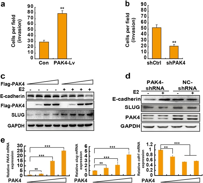
Figure 4.
Nuclear PAK4 promotes breast cancer cell invasion via downregulation of E-cadherin. a PAK4 promotes the invasion of breast cancer cells with E2 stimulation. Transwell invasion assays of MCF-7 cells that were infected with lentivirus Con or PAK4. The number of cells was counted in 16 independent symmetrical microscopic visual fields (× 400 original magnification). The bar chart shows values of the means ± s.d. from three independent experiments. **P < 0.01. b Knockdown of PAK4 suppresses the invasion of human breast cancer cells with E2 stimulation. Transwell invasion assay of shCtrl or shPAK4 MCF-7 cells. The data are shown as the means ± s.d. from triplicate experiments. **P < 0.01 according to Student’s t-test. c PAK4 regulates E-cadherin and Slug protein expression in breast cancer cells. MCF-7 cells were transiently transfected with control or increasing amounts of Flag-PAK4 expression plasmid with or without E2 (10-9 M) for 48 h. E-cadherin, Slug and PAK4 were detected by western blotting. GAPDH was used as a loading control. d MCF-7 cells infected with shPAK4 lentivirus or control shRNA (shCtrl) for 3 d and were either untreated or treated with E2 for 24 h. Western blot analysis revealed PAK4, E-cadherin and Slug protein levels. e PAK4 overexpression resulted in the suppression of cdh1 and promotion of slug gene expression. MCF-7 cells were transiently transfected with control or increasing amounts of Flag-PAK4 expression plasmids and treated with E2 (10−9 M) for 24 h. The cells were then harvested and analyzed for PAK4, slug, and cdh1 mRNA using quantitative real-time PCR assays. Real-time PCR values were normalized to the housekeeping gene β-actin. Experiments were performed three times, each with technical duplicates in the quantitative RT-PCR assays, and the data are presented as the means ± s.d. **P < 0.01; ***P < 0.001 according to Student’s t-test

