Summary
Third-degree burns of the lower extremities are among the most difficult burn injuries to treat as they frequently expose bone, tendons or articular surfaces. Coverage with a flap is the ideal treatment, but local tissue is often unavailable, and free flaps require sophisticated equipment and perfect microsurgical technique. We demonstrate a treatment option to obtain a stable cutaneous coverage for this kind of injury, consisting in an association of skin grafts, amniotic membrane and bilaminar dermal matrix templates. This combined treatment proved to be an excellent option to cover a wide area of tibial exposure with low donor site morbidity and good functional and aesthetic results. This shows that artificial dermis is a good alternative for treating bone exposure, especially in patients for whom a classic flap reconstruction is not suitable.
Keywords: bone exposure, dermal matrix, skin graft, amniotic membrane, deep burn
Abstract
Les brûlures du troisième degré distales des membres inférieurs sont parmi les plus difficiles à traiter en raison de la fréquence des expositions osseuses, tendineuses ou articulaires. Bien que l’utilisation de lambeaux soit le traitement idéal de ces expositions, les tissus adjacents sont souvent inutilisables et les lambeaux libres requièrent un équipement spécifique et une maitrise des techniques microchirurgicales. Nous soumettons une option thérapeutique permettant d’obtenir une couverture cutanée stable pour ce type de brûlures : association de greffe cutanée, de membrane amniotique et de matrice dermique double couche. Ce traitement combiné s’est révélé être un excellent choix thérapeutique pour couvrir de larges expositions tibiales avec peu de morbidité de site donneur et un bon résultat fonctionnel et esthétique. Cela démontre que le derme artificiel peut être une alternative thérapeutique pour les expositions osseuses, surtout lorsque qu’une couverture par lambeau n’est pas possible.
Introduction
Third-degree burns of the lower extremities are among the most difficult burn injuries to treat as they frequently expose bone, tendons or articular surfaces, and they usually cannot be treated with skin grafts or local flaps.1 In some cases, extensive bone necrosis and osteomyelitis may warrant early amputation to preclude a systemic infection.2 A stable cutaneous coverage is the main goal of treatment, and early treatment is advocated. Local flaps or free flaps are the preferred choice for most authors, but donor site morbidity and patient comorbidities must be considered.2,3 We hereby present a clinical case that highlights the challenge of treating these injuries and the importance of planning the reconstructive approach.
Case presentation
We present the case of a 41-year-old male who suffered an extensive deep burn on his lower limbs caused by a fire in his apartment. His comorbidities included chronic hepatic disease (hepatitis C virus infection [HCV] and alcoholism), and he attended a rehabilitation program (with methadone) because of previous drug abuse.
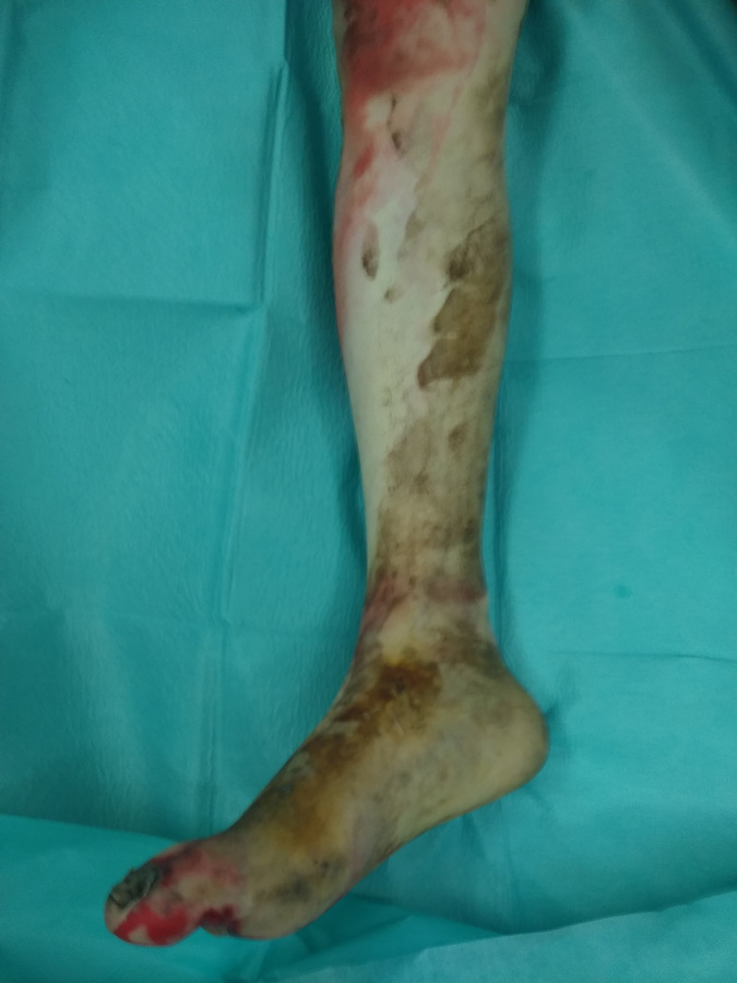
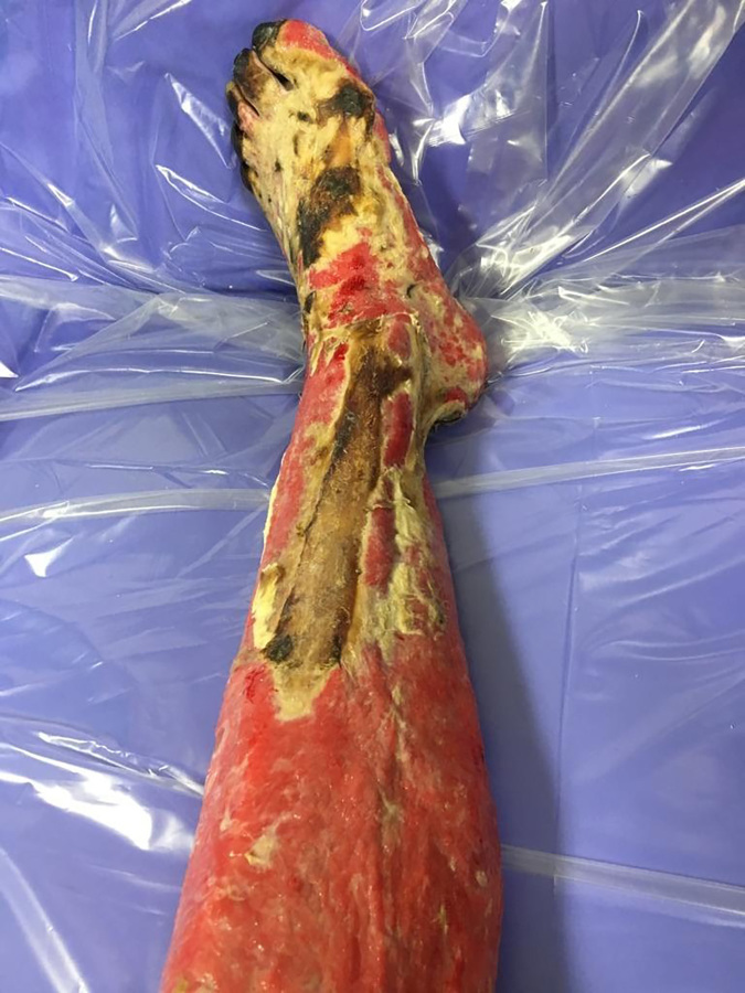
He was found unconscious and was promptly intubated. He was then transported to a tertiary care hospital with a dedicated burn unit and an emergency plastic surgery team. The trip took approximately 2 hours. The patient had deep second- and thirddegree burns of the entire legs and feet, circumferential, and the postero-medial surface of both thighs (total body surface area burned [TBSA] approximately 30%). The physical examination showed that intracompartmental pressure of both legs and feet was higher than normal. The team performed emergency escharotomies (Figs. 1, 2 and 3) on the medial and lateral side of the legs and dorsum of the feet. Perfusion was assessed again and both extremities were well vascularized.
Fig. 1. Right limb burn.
Fig. 2. Left limb: medial leg escharotomies.
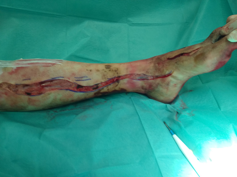
Fig. 3. Left limb: lateral leg escharotomies and dorsum of the foot.

After fluid resuscitation and hemodynamic stabilization, the patient was then submitted to serial debridement of all the burn eschar. He had lost all skin coverage on both legs, dorsum of the feet and part of the plantar surface. He had bone exposure of the whole lower half of both tibias (antero-medial surface) below knee amputation was considered. Nonetheless, since the patient was young and a bilateral amputation would cause significant morbidity, we opted to pursue a reconstructive approach.
Firstly, most of the burnt areas with no bone exposure (thighs, upper legs and feet) were treated with a meshed (1:6) split thickness skin graft (STSG) covered with amniotic membrane. The healing was uneventful with stable cicatrisation 18 days later (Fig. 5).
Fig. 4. Left limb after debridement: extensive bone exposure.
Fig. 5. Right limb after STSG, showing significant tibial exposure.
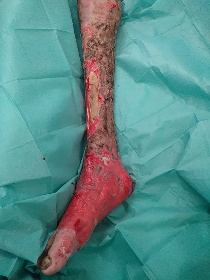
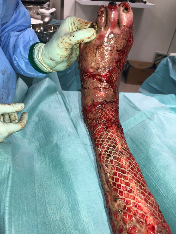
Secondly, we performed bilateral removal of the outer layer of the anterior cortex of both tibias causing them to bleed, as well as a superficial curettage of the exposed bone on the left foot. In the same procedure and after careful haemostasis, we covered the exposed areas with a bilaminar dermal matrix template (Integra®, Integra LifeSciences Corp., Plainsboro, N.J.), fixed with staples and with an octinide-based dressing. The remaining non-healing wounds were covered with a split thickness skin graft (Figs. 6, 7 and 8).
Fig. 6. Placement of Integra dermal matrix over both tibias and the dorsum of the left foot.

Fig. 7. Placement of Integra dermal matrix over both tibias and the dorsum of the left foot.
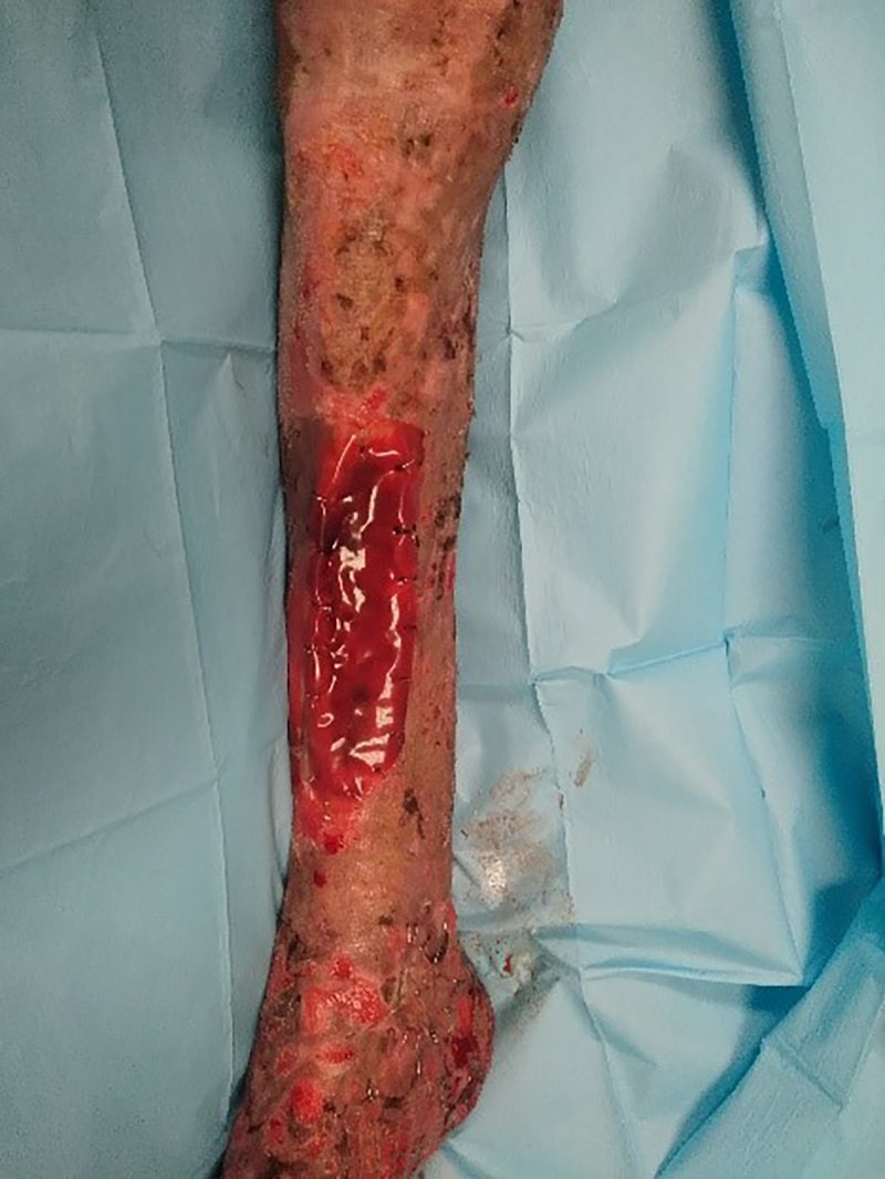
Fig. 8. Placement of Integra dermal matrix over both tibias and the dorsum of the left foot.
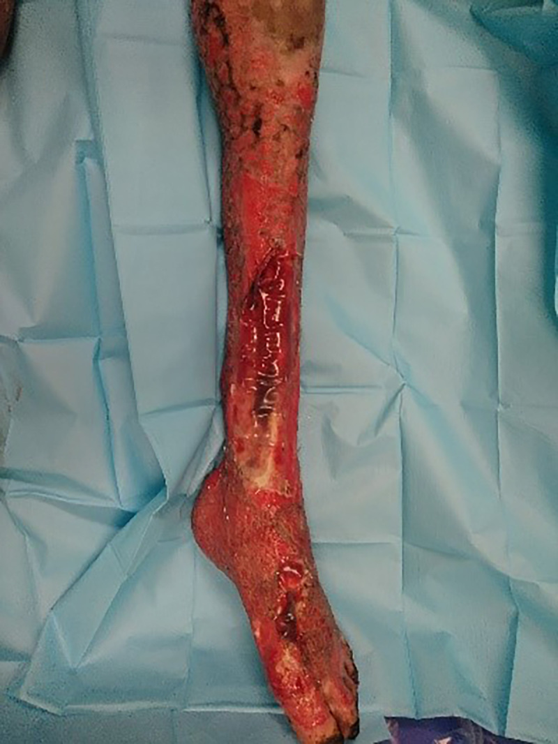
Three weeks later, the silicone layer of the template on the right leg was peach-coloured and ready to be removed. We covered the wound with a meshed (1:3) split thickness skin graft (Fig. 9). In the same procedure, we performed an amputation of the 4th and 5th left toes since they had extensive necrotic bone. One week later (4 weeks after implantation), the dermal matrix templates of the left lower limb were submitted to the same procedure (Fig. 10).
Fig. 9. Right limb: removal of the silicone layer and coverage with STSG.
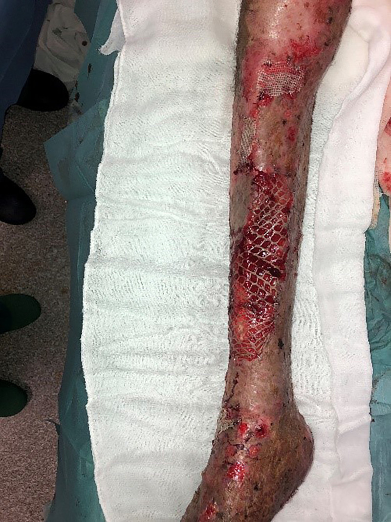
Fig. 10. Left limb: removal of the silicone layer and coverage with STSG.
The dressings were changed three times per week. The evolution of the wounds was very positive, with a graft take of around 95% and no concurrent infections. One month after the last surgery, the patient had only a few residual non-epithelized areas (for secondary healing) with stable coverage over both tibias (Figs. 11 and 12). He was transferred to a rehabilitation hospital where he could continue his functional recovery.
Fig. 11. One month post-op: left limb.
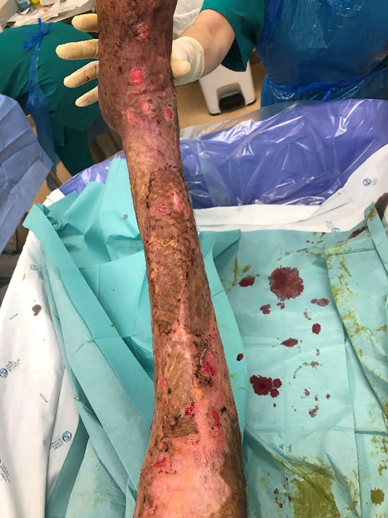
Fig. 12. One month post-op: right limb.
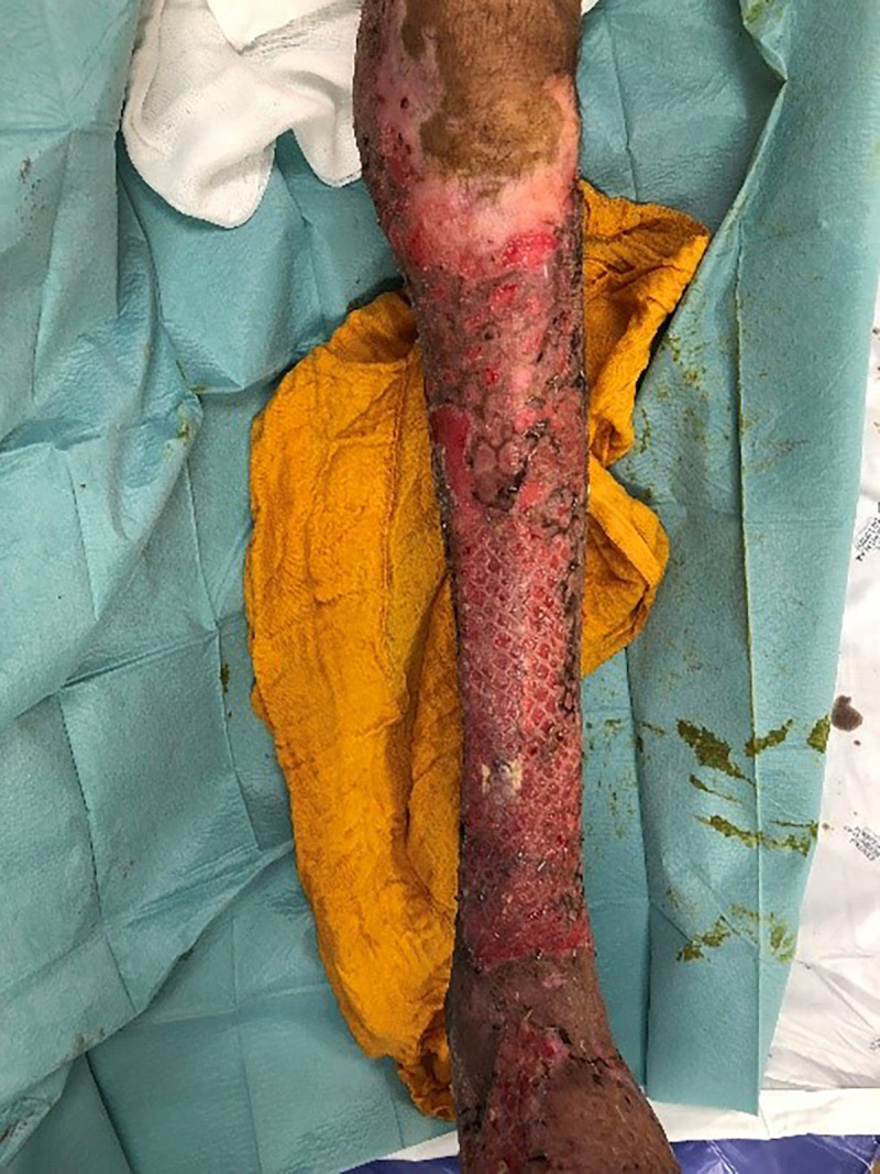
Discussion
In this clinical case, the most critical point was the early decision between amputation and reconstructive treatment. The patient was young, and his hepatic disease was not terminal. He ambulated and had no osteo-articular disabilities. The reconstructive option would allow him to maintain plantar sensation and recover, even if partially, the function of both legs. Amputation would have probably significantly decreased the immediate risk of infection, third space losses and length of stay in the hospital, and accelerated the overall recovery of the patient. On the other hand, since the injuries were bilateral and reached the middle thigh, bilateral amputation would have had to be done just below the knee or even above it. This would have caused significant morbidity. Additionally, this option would have still been available at a later stage if the reconstructive efforts were not effective. After taking into account all the above-mentioned factors, the team decided to proceed with a reconstructive treatment.
The patient had a very large tegumentary deficit with three considerable zones of bone exposure. The areas where no important structures were exposed were treated with a meshed split thickness skin graft (1:6) covered with an amniotic membrane dressing. We used this biological dressing because it significantly improves rate of complete graft take and reduces the time to complete cicatrisation.4,5,6
Regarding the coverage of exposed bone areas, treatment options were scarce. Most authors agree that early reconstruction with a flap is the best way to treat bone exposure after a burn, as it provides stable soft tissue coverage and improves bone viability.2 In this case, local/loco-regional flaps were out of the equation since the surrounding zone was burnt as well. Free tissue transfer would have been a possible solution. Nonetheless, this would have required three free flaps and highly technical skills. Additionally, finding suitable (healthy) receptor vessels would have been a problem as again, all the surrounding area was burnt.
Another option was to perform an extensive debridement of the bone cortical and drill until bleeding, followed by a skin graft. The trephination would allow vascular growth from the bone surface. 7,8 Nonetheless, this kind of procedure has a modest success rate and even if the graft does heal, the skin coverage is usually very prone to breakdown since it misses the dermal substratum.1,9,10
After analysing all the options available, we opted to proceed with debridement of necrotic bone (decortication) and coverage with a dual layer dermal matrix template. It served as temporary coverage and as a scaffold for the ingrowth of a “neodermis” that later received a skin graft.1,9 The result was a stable coverage with no infections and minimal donor site morbidity. The patient was also spared lengthy procedures (as is the case with free flaps).
In conclusion, circumferential third-degree burns of the lower limbs pose an extreme challenge to all plastic surgeons. Sometimes the damage is insurmountable, and amputation may be the best solution. Nonetheless, as this case report shows, there are certain occasions when a reconstructive approach should be attempted to achieve the best possible outcome. Preservation of a sensate, normal-length limb with stable cutaneous coverage is the ultimate goal.
Acknowledgments
Consent / Conflict of interest. The Authors declare that there is no conflict of interest. Oral and written informed consent for medical photographs and medical records was obtained from the patient.
Funding. This work received no specific grant from any funding agency in the public, commercial or not-for-profit sectors.
References
- 1.Lee LF, Porch JV, Spenler W, Garner WL. Integra in lower extremity reconstruction after burn injury. Plastic Reconstr Surg. 2008;121(4):1256–1262. doi: 10.1097/01.prs.0000304237.54236.66. [DOI] [PubMed] [Google Scholar]
- 2.Verbelen J, Hoeksema H, Pirayesh A, Van Landuyt K, Monstrey S. Exposed tibial bone after burns: flap reconstruction versus dermal substitute. Burns. 2016;42(29:e31–e37. doi: 10.1016/j.burns.2015.08.013. [DOI] [PubMed] [Google Scholar]
- 3.Shamian B, Hinds RM, Capo JT. Novel use of synthetic acellular dermal matrix for coverage of a tibial defect following resection of an osteochondroma: a case report. Int J Low Extrem Wounds. 2016;15(1):82–85. doi: 10.1177/1534734615604279. [DOI] [PubMed] [Google Scholar]
- 4.Lavor MA, Michael GM, Tamire YG, Dorofee ND. Meshed splitthickness autograft with a viable cryopreserved placental membrane overlay for lower-extremity recipient sites with increased risk of graft failure. Eplasty. 2018;18 [PMC free article] [PubMed] [Google Scholar]
- 5.Mohammadi AA, Jafari SMS, Kiasat M. Effect of fresh human amniotic membrane dressing on graft take in patients with chronic burn wounds compared with conventional methods. Burns. 2013;39(2):349–353. doi: 10.1016/j.burns.2012.07.010. [DOI] [PubMed] [Google Scholar]
- 6.Mohammadi AA, Johari HG, Eskandari S. Effect of amniotic membrane on graft take in extremity burns. Burns. 2013;39(6):1137–1141. doi: 10.1016/j.burns.2013.01.017. [DOI] [PubMed] [Google Scholar]
- 7.Yeong EK, Chen SH, Tang YB. The treatment of bone exposure in burns by using artificial dermis. Ann Plast Surg. 2012;9(6)X:607–610. doi: 10.1097/SAP.0b013e318273f845. [DOI] [PubMed] [Google Scholar]
- 8.Singh M, Godden D, Farrier J, Ilankovan V. Use of a dermal regeneration template and full-thickness skin grafts to reconstruct exposed bone in the head and neck. Br Journal Oral Maxillofac Surg. 2016;54(10):1123–1125. doi: 10.1016/j.bjoms.2016.03.004. [DOI] [PubMed] [Google Scholar]
- 9.Kahn SA, Beers BR, Lentz CW. Use of acellular dermal replacement in reconstruction of nonhealing lower extremity wounds. J Burn Care Res. 2011;32(1)X:124–128. doi: 10.1097/BCR.0b013e318204b327. [DOI] [PubMed] [Google Scholar]
- 10.Canonico S, Campitiello F, Della Corte A, Fattopace A. The use of a dermal substitute and thin skin grafts in the cure of “complex” leg ulcers. Dermatologic Surgery. 2009;35(2):195–200. doi: 10.1111/j.1524-4725.2008.34409.x. [DOI] [PubMed] [Google Scholar]


