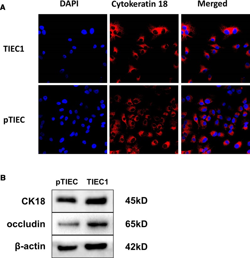
Fig. 3.
Biochemical characteristics of TIEC1s and pTIECs. a Immunofluorescence staining of TIEC1s and pTIECs. The nucleus was stained with DAPI (blue) and cytokeratin 18 was stained with anti-cytokeratin 18 antibodies (red). b Detection of proteins by western blot. Cytokeratin 18 and occludin were both expressed in TIEC1s and pTIECs

