

A 32-year-old male, known case of chronic myeloid leukemia (CML) on Imatinib with irregular compliance for 6 years, presented with painless swellings over the scalp, face and trunk for 3-weeks. He had non-tender, purplish nodules and plaques of varying sizes over the scalp, face and trunk giving “blueberry muffin” appearance (Fig. 1a). Hemoglobin was 8.3 g/dL; TLC 66,900/mm3 and platelets 70,000/mm3. Biopsy from skin nodule was suggestive of leukemia cutis (Fig. 2a, b). Bone marrow showed CML in chronic phase (Fig. 2c, d). Because of poor finances, he was counseled regarding compliance and imatinib dose was increased to 800 mg/day. Six weeks later, the complete hematological response was achieved, and skin lesions gradually resolved to hyperpigmented macules (Figure 1b).
Fig. 1.
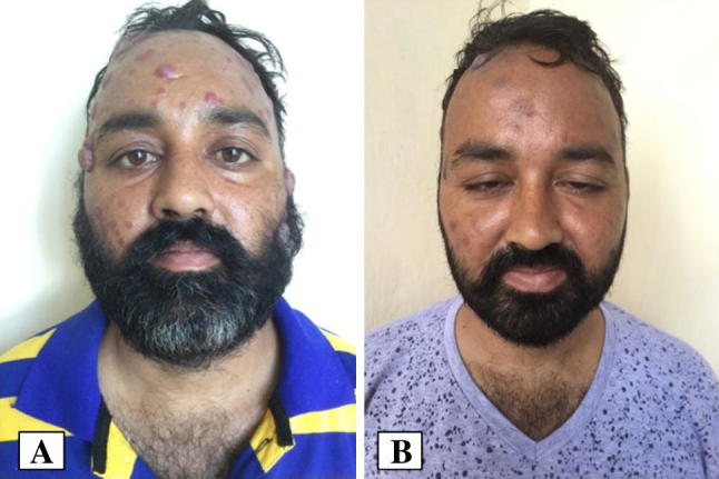
a Clinical photograph showing purplish nodules and plaques of varying sizes over face giving “blue berry muffin” appearance. b Clinical photograph after 3 weeks of TKI showing near complete resolution of facial lesions
Fig. 2.

a Photomicrograph from the skin biopsy shows presence of leukaemic cells in the dermis (H&E × 20). b Photomicrograph shows leukaemic cells positive for CD34 (IHCX20). c Bone marrow aspirate smear shows markedly hypercellular marrow spaces with myeloid buldge (May Grunwald Giemsa × 400). d Bone marrow biopsy shows markedly hypercellular marrow spaces with granulocytic and megakaryocytic hyperplasia (Hematoxylin and eosin stain × 200)
Leukemia cutis (LC) refers to cutaneous lesions resulting from infiltration by neoplastic leucocytes. Morphologically similar conditions include mycosis fungoides, scleromyxedema, vasculitis, drug rash, and Kaposi’s sarcoma, necessitating diagnosis confirmation by histopathological examination and immunophenotyping [1]. The incidence of LC in CML is 2–8% and portends disease progression and poor prognosis (median survival 9.4 months) [2]. The index case neither had any hepatosplenomegaly nor the evidence of accelerated or blast phase. The cutaneous lesions responded to Imatinib although aggressive clinical profile warranted a second-line tyrosine kinase inhibitor.
Conflict of interest
There is no conflict of interest between the authors.
Ethical Approval
All procedures performed in studies involving human participants were in accordance with the ethical standards of the institutional and/or national research committee and with the 1964 Helsinki declaration and its later amendments or comparable ethical standards.
Human and Animals Rights
No animals were involved in the study.
Informed Consent
Informed signed written consent was taken from the patient involved.
References
- 1.Behera B, Kumari R, Manobalan K, et al. Leukemia cutis presenting as scaly plaques in a Christmas tree distribution in a patient with atypical chronic myeloid leukemia. Indian J Dermatol Venereol Leprol. 2017;83:236–238. doi: 10.4103/0378-6323.193625. [DOI] [PubMed] [Google Scholar]
- 2.Kaddu S, Zenahlik P, Beham-Schmid C, et al. Specific cutaneous infiltrates in patients with myelogenous leukemia: a clinicopathologic study of 26 patients with assessment of diagnostic criteria. J Am Acad Dermatol. 1999;40:966–978. doi: 10.1016/S0190-9622(99)70086-1. [DOI] [PubMed] [Google Scholar]