

Abstract
In contrast to the skin, aging of the hair has seemingly only recently found the attention of dermatological meetings, mainly promoted by the cosmetic industry for marketing purposes. In fact, basic scientists interested in the biology of hair growth and pigmentation have for some time already exposed the hair follicle as a highly accessible model with unique opportunities for the study of age-related effects. As a result, the science of hair aging focuses on two main streams of interest: the esthetic problem of aging hair and its management, in terms of age-related effects on hair color, quantity, and quality; and the biological problem of aging hair, in terms of microscopic, biochemical, and molecular changes underlying the aging process. Ultimately, the aim of hair anti-aging is to delay, lessen, or reverse the effects of aging on hair. According to the complex nature of the aging process, the treatment for lifetime scalp and hair health has to be holistic to include the multitude of contributing factors in a polyhedral and patient-specific manner. It comprises both medical treatments and hair cosmetics. Accordingly, the discovery of pharmacological targets and the development of safe and effective drugs for treatment of hair loss indicate strategies of the drug industry for maintenance of hair growth and quantity, while the hair care industry has become capable of delivering active compounds directed toward meeting the consumer demand for maintenance of hair cosmesis and quality. “Where there's life, there's hope” (Ecclesiastes 9:3-5).
Key words: Hair aging, hair anti-aging, hair fiber quality, pattern hair loss, senescent alopecia
INTRODUCTION
“The problems are solved, not by giving new information, but by arranging what we have known since long.”
–Ludwig Wittgenstein (1889–1951)
In contrast to the skin, aging of the hair has seemingly only recently found the attention of major dermatological meetings, mainly promoted by the cosmetic industry for global marketing purposes.[1] Since the appearance of hair plays an important role in people's overall physical appearance and self-perception, and aging of the hair is particularly visible, the hair care industry has a substantial interest in delivering products that are directed toward meeting this consumer demand. With today's increasing life-expectation and quality of life, the desire for beautiful, healthy looking hair plays a bigger role than ever. This attention reflects a hair care market that is a multibillion dollar enterprise worldwide.
Meanwhile, basic scientists interested in the biology of hair growth and pigmentation have for some time already exposed the hair follicle as a highly accessible model with unique opportunities for the study of the age-related effects:[2] the hair follicle's complex multicell type interaction system involving epithelium, mesenchyme, and neuroectoderm, and its unique cyclical activity of growth, regression, rest, and regrowth provide the investigator with a range of stem, differentiating, mitotic, and postmitotic terminally differentiated cells, including cells with variable susceptibility to apoptosis, for study.
As a result, the science of hair aging focuses on two main streams of interest: the esthetic problem of aging hair and its management, regarding age-related effects on hair color, quantity, and quality; and the biological problem of aging hair, regarding microscopic, biochemical, and molecular changes underlying the aging process.[3]
It is the aim of this paper to supply a comprehensive comment of what has been known and what is new in the science of hair aging, and its relevance for the research and development of effective strategies for maintenance of beautiful and healthy hair in the young and old.
FIRST ATTEMPTS INTO AN UNDERSTANDING OF HAIR AGING
The original studies into hair aging were performed in the 1980's by Pinkus,[4] Ebling,[5] and Kligman.[6] Ebling's estimation of the diameters of plucked scalp hairs is the first credible attempt to measurement in the literature. He found that regardless of age, hairs from the same individual showed a wide diversity of diameters, with a shift toward smaller diameters with old age.[5] While Pinkus originally coined the term senile alopecia,[4] Kligman performed the first comparative histopathology of male pattern baldness and senescent alopecia with the conclusion that male pattern baldness and senescent alopecia are clearly different processes:[6] while he found that hair follicle miniaturization, inflammation, and fibrosis are the hallmarks of male pattern baldness, senescent alopecia was characterized by a modest reduction in the size of follicles that were otherwise normal. Therefore, he considered what Pinkus had described as a “fibrosing alopecia, the result of a prominent increase in collagen which choked the epithelium of the follicle till it disappeared, leaving only a collapsed fibrous sheath”[4] rather to be a late stage of male pattern baldness in the elderly than senescent alopecia. Since streamer fibrosis was not a feature of senescent alopecia, he considered that “theoretically at least, hair growth in senescent alopecia could be stimulated by pharmacological means, because scarring does not stand in the way.”[6]
In 1995, Courtois et al.[7] studied the duration of hair cycles in balding and nonbalding male subjects by observations at monthly intervals over a period of 8–14 years to characterize the effects of aging on the hair of these subjects. They found a reduction in the duration of hair growth and in the diameter of hair shafts, and a prolongation of the interval between the shedding of a hair in telogen and the emergence of a replacement hair in anagen. Interestingly, aging did not appear to follow a perfectly regular course over time. Periods of stability, or even partial remission, alternated with periods of more marked evolution, reflecting perhaps the influence of individual factors such as the individual's general health, lifestyle, and risk factors for aging.
At the turn of the millennium, some controversy arose with regard to the concept of senescent alopecia specifically whether pattern hair loss and senescent alopecia represented two distinct entities: in a study performed in 2001, Price et al.[8] compared men aged 60 years or older with presumed senescent alopecia with younger men with typical male pattern hair loss and found in scalp biopsies from patients with senescent alopecia follicular miniaturization that was indistinguishable from male pattern baldness. The histopathology of aging hair was further scrutinized in 2003 by Sperling,[9] in 2005 by Sinclair and Chapman,[10] and in 2011 by Whiting,[11] who all found modest changes in total hair counts, anagen-to-telogen-ratios, and terminal-to-vellus hair ratios in senescent alopecia and a significant overlap with pattern hair loss. Furthermore, inflammation was not a feature. Ultimately, Whiting concluded that old age was not a significant cause of hair loss.[11]
VENTURE INTO THE COSMETIC PROPERTIES OF AGING HAIR
Aging of hair affects the hair color, the hair production, and the structural properties of the hair fiber with its consequence for manageability and overall appearance of the hair. While most dermatological literature on age-related hair changes has focused on hair loss, it is equally important that the hair fibers that emerge from the scalp exhibit significant age-related changes that have an equal impact on the overall cosmetic properties of the hair.[12] Depending on the individual's original hair color, quantity, quality, and hair care habits, there is a great variability in the age of onset of first signs of hair aging.
Age-related deterioration of cosmetic properties of hair is related to changes in hair pigmentation (graying), diameter, curvature, structural properties (stretching, bending, torsional rigidity), lipid composition, and the interdependence of these changes. It is these changes that are ultimately experienced by subjects that have retained their hair as they age.[12]
Hair diameter changes with age are likely to impart the largest impact on overall perception of hair aging. The largest study on female hair diameter in relation to age was performed in 1988 by Otsuka and Nemoto[13] on >18'000 Japanese females aged 10–60 years. The study showed that hair diameter versus age does not represent a linear relationship, but rather a curvature that increases to a maximum near the age of 40 and thereafter decreases. The second largest study performed more recently by Robbins et al.[14] on 1099 Caucasian females aged 18–66 years with perceived hair loss revealed the age for maximum diameter to be 43–46 years. Several smaller studies on age versus diameter were in reasonable agreement with the conclusions of these two large studies, indicating that the age of maximum diameter for females is near the forties. One exception is the study of Birch et al.[15] on >300 Caucasian females concluding that the age for maximum hair diameter was near the thirties. Postmenopausal women were shown to have significantly lower hair fiber diameters (lower frontal scalp hair density, and lower growth rates) than premenopausal women for the frontal but not the occipital scalp region. This effect was independent of age, together with the colocalization with pattern hair loss, suggesting an impact of the hormonal effects of menopause on hair diameter.
In contrast to females, Otsuko and Nemoto[13] found in Japanese men scalp hair fiber diameter to increase to a maximum in the late teenage years and then to decrease relatively rapidly with increasing age. In their study of 10 male subjects aged 25–49, Courtois et al.[7] demonstrated that the diameter of hair shafts decreased with age beginning at age 25.
Changes in hair fiber curvature with age have an important effect on almost all important cosmetic properties. Nagase et al.[16] studied hair curvature of hair from 132 Japanese females aged 10–70 years, and found an increase in curvature with age. In a different publication by the same authors,[17] frizziness was explained as a lack of synchronization in the curvature of neighboring hair fibers in an assembly of hair.
Changes in hair fiber diameter and curvature with age also affect structural properties of hairs increasing combing forces and therefore breakage.
Robbins et al.[14] described the effects of the diameter and hair density in relation to age, and proposed a new metric relative scalp coverage for the perception of the amount of hair on one's head. This metric is defined as a two-dimensional parameter as the average of fiber cross-sectional area multiplied by the number of hair fibers per square centimeter. Considering diameter and density, relative scalp coverage was found to peak at age 35, as a result of hair diameter increasing until about age 45, and density peaking in the late twenties. Robbins et al.[14] further proposed that when additional relevant parameters are taken into account for relative scalp coverage, it will provide a multidimensional parameter involving: diameter, density, fiber curvature, and color. As the color of hair changes with age, the perception of scalp coverage will change. In addition, graying will decrease hair luster.
Age-related lipid changes affect hair greasiness, shine, softness, and smoothness. The two major sources of hair lipids are the hair matrix cells (cholesterol, cholesterol sulfate, ceramides, covalent fatty acids, 18-methyl eicosanoic acid) and the hair follicle-associated sebaceous glands (squalene, wax esters, triglycerides, and total fatty acids). The amount of sebum produced varies with the size of sebaceous glands, being low before puberty, rapidly increasing at puberty, and remaining at a high level until 45–50 where it declines. The decline is greater in females than in males.[18]
Finally, weathering represents the wear and tear that mainly affects the free end of the growing hair fiber. Once the hair shaft leaves the skin and grows, it undergoes some degree of degeneration depending on the extent of environmental and cosmetic damage. Since scalp hair has the longest hair growing phase, it is subject to more damage than hairs of other body sites. In normal hair, the damage is most prominent near the tip of scalp hair that often appears lusterless and paler than the more proximal growth, with varying degrees of split ends (trichoptilosis). The hair fiber with its normal surface structure of overlapping cuticular cells is potentially susceptible to friction damage from excessive combing and brushing, particularly when wet. Gray hair has been found to have increased sensitivity to weathering,[19] increased cysteic acid residues and decreased cystine, and increased fiber reactivity to reducing and oxidizing agents. Moreover, gray hair is more sensitive to ultraviolet radiation (UVR). Photochemical impairment of the hair includes degradation and loss of hair proteins as well as degradation of hair pigment. UV-B radiation is responsible for hair protein loss, and UV-A radiation is responsible for hair color changes. Absorption of radiation in photosensitive amino acids of the hair and their photochemical degradation is producing free radicals. They have adverse impact on the hair proteins, especially keratin, while melanin can partially immobilize free radicals and block their entrance in the keratin matrix.
ESTABLISHMENT OF GERONTOBIOLOGY OF THE HAIR FOLLICLE AS A SCIENCE
On the basis of their investigations into hair graying, Tobin and Paus[20] ultimately established the study of the aging hair follicle as a science they called “gerontobiology of the hair follicle”. As opposed to the continuous cutaneous melanogenesis, the coupling of hair follicle melanogenesis to the hair growth cycle is a distinguishing feature of follicular melanogenesis. This cycle involves periods of melanocyte proliferation and maturation during anagen, and melanocyte death through apoptosis during catagen. Each hair cycle is associated with the reconstruction of an intact hair follicle pigmentary unit at least for the first 10 cycles, thereafter white hairs appear suggesting age-related exhaustion of the pigmentary potential in the individual hair follicle.
So far, the process of hair graying has been attributed to the loss of the pigment-forming melanocytes from the aging hair follicle.[21] The net effect of this reduction is that fewer melanosomes are incorporated into cortical keratinocytes of the hair shaft. In addition, there appears also to be a defect of melanosome transfer, as keratinocytes may not contain melanin despite their proximity to melanocytes with remaining melanosomes. This defect is further corroborated by the observation of melanin debris in and sometimes around the graying hair bulb. This anomaly is due to either defective melanosomal transfer to the cortical keratinocytes or melanin incontinence due to melanocyte degeneration. Eventually, no melanogenic melanocytes remain in the hair bulb. This decrease of melanin synthesis is associated with a decrease in tyrosinase activity.
Ultrastructural studies have shown that remaining melanocytes not only contain fewer melanosomes; however, the residual melanosomes may be packaged within autophagolysosomes. This removal of melanosomes into autophagolysosomes suggests that they are defective, possibly with reactive melanin metabolites. This interpretation is supported by the observation that melanocytes in graying hair bulbs are frequently highly vacuolated, a common cellular response to increased oxidative stress.[20]
By analogy to Harman's[22] original free radical theory of aging, Arck et al.[23] proposed a “free radical theory of graying:” the extraordinary melanogenic activity of pigmented bulbar melanocytes, continuing for up to 10 years in some hair follicles, is likely to generate large amounts of reactive oxygen species through the hydroxylation of tyrosine and the oxidation of DOPA to melanin. If not adequately removed by an efficient antioxidant system, an accumulation of these reactive oxidative species will generate significant oxidative stress. It is possible that the antioxidant system becomes impaired with age leading to damage to the melanocyte itself from its own melanogenesis-related oxidative stress. Since mutations occur at a higher rate in tissue exposed to high levels of oxidative stress, and these accumulate with age, the induction of replicative senescence with apoptosis is likely to be an important protective mechanism against cell transformation. Wood et al.[24] originally demonstrated that human white scalp hair shafts accumulate hydrogen peroxide (H2O2) in millimolar concentrations and almost absent catalase and methionine sulfoxide reductase (MSR) protein expression in association with functional loss of methionine sulfoxide repair in the entire gray hair follicle. Accordingly, methionine sulfoxide formation of methionine residues (Met), including Met 374 in the active site of tyrosinase, the key enzyme in melanogenesis, limits enzyme functionality, which eventually leads to loss of hair color. While the entire hair follicle is subject to H2O2-mediated stress, it is tempting to assume that, besides tyrosinase and MSR, other proteins and peptides, including anti-apoptotic Bcl-2 protein, are targets for oxidation, which in turn could explain melanocyte apoptosis in the gray hair follicle.
Since the discovery of unpigmented melanocyte stem cells located within the hair follicle by Nishimura et al.,[25] the question arose whether the process underlying hair graying arises specifically from changes in differentiated, pigmented melanocytes or the unpigmented progenitors which provide them. Utilizing melanocyte-tagged transgenic mice and aging human hair follicles, Nishimura et al.[26] demonstrated that hair graying is caused by defective self-maintenance of melanocyte stem cells, and not of differentiated melanocytes.
Now, what does the aging of the hair follicle pigmentary unit teach us about aging of the hair follicle? Ultimately, Nishimura.[27] also found mouse hair follicles to age through defective renewal of hair follicle stem cells in the same manner as maintenance of melanocyte stem cells becomes incomplete with aging. Using genomic instability syndromes and exposure to ionizing radiation as models, Nishimura proposed an accumulation of DNA damage to be involved in the aging process of the hair follicle. Hair production is fueled by stem cells, which transition between cyclical bouts of rest and activity. Aged hair follicle stem cells exhibit enhanced resting and abbreviated growth phases and are delayed in response to tissue-regenerating cues. Ultimately, aged hair follicle stem cells are poor at initiating proliferation and show diminished self-renewing capacity on extensive use.
SENESCENT ALOPECIA AND PATTERN HAIR LOSS: SIMILARITIES AND DIFFERENCES
Senescent alopecia has been defined as non-androgen-dependent hair thinning found in those over 60 years of age with a negative family history for common baldness. Much like pattern hair loss, it involves a progressive decrease in the number of anagen follicles and hair diameter. And yet, data comparing senescent alopecia with pattern hair loss using microarray analysis have demonstrated significant differences in the gene expression profiles suggesting that they represent different entities:[28] In senescent alopecia, genes involved in epithelial signal to dermal papilla (FGF5), actin cytoskeleton (DST, ACTN2, TNNI3, PARVB) and mitochondrial function (JAK2, PRKD3, AK2, TRAP1, TRIO, ATP12A, MLL4, STK22B) were downregulated, while oxidative stress and inflammatory response genes were upregulated. In pattern hair loss, genes required for anagen onset (Wnt-beta-catenin, TGF-alpha, TGF-beta, Stat-3, Stat-1), epithelial signal to dermal papilla (PPARd, insulin-like growth factor-1 [IGF-1]), hair shaft differentiation (Notch, Msx2, KRTs, KAPs), and anagen maintenance (Msx2, Activin, IGF-1) were downregulated, while genes for catagen (BDNF, BMP2, BMP7, VDR, IL-1, ER) and telogen induction and maintenance (VDR, RAR) were upregulated.
In pattern hair loss, major advances have been achieved in the understanding of peculiarities of the androgen metabolism involved.[29] Since many extrinsic hair growth-modulatory factors, such as androgens,[30] apparently operate at least in part through the dermal papilla, research is focused on identifying androgen-regulated factors deriving from dermal papilla cells. Of the factors that have been suggested to play a role in hair growth, IGF-1 has been reported as altered in vitro by androgens,[31] and stem cell factor (SCF) has been found to be produced in higher amounts by androgen-dependent beard cells than in control nonbalding scalp cells, presumably also in response to androgens.[32] Since SCF is the ligand for the cell surface receptor c-kit on melanocytes, this may also play a role for hair pigmentation. Nevertheless, the limited success rate of treatment of pattern hair loss with modulators of androgen metabolism, such as the 5alpha-reductase inhibitors, means that further pathogenic pathways must be taken into account with oxidative stress and follicular microinflammation and fibrosis being the focus of current investigations.
Naito et al.[33] analyzed the effect of lipid peroxides on hair follicles and observed that the topical application of linolein hydroperoxides, one of the lipid peroxides, lead to the early onset of the catagen phase in murine hair cycles. Furthermore, they found that lipid peroxides induced apoptosis of hair follicle cells. They also induced apoptosis in human epidermal keratinocytes by upregulating apoptosis-related genes. These results indicate that lipid peroxides, which can cause free radicals, induce the apoptosis of hair follicle cells, and this is followed by early onset of the catagen phase.
Finally, Bahta et al.[34] al cultured dermal hair papilla cells (DPC) from balding and nonbalding scalp and originally demonstrated that balding DPCs grow slower in vitro than nonbalding DPCs. Loss of proliferative capacity of balding DPCs was associated with changes in cell morphology, expression of senescence-associated beta-galactosidase, decreased expression of proliferating cell nuclear antigen and Bmi-1, upregulation of p16 (INK4a)/pRb and nuclear expression of markers of oxidative stress and DNA damage including heat shock protein-27, superoxide dismutase catalase, ataxia-telangiectasia-mutated (ATM) kinase, and ATM-and Rad3-related protein. The finding of premature senescence of balding DPC in vitro in association with expression of p16 (INK4a)/pRB suggests that balding DPCs are particularly sensitive to environmental stress.
In view of this role of oxidative stress, the question arises whether pattern hair loss should not be redefined from a genetically determined, androgen-induced, age-dependent, progressive hair loss with sex-dependent differences in incidence, pattern and severity to a genetically determined, organ-specific (hair follicle), accelerated aging process with increased sensitivity to internal and external factors of hormonal, environmental, inflammatory, vascular, and dietary origin, with its implications for treatment strategies beyond minoxidil and the 5alpha-reductase inhibitors.[3]
The implication of a microscopic follicular inflammation in pattern hair loss has emerged from several studies.[35,36,37] Mahé et al.[36] proposed the term “microinflammation” because the process involves a slow, subtle, and indolent course, in contrast to the inflammatory and destructive process in the classical inflammatory scarring alopecias. The significance of these findings is underscored by the morphometric studies performed by Whiting,[37] who showed that a smaller proportion of male pattern hair loss patients with evidence of microinflammation and fibrosis had regrowth in response to treatment in comparison to those without. An important question is how the inflammatory reaction is generated around the individual hair follicle. Inflammation is regarded a multistep process which may start from a primary event. Eichmüller et al.[38] proposed that alopecia may result from cumulative physiological degeneration of selected hair follicles: they described in healthy murine skin clusters of perifollicular macrophages as perhaps indicating the existence of a physiological program of immunologically controlled hair follicle degeneration by which malfunctioning follicles are removed by programmed organ deletion, and suggested that perhaps an exaggerated form of this process might underlie some clinical forms of primary scarring alopecia, specifically fibrosing alopecia in a pattern distribution[39] or cicatricial pattern hair loss.[40]
HAIR FIBER QUALITY OF LIFE FROM SCALP TO TIPS
The medical focus has traditionally been on either hair loss or on specific dermatologic conditions of the scalp. Indeed, the adjoining structural arrangement of the scalp and hair leads to an interdependent relationship between the two. The protective benefits of the hair to the scalp, such as UVR screening, moisture retention, and mechanical shielding, are apparent, while the role of the scalp as an incubatory environment for the pre-emergent hair fiber is often disregarded.[41] However, there is a wealth of observational data on specific dermatological conditions of the scalp, such as dandruff and seborrheic dermatitis,[42,43,44,45,46,47,48] atopic dermatitis,[44,49] and psoriasis,[50,51,52,53,54,55,56,57,58,59] providing the evidence for the role of the scalp condition in supporting the production of healthy hair. Again, oxidative stress is prevalent in all of these skin conditions,[60,61,62,63,64,65,66,67,68,69,70,71,72] just as in normal skin aging and related to the specific microbiome of the scalp and to the environmental exposures.
The scalp is the anatomical area bordered by the face at the front, and by the neck at the sides and back, with a usually high density of terminal hair growth and numerous sebaceous glands. These contribute to a specific microenvironment. Explicitly, sebaceous areas have greater species richness than dry ones, with implications for both skin physiology and pathologic conditions.[73] Specifically, the scalp commensal lipophilic yeast Malassezia spp. normally inhabits the scalp, making up about 45% of its resident microflora, while in patients with dandruff, it is the predominant yeast type with about 75%, and in seborrheic dermatitis with about 83% of the scalp's resident microflora.[74] The inflammatory process is believed to be mediated by fungal metabolites, specifically free fatty acids released from sebaceous triglycerides, while it has been recognized to be a source of oxidative stress.[75]
In summary, the condition of the hair fiber must be viewed as the result of a combination of preemergent and of postemergent factors.[76] Sources of oxidative stress with impact on the preemergent fiber include oxidative metabolism,[77] smoking,[78] UVR,[79] inflammation from microbial, pollutant, or irritant origins, and oxidized scalp lipids. Sources of oxidative stress with impact on the postemergent fiber include again UVR enhanced by the presence of copper,[80] and the chemical insults from oxidizing hair colorants and pollutants.
HAIR ANTI-AGING STRATEGIES
As in the rest of anti-aging medicine, the aim of hair anti-aging is to delay, lessen or reverse the effects of aging on hair. According to the complex nature of the aging process, the treatment for lifetime scalp and hair health has to be holistic to include the multitude of contributing factors in a polyhedral and patient-specific manner. It comprises both medical treatments and hair cosmetics.[81]
Accordingly, the discovery of pharmacological targets and the development of safe and effective drugs for the treatment of hair loss indicate strategies of the drug industry for maintenance of hair growth and quantity, while the hair care industry has become capable of delivering active compounds directed toward meeting the consumer demand for maintenance of hair cosmesis and quality.
Medical treatments include specific pharmacological treatments of patterned hair loss, such as topical minoxidil and 5alpha-reductase inhibition, pharmacological treatments of specific dermatological conditions of the scalp, such as dandruff and seborrheic dermatitis, and management of age-related general health problems affecting the condition of the hair: nutritional, endocrine, psychological, drug-related, substance abuse (including smoking), and multimorbidity.
Since there is a decrease in 5alpha-reductase activity in senescent alopecia,[8] efficacy of the 5alpha-reductase inhibitors decreases in men after the age of 60 years. Given that minoxidil exerts its effect independent of androgen metabolism, and fibrosis is not a feature of senescent alopecia, minoxidil works well in these men who have retained some hair [Figure 1a and b]. Moreover, minoxidil is effective in women irrespective of estrogen levels, and therefore, retains its efficacy in women after menopause [Figure 2a and b], and in women undergoing endocrine therapy for breast cancer.[82]
Figure 1.

Successful treatment of senescent alopecia in a 70-year old male with 5% topical minoxidil solution b. i. d. (a) before, and (b) after 6 months treatment
Figure 2.
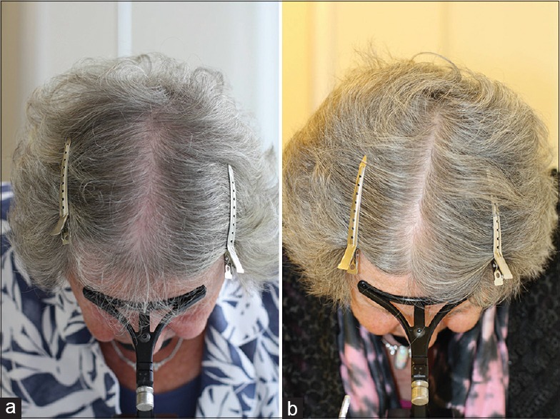
Successful treatment of senescent alopecia in an 83-year old female with 2% topical minoxidil solution b. i. d. (a) before, and (b) after 6 months treatment
The value of the special ingredients shampoos cannot be underscored as additional elements to enhance the action of the pharmaceutical agents, either topical or systemic, by cleansing the scalp and contributing to a healthy scalp microbiome. The use of appropriate surfactants, combined with ingredients such as zinc pyrithione, ciclopirox, salicylic acid,[83,84,85,86] copper chelating agents, such as N, N'-ethylene diamine disuccinic acid and histidine,[87] and others, contribute to maintain Malassezia spp. and oxidative stress under control.
Cosmetic treatments include appropriate hair grooming habits, appropriate shampoo selection, conditioner use, hair coloring, hair styling aids, and hair and scalp photoprotection. The efficacy of hair growth promoting agents or anti-aging active ingredients in shampoo form is questionable given their dilution with water and short contact time, unless they can be absorbed in effective quantities. Nevertheless, cosmetic products represent a key integral part of management of aging hair since they may significantly improve the condition of the hair fiber with more immediate effects than the pharmacological agents. Specifically, a leave-on technology combination of caffeine, niacinamide, panthenol, dimethicone and an acrylate polymer (CNPDA) has been demonstrated to affect both the diameter and the behavior of individual terminal scalp hair fibers. Beyond the diameter increase, the CNPDA-thickened fibers also demonstrated increased suppleness/pliability (decreased shear modulus) and better ability to withstand force without breaking (increased break stress). Although cosmetic treatments will not reverse the process of hair aging, such technologies help mitigate the effects of age-related thinning of hair.[88]
Finally, there are a number of botanicals with alleged anti-aging effects from both traditional Chinese and Ayurvedic medicine. Asia, Korea, China, and Japan have legally adopted the traditional Oriental medical system along with Western medicine. A number of traditional herbal drugs, including the polypharmacy type of prescription, i.e., the combination of multiple herbs, are available and widely dispensed. This polypharmacy type of herbal therapy allegedly exhibits holistic effectiveness by exerting multitargeted effects. The Traditional Oriental Medicine Database (Tradi Med 2000 DB) represents a database containing specific information such as: formulae, chemical information on ingredients, botanical information on herbal materials, and a dictionary of disease classifications. Using the TradMed 2000 DB, Chang[89] identified more than a dozen herbs with anti-aging effects used in traditional Oriental herbal therapy, of which Polygonum multiflorum and Panacis ginseng radix [Figure 3] probably represent the commercially most utilized in hair cosmetics, also on the Western market. The former is also referred to as “Ho Shou Wu”, literally meaning “Mr. Ho's hair is black”. The name refers to the story of a 58-year-old man named Ho, whose gray hair returned to its black color after taking the herb. He also became more youthful and was able to father several children. Supposedly, he lived to become 160, retaining his black hair. The English word ginseng derives from the Hokkien Chinese jîn-sim (人蔘), with the first character 人 meaning “person” and the second 蔘 “plant root”, referring to the root's peculiar anthropomorphic shape. Although the effective constituents of the traditional herbal remedies have not been fully elucidated, and for many, there is little clinical evidence for their health benefits, there is an emerging interest to study them systematically with respect to current research trends in popular anti-aging medicine.
Figure 3.
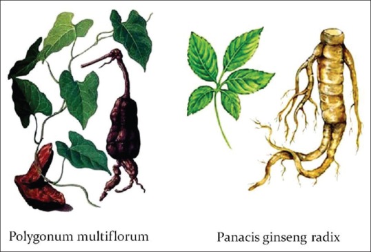
Traditional Chinese herbal remedies with alleged anti-aging properties: Polygonum multiflorum or “Ho Shou Wu,” literally meaning “Mr. Ho's hair is black” (left), and Panacis ginseng radix praised for its anthropomorphic form (right)
Declaration of patient consent
The authors certify that they have obtained all appropriate patient consent forms. In the form the patient(s) has/have given his/her/their consent for his/her/their images and other clinical information to be reported in the journal. The patients understand that their names and initials will not be published and due efforts will be made to conceal their identity, but anonymity cannot be guaranteed.
Financial support and sponsorship
Nil.
Conflicts of interest
There are no conflicts of interest.
REFERENCES
- 1.Blume-Peytavi U. International Forum of Dermatology (sponsored by Pierre Fabre) Barcelona Spain: Oral communication; 2018. Senescent alopecia: Myth or reality? [Google Scholar]
- 2.Trüeb RM, Tobin DH, editors. Aging Hair: Springer Berlin Heidelberg; 2010. [Google Scholar]
- 3.Trüeb RM. 10th World Congress of Hair Research. Kyoto, Japan: Oral communication; 2017. Medical and cosmetic treatments for aging male and female hair. [Google Scholar]
- 4.Pinkus H. Alopecia. Clinicopathologic correlations. Int J Dermatol. 1980;19:245–53. doi: 10.1111/j.1365-4362.1980.tb00318.x. [DOI] [PubMed] [Google Scholar]
- 5.Ebling FJ. Age changes in cutaneous appendages. J Appl Cosmetol. 1985;3:243–50. [Google Scholar]
- 6.Kligman AM. The comparative histopathology of male-pattern baldness and senescent baldness. Clin Dermatol. 1988;6:108–18. doi: 10.1016/0738-081x(88)90074-0. [DOI] [PubMed] [Google Scholar]
- 7.Courtois M, Loussouarn G, Hourseau C, Grollier JF. Ageing and hair cycles. Br J Dermatol. 1995;132:86–93. doi: 10.1111/j.1365-2133.1995.tb08630.x. [DOI] [PubMed] [Google Scholar]
- 8.Price VH, Sawaya ME, Headington JT. Histology and hormonal activity in senescent thinning in men. Presentation at SID, Annual Meeting; Washington, DC. 2001. [Google Scholar]
- 9.Sperling LC. Senescent balding (“senile alopecia”) In: Sperling LC, editor. An Atlas of Hair Pathology with Clinical Correlations. New York: Parthenon Publishing; 2003. pp. 35–6. [Google Scholar]
- 10.Sinclair R, Chapman A, Magee J. The lack of significant changes in scalp hair follicle density with advancing age. Br J Dermatol. 2005;152:646–9. doi: 10.1111/j.1365-2133.2005.06310.x. [DOI] [PubMed] [Google Scholar]
- 11.Whiting DA. How real is senescent alopecia? A histopathologic approach. Clin Dermatol. 2011;29:49–53. doi: 10.1016/j.clindermatol.2010.07.007. [DOI] [PubMed] [Google Scholar]
- 12.Trüeb RM. 10th World Congress of Hair Research. Kyoto, Japan: Oral communication; 2017. Global view on extrinsic and intrinsic factors affecting hair fiber quality of life. [Google Scholar]
- 13.Otsuka H, Nemoto T. Study on Japanese hair. Koshkaischi. 1988;12:192–7. [Google Scholar]
- 14.Robbins C, Mirmirani P, Messenger AG, Birch MP, Youngquist RS, Tamura M, et al. What women want – Quantifying the perception of hair amount: An analysis of hair diameter and density changes with age in Caucasian women. Br J Dermatol. 2012;167:324–32. doi: 10.1111/j.1365-2133.2012.11010.x. [DOI] [PubMed] [Google Scholar]
- 15.Birch MP, Messenger JF, Messenger AG. Hair density, hair diameter and the prevalence of female pattern hair loss. Br J Dermatol. 2001;144:297–304. doi: 10.1046/j.1365-2133.2001.04018.x. [DOI] [PubMed] [Google Scholar]
- 16.Nagase S, Kajiura Y, Mamada A, Abe H, Shibuichi S, Satoh N, et al. Changes in structure and geometric properties of human hair by aging. J Cosmet Sci. 2009;60:637–48. [PubMed] [Google Scholar]
- 17.Nagase S, Tsuchiya M, Matsui T, Shibuichi S, Tsujimura H, Masukawa Y, et al. Characterization of curved hair of Japanese women with reference to internal structures and amino acid composition. J Cosmet Sci. 2008;59:317–32. [PubMed] [Google Scholar]
- 18.Nicolaides N, Rothman S. Studies on the chemical composition of human hair fat. II. The overall composition with regard to age, sex and race. J Invest Dermatol. 1953;21:9–14. doi: 10.1038/jid.1953.63. [DOI] [PubMed] [Google Scholar]
- 19.Hollfelder B, Blankenburg G, Wolfram LJ, Höcker H. Chemical and physical properties of pigmented and non-pigmented hair ('grey hair') Int J Cosmet Sci. 1995;17:87–9. doi: 10.1111/j.1467-2494.1995.tb00112.x. [DOI] [PubMed] [Google Scholar]
- 20.Tobin DJ, Paus R. Graying: Gerontobiology of the hair follicle pigmentary unit. Exp Gerontol. 2001;36:29–54. doi: 10.1016/s0531-5565(00)00210-2. [DOI] [PubMed] [Google Scholar]
- 21.Commo S, Gaillard O, Bernard BA. Human hair greying is linked to a specific depletion of hair follicle melanocytes affecting both the bulb and the outer root sheath. Br J Dermatol. 2004;150:435–43. doi: 10.1046/j.1365-2133.2004.05787.x. [DOI] [PubMed] [Google Scholar]
- 22.Harman D. Aging: A theory based on free radical and radiation chemistry. J Gerontol. 1956;11:298–300. doi: 10.1093/geronj/11.3.298. [DOI] [PubMed] [Google Scholar]
- 23.Arck PC, Overall R, Spatz K, Liezman C, Handjiski B, Klapp BF, et al. Towards a “free radical theory of graying”: Melanocyte apoptosis in the aging human hair follicle is an indicator of oxidative stress induced tissue damage. FASEB J. 2006;20:1567–9. doi: 10.1096/fj.05-4039fje. [DOI] [PubMed] [Google Scholar]
- 24.Wood JM, Decker H, Hartmann H, Chavan B, Rokos H, Spencer JD, et al. Senile hair graying: H2O2-mediated oxidative stress affects human hair color by blunting methionine sulfoxide repair. FASEB J. 2009;23:2065–75. doi: 10.1096/fj.08-125435. [DOI] [PubMed] [Google Scholar]
- 25.Nishimura EK, Jordan SA, Oshima H, Yoshida H, Osawa M, Moriyama M, et al. Dominant role of the niche in melanocyte stem-cell fate determination. Nature. 2002;416:854–60. doi: 10.1038/416854a. [DOI] [PubMed] [Google Scholar]
- 26.Nishimura EK, Granter SR, Fisher DE. Mechanisms of hair graying: Incomplete melanocyte stem cell maintenance in the niche. Science. 2005;307:720–4. doi: 10.1126/science.1099593. [DOI] [PubMed] [Google Scholar]
- 27.Nishimura EK. 7th World Congress for Hair Research. Edinburgh, Scotland: Oral communication; 2013. Oral Communication. [Google Scholar]
- 28.Karnik P, Shah S, Dvorkin-Wininger Y, Oshtory S, Mirmirani P. Microarray analysis of androgenetic and senescent alopecia: Comparison of gene expression shows two distinct profiles. J Dermatol Sci. 2013;72:183–6. doi: 10.1016/j.jdermsci.2013.06.017. [DOI] [PMC free article] [PubMed] [Google Scholar]
- 29.Kaufman KD. Androgen metabolism as it affects hair growth in androgenetic alopecia. Dermatol Clin. 1996;14:697–711. doi: 10.1016/s0733-8635(05)70396-x. [DOI] [PubMed] [Google Scholar]
- 30.Randall VA, Thornton MJ, Messenger AG. Cultured dermal papilla cells from androgen-dependent human hair follicles (e.g. Beard) contain more androgen receptors than those from non-balding areas of scalp. J Endocrinol. 1992;133:141–7. doi: 10.1677/joe.0.1330141. [DOI] [PubMed] [Google Scholar]
- 31.Itami S, Kurata S, Takayasu S. Androgen induction of follicular epithelial cell growth is mediated via insulin-like growth factor-I from dermal papilla cells. Biochem Biophys Res Commun. 1995;212:988–94. doi: 10.1006/bbrc.1995.2067. [DOI] [PubMed] [Google Scholar]
- 32.Hibberts NA, Messenger AG, Randall VA. Dermal papilla cells derived from beard hair follicles secrete more stem cell factor (SCF) in culture than scalp cells or dermal fibroblasts. Biochem Biophys Res Commun. 1996;222:401–5. doi: 10.1006/bbrc.1996.0756. [DOI] [PubMed] [Google Scholar]
- 33.Naito A, Midorikawa T, Yoshino T, Ohdera M. Lipid peroxides induce early onset of catagen phase in murine hair cycles. Int J Mol Med. 2008;22:725–9. [PubMed] [Google Scholar]
- 34.Bahta AW, Farjo N, Farjo B, Philpott MP. Premature senescence of balding dermal papilla cells in vitro is associated with p16(INK4a) expression. J Invest Dermatol. 2008;128:1088–94. doi: 10.1038/sj.jid.5701147. [DOI] [PubMed] [Google Scholar]
- 35.Jaworsky C, Kligman AM, Murphy GF. Characterization of inflammatory infiltrates in male pattern alopecia: Implications for pathogenesis. Br J Dermatol. 1992;127:239–46. doi: 10.1111/j.1365-2133.1992.tb00121.x. [DOI] [PubMed] [Google Scholar]
- 36.Mahé YF, Michelet JF, Billoni N, Jarrousse F, Buan B, Commo S, et al. Androgenetic alopecia and microinflammation. Int J Dermatol. 2000;39:576–84. doi: 10.1046/j.1365-4362.2000.00612.x. [DOI] [PubMed] [Google Scholar]
- 37.Whiting DA. Diagnostic and predictive value of horizontal sections of scalp biopsy specimens in male pattern androgenetic alopecia. J Am Acad Dermatol. 1993;28:755–63. doi: 10.1016/0190-9622(93)70106-4. [DOI] [PubMed] [Google Scholar]
- 38.Eichmüller S, van der Veen C, Moll I, Hermes B, Hofmann U, Müller-Röver S, et al. Clusters of perifollicular macrophages in normal murine skin: Physiological degeneration of selected hair follicles by programmed organ deletion. J Histochem Cytochem. 1998;46:361–70. doi: 10.1177/002215549804600310. [DOI] [PubMed] [Google Scholar]
- 39.Zinkernagel MS, Trüeb RM. Fibrosing alopecia in a pattern distribution: Patterned lichen planopilaris or androgenetic alopecia with a lichenoid tissue reaction pattern? Arch Dermatol. 2000;136:205–11. doi: 10.1001/archderm.136.2.205. [DOI] [PubMed] [Google Scholar]
- 40.Olsen EA. Female pattern hair loss and its relationship to permanent/cicatricial alopecia: A new perspective. J Investig Dermatol Symp Proc. 2005;10:217–21. doi: 10.1111/j.1087-0024.2005.10109.x. [DOI] [PubMed] [Google Scholar]
- 41.Schwartz JR, Henry JP, Kerr KM, Flagler MJ, Page SH, Redman-Furey N, et al. Incubatory environment of the scalp impacts pre-emergent hair to affect post-emergent hair cuticle integrity. J Cosmet Dermatol. 2018;17:105–11. doi: 10.1111/jocd.12355. [DOI] [PubMed] [Google Scholar]
- 42.Schwartz JR, Henry JP, Kerr KM, Mizoguchi H, Li L. The role of oxidative damage in poor scalp health: Ramifications to causality and associated hair growth. Int J Cosmet Sci. 2015;37(Suppl 2):9–15. doi: 10.1111/ics.12289. [DOI] [PubMed] [Google Scholar]
- 43.Sinclair RD, Schwartz JR, Rocchetta HL, Dawson TL, Jr, Fisher BK, Meinert K, et al. Dandruff and seborrheic dermatitis adversely affect hair quality. Eur J Dermatol. 2009;19:410–1. doi: 10.1684/ejd.2009.0708. [DOI] [PubMed] [Google Scholar]
- 44.Kim KS, Shin MK, Park HK. Effects of scalp dermatitis on chemical property of hair keratin. Spectrochim Acta A Mol Biomol Spectrosc. 2013;109:226–31. doi: 10.1016/j.saa.2013.02.009. [DOI] [PubMed] [Google Scholar]
- 45.Piérard-Franchimont C, Xhauflaire-Uhoda E, Loussouarn G, Saint Léger D, Piérard GE. Dandruff-associated smouldering alopecia: A chronobiological assessment over 5 years. Clin Exp Dermatol. 2006;31:23–6. doi: 10.1111/j.1365-2230.2005.02026.x. [DOI] [PubMed] [Google Scholar]
- 46.Piérard-Franchimont C, Xhauflaire-Uhoda E, Piérard GE. Revisiting dandruff. Int J Cosmet Sci. 2006;28:311–8. doi: 10.1111/j.1467-2494.2006.00326.x. [DOI] [PubMed] [Google Scholar]
- 47.Pitney L, Weedon D, Pitney M. Is seborrhoeic dermatitis associated with a diffuse, low-grade folliculitis and progressive cicatricial alopecia? Australas J Dermatol. 2016;57:e105–7. doi: 10.1111/ajd.12289. [DOI] [PubMed] [Google Scholar]
- 48.Kim KS, Shin MK, Ahn JJ, Haw CR, Park HK. Investigation of hair shaft in seborrheic dermatitis using atomic force microscopy. Skin Res Technol. 2011;17:288–94. doi: 10.1111/j.1600-0846.2010.00495.x. [DOI] [PubMed] [Google Scholar]
- 49.Kim KS, Shin MK, Kim JH, Kim MH, Haw CR, Park HK, et al. Effects of atopic dermatitis on the morphology and water content of scalp hair. Microsc Res Tech. 2012;75:620–5. doi: 10.1002/jemt.21101. [DOI] [PubMed] [Google Scholar]
- 50.Kim KS, Shin MK, Ahn JJ, Haw CR, Park HK. A comparative study of hair shafts in scalp psoriasis and seborrheic dermatitis using atomic force microscopy. Skin Res Technol. 2013;19:e60–4. doi: 10.1111/j.1600-0846.2011.00608.x. [DOI] [PubMed] [Google Scholar]
- 51.Shuster S. Psoriatic alopecia. Arch Dermatol. 1990;126:397. doi: 10.1001/archderm.1990.01670270129024. [DOI] [PubMed] [Google Scholar]
- 52.Runne U, Kroneisen-Wiersma P. Psoriatic alopecia: Acute and chronic hair loss in 47 patients with scalp psoriasis. Dermatology. 1992;185:82–7. doi: 10.1159/000247418. [DOI] [PubMed] [Google Scholar]
- 53.Wyatt E, Bottoms E, Comaish S. Abnormal hair shafts in psoriasis on scanning electron microscopy. Br J Dermatol. 1972;87:368–73. doi: 10.1111/j.1365-2133.1972.tb07424.x. [DOI] [PubMed] [Google Scholar]
- 54.Headington JT, Gupta AK, Goldfarb MT, Nickoloff BJ, Hamilton TA, Ellis CN, et al. A morphometric and histologic study of the scalp in psoriasis. Paradoxical sebaceous gland atrophy and decreased hair shaft diameters without alopecia. Arch Dermatol. 1989;125:639–42. [PubMed] [Google Scholar]
- 55.Plozzer C, Coletti C, Kokelj F, Trevisan G. Scanning electron microscopy study of hair shaft disorders in psoriasis. Acta Derm Venereol Suppl (Stockh) 2000;211:9–11. doi: 10.1080/00015550050500031. [DOI] [PubMed] [Google Scholar]
- 56.Kumar B, Soni A, Saraswat A, Kaur I, Dogra S. Hair in psoriasis: A prospective, blinded scanning electron microscopic study. Clin Exp Dermatol. 2008;33:491–4. doi: 10.1111/j.1365-2230.2008.02740.x. [DOI] [PubMed] [Google Scholar]
- 57.Schoorl WJ, van Baar HJ, van de Kerkhof PC. The hair root pattern in psoriasis of the scalp. Acta Derm Venereol. 1992;72:141–2. [PubMed] [Google Scholar]
- 58.Denizli H, Güler Özden M, Sentürk N, Bek Y. Trichogram findings in psoriasis and seborrheic dermatitis patients. Turk Klinikleri J Dermatol. 2017;27:61–8. [Google Scholar]
- 59.Stanimirović A, Skerlev M, Stipić T, Beck T, Basta-Juzbasić A, Ivanković D, et al. Has psoriasis its own characteristic trichogram? J Dermatol Sci. 1998;17:156–9. doi: 10.1016/s0923-1811(97)00082-0. [DOI] [PubMed] [Google Scholar]
- 60.Bickers DR, Athar M. Oxidative stress in the pathogenesis of skin disease. J Invest Dermatol. 2006;126:2565–75. doi: 10.1038/sj.jid.5700340. [DOI] [PubMed] [Google Scholar]
- 61.Ozturk P, Arican O, Belge Kurutas E, Karakas T, Kabakci B. Oxidative stress in patients with scalp seborrheic dermatitis. Acta Dermatovenerol Croat. 2013;21:80–5. [PubMed] [Google Scholar]
- 62.Yile W, Yili W, Xunchun W. Oxidative stress level in patients with facial seborrheic dermatitis. Guongdong Med J. 2013;34:3620–1. [Google Scholar]
- 63.Emre S, Metin A, Demirseren DD, Akoglu G, Oztekin A, Neselioglu S, et al. The association of oxidative stress and disease activity in seborrheic dermatitis. Arch Dermatol Res. 2012;304:683–7. doi: 10.1007/s00403-012-1254-0. [DOI] [PubMed] [Google Scholar]
- 64.Trznadel-Grodzka E, Kaczorowska A, Rotsztejn H. Oxygen metabolism in seborrhoeic dermatitis. Centr Europ J Immunol. 2011;36:248–53. [Google Scholar]
- 65.Tsuboi H, Kouda K, Takeuchi H, Takigawa M, Masamoto Y, Takeuchi M, et al. 8-hydroxydeoxyguanosine in urine as an index of oxidative damage to DNA in the evaluation of atopic dermatitis. Br J Dermatol. 1998;138:1033–5. doi: 10.1046/j.1365-2133.1998.02273.x. [DOI] [PubMed] [Google Scholar]
- 66.Nakai K, Yoneda K, Maeda R, Munehiro A, Fujita N, Yokoi I, et al. Urinary biomarker of oxidative stress in patients with psoriasis vulgaris and atopic dermatitis. J Eur Acad Dermatol Venereol. 2009;23:1405–8. doi: 10.1111/j.1468-3083.2009.03327.x. [DOI] [PubMed] [Google Scholar]
- 67.Omata N, Tsukahara H, Ito S, Ohshima Y, Yasutomi M, Yamada A, et al. Increased oxidative stress in childhood atopic dermatitis. Life Sci. 2001;69:223–8. doi: 10.1016/s0024-3205(01)01124-9. [DOI] [PubMed] [Google Scholar]
- 68.Niwa Y, Sumi H, Kawahira K, Terashima T, Nakamura T, Akamatsu H, et al. Protein oxidative damage in the stratum corneum: Evidence for a link between environmental oxidants and the changing prevalence and nature of atopic dermatitis in Japan. Br J Dermatol. 2003;149:248–54. doi: 10.1046/j.1365-2133.2003.05417.x. [DOI] [PubMed] [Google Scholar]
- 69.Kadam DP, Suryakar AN, Ankush RD, Kadam CY, Deshpande KH. Role of oxidative stress in various stages of psoriasis. Indian J Clin Biochem. 2010;25:388–92. doi: 10.1007/s12291-010-0043-9. [DOI] [PMC free article] [PubMed] [Google Scholar]
- 70.Yazici C, Köse K, Utaş S, Tanrikulu E, Taşlidere N. A novel approach in psoriasis:First usage of known protein oxidation markers to prove oxidative stress. Arch Dermatol Res. 2016;308:207–12. doi: 10.1007/s00403-016-1624-0. [DOI] [PubMed] [Google Scholar]
- 71.Dimon-Gadal S, Gerbaud P, Thérond P, Guibourdenche J, Anderson WB, Evain-Brion D, et al. Increased oxidative damage to fibroblasts in skin with and without lesions in psoriasis. J Invest Dermatol. 2000;114:984–9. doi: 10.1046/j.1523-1747.2000.00962.x. [DOI] [PubMed] [Google Scholar]
- 72.Bayer M, Mosandl A, Thaçi D. Improved enantioselective analysis of polyunsaturated hydroxy fatty acids in psoriatic skin scales using high-performance liquid chromatography. J Chromatogr B Analyt Technol Biomed Life Sci. 2005;819:323–8. doi: 10.1016/j.jchromb.2005.02.008. [DOI] [PubMed] [Google Scholar]
- 73.Reichel M, Heisig P, Kampf G. Identification of variables for aerobic bacterial density at clinically relevant skin sites. J Hosp Infect. 2011;78:5–10. doi: 10.1016/j.jhin.2011.01.017. [DOI] [PubMed] [Google Scholar]
- 74.McGinley KJ, Leyden JJ, Marples RR, Kligman AM. Quantitative microbiology of the scalp in non-dandruff, dandruff, and seborrheic dermatitis. J Invest Dermatol. 1975;64:401–5. doi: 10.1111/1523-1747.ep12512335. [DOI] [PubMed] [Google Scholar]
- 75.Kurutas EB, Ozturk P. The evaluation of local oxidative/nitrosative stress in patients with pityriasis versicolor: A preliminary study. Mycoses. 2016;59:720–5. doi: 10.1111/myc.12522. [DOI] [PubMed] [Google Scholar]
- 76.Trüeb RM. The impact of oxidative stress on hair. Int J Cosmet Sci. 2015;37(Suppl 2):25–30. doi: 10.1111/ics.12286. [DOI] [PubMed] [Google Scholar]
- 77.Trüeb RM. Oxidative stress in ageing of hair. Int J Trichology. 2009;1:6–14. doi: 10.4103/0974-7753.51923. [DOI] [PMC free article] [PubMed] [Google Scholar]
- 78.Trüeb RM. Association between smoking and hair loss: Another opportunity for health education against smoking? Dermatology. 2003;206:189–91. doi: 10.1159/000068894. [DOI] [PubMed] [Google Scholar]
- 79.Trüeb RM. Is androgenetic alopecia a photoaggravated dermatosis? Dermatology. 2003;207:343–8. doi: 10.1159/000074111. [DOI] [PubMed] [Google Scholar]
- 80.Grosvenor AJ, Marsh J, Thomas A, Vernon JA, Harland DP, Clerens S, et al. Oxidative modification in human hair: The effect of the levels of cu (II) ions, UV exposure and hair pigmentation. Photochem Photobiol. 2016;92:144–9. doi: 10.1111/php.12537. [DOI] [PubMed] [Google Scholar]
- 81.Trüeb RM. Aging of hair. J Cosmet Dermatol. 2005;4:60–72. doi: 10.1111/j.1473-2165.2005.40203.x. [DOI] [PubMed] [Google Scholar]
- 82.Trüeb RM. Minoxidil for endocrine therapy-induced alopecia in women with breast cancer-Saint Agatha's blessing? JAMA Dermatol. 2018;154:656–8. doi: 10.1001/jamadermatol.2018.0453. [DOI] [PubMed] [Google Scholar]
- 83.Trüeb RM. Dermocosmetic aspects of hair and scalp. J Investig Dermatol Symp Proc. 2005;10:289–92. doi: 10.1111/j.1087-0024.2005.10118.x. [DOI] [PubMed] [Google Scholar]
- 84.Trüeb RM. Shampoos: Ingredients, efficacy and adverse effects. J Dtsch Dermatol Ges. 2007;5:356–65. doi: 10.1111/j.1610-0387.2007.06304.x. [DOI] [PubMed] [Google Scholar]
- 85.Gavazzoni Dias MF. Hair cosmetics: An overview. Int J Trichology. 2015;7:2–15. doi: 10.4103/0974-7753.153450. [DOI] [PMC free article] [PubMed] [Google Scholar]
- 86.Gavazzoni Dias MF, de Almeida AM, Cecato PM, Adriano AR, Pichler J. The shampoo pH can affect the hair: myth or reality? Int J Trichology. 2014;6(3):95–9. doi: 10.4103/0974-7753.139078. [DOI] [PMC free article] [PubMed] [Google Scholar]
- 87.Marsh JM, Davis MG, Lucas RL, Reilman R, Styczynski PB, Li C, et al. Preserving fibre health: Reducing oxidative stress throughout the life of the hair fibre. Int J Cosmet Sci. 2015;37(Suppl 2):16–24. doi: 10.1111/ics.12285. [DOI] [PubMed] [Google Scholar]
- 88.Davis MG, Thomas JH, van de Velde S, Boissy Y, Dawson TL, Jr, Iveson R, et al. A novel cosmetic approach to treat thinning hair. Br J Dermatol. 2011;165(Suppl 3):24–30. doi: 10.1111/j.1365-2133.2011.10633.x. [DOI] [PubMed] [Google Scholar]
- 89.Chang IM. Anti-aging and health-promoting constituents derived from traditional oriental herbal remedies: Information retrieval using the TradiMed 2000 DB. Ann N Y Acad Sci. 2001;928:281–6. doi: 10.1111/j.1749-6632.2001.tb05657.x. [DOI] [PubMed] [Google Scholar]