Abstract
The Drosophila protocadherins Dachsous and Fat regulate growth and tissue polarity by modulating the levels, membrane localization and polarity of the atypical myosin Dachs. Localization to the apical junctional membrane is critical for Dachs function, and the adapter protein Vamana/Dlish and palmitoyl transferase Approximated are required for Dachs membrane localization. However, how Dachs levels are regulated is poorly understood. Here we identify the early girl gene as playing an essential role in Fat signaling by limiting the levels of Dachs protein. early girl mutants display overgrowth of the wings and reduced cross vein spacing, hallmark features of mutations affecting Fat signaling. Genetic experiments reveal that it functions in parallel with Fat to regulate Dachs. early girl encodes an E3 ubiquitin ligase, physically interacts with Dachs, and regulates its protein stability. Concomitant loss of early girl and approximated results in accumulation of Dachs and Vamana in cytoplasmic punctae, suggesting that it also regulates their trafficking to the apical membrane. Our findings establish a crucial role for early girl in Fat signaling, involving regulation of Dachs and Vamana, two key downstream effectors of this pathway.
Author summary
During development, organs grow to achieve a consistent final size. The evolutionarily conserved Hippo signaling network plays a central role in organ size control, and when dysregulated can be associated with cancer and other diseases. Fat signaling is one of several upstream pathways that impinge on Hippo signaling to regulate organ growth. We describe here identification of the Drosophila early girl gene as a new component of the Fat signaling pathway. We show that Early girl controls Fat signaling by regulating the levels of the Dachs protein. However Early girl differs from other Fat signaling regulators in that it doesn’t influence planar cell polarity or control the polarity of Dachs localization. early girl encodes a conserved protein that is predicted to influence protein stability, and it can physically associate with Dachs. We also discovered that Early girl acts together with another protein, called Approximated, to regulate the sub-cellular localization of Dachs and a Dachs-interacting protein called Vamana. Altogether, our observations establish Early girl as an essential component of Fat signaling that acts to regulate the levels and localization of Dachs and Vamana.
Introduction
Precise coordination of growth and morphogenesis during development is critical for formation of organs of correct proportions and optimal function. This is achieved through the cumulative effect of biochemical signaling through morphogens and biomechanical signals mediated through the actomyosin network. The protocadherins Dachsous (Ds) and Fat initiate a signaling cascade (Fat signaling), which functions to restrict growth by activating the Hippo signaling pathway [1, 2] and influences morphogenesis by modulating planar cell polarity (PCP) [3–6]. Fat signaling regulates the Hippo pathway and influences PCP by modulating membrane localization of the atypical myosin Dachs. Multiple studies have provided insight both into the mechanisms by which Dachs influences the Hippo pathway and PCP, and into how Fat regulates Dachs [7–18]. However, the mechanisms that control Dachs levels and membrane localization are still not completely understood.
Ds and Fat are protocadherins with large extracellular domains (ECD) and small intracellular domains (ICD) and localize to the apico-lateral membrane just apical to the adherens junctions. The ECDs of Ds and Fat interact with each other across the cell-cell junctions in a heterophilic manner and the Golgi resident kinase, Four-jointed (Fj), modulates this interaction by phosphorylating their ECDs [19–22]. In most developing tissues Ds and Fj are expressed in opposite gradients under the influence of morphogens [2]. The heterophilic interaction between Ds and Fat and the graded expression of Ds and Fj contributes to planar polarized localization of Ds and Fat within cells, where in the developing Drosophila wing Fat is preferentially enriched on the proximal side while Ds is enriched on the distal side [23–25]. Ds and Fat then regulate the levels and polarity of Dachs at cell membranes to influence Hippo signaling and PCP. In absence of Fat, increased amounts of Dachs accumulate around the entire circumference of cells [8, 24]. Conversely, overexpression of Fat or just the Fat ICD displaces Dachs from the membrane into the cytoplasm [8, 16].
The Hippo signaling pathway plays a central role in growth regulation and includes Hippo and its cofactor Salvador and Warts (Wts) and the Warts cofactor Mats [26]. Hippo phosphorylates and activates Wts, which in turn phosphorylates the transcriptional co-activator Yorkie (Yki). Phosphorylated Yki is sequestered in the cytoplasm. When the pathway is inactive, unphosphorylated Yki translocates into the nucleus, where it associates with Scalloped and induces the transcription of downstream target genes such as expanded (ex), bantam (ban), and cyclin E, which modulate Hippo pathway activity, stimulate growth, and regulate cell fate. Multiple upstream regulators impinge on Wts to regulate Hippo signaling, including Fat signaling. Fat affects the membrane localization of Expanded, the levels of Wts and the interaction of Wts with Mats [7, 11, 27–30]. Dachs is required for each of these effects on Hippo signaling. Similarly, Fat signaling influences PCP signaling partly by regulating the Spiny legs isoform of the prickle locus [12, 13] and partly through an influence on junctional tension [31], and Dachs is involved in both of these processes.
Dachs localizes to the apico-lateral plasma membrane in a planar polarized manner [8]. In the developing wing discs Dachs localizes to the distal side of the cell [8, 23, 24, 32]. Several proteins that influence membrane localization of Dachs have been identified as components of the Fat signaling pathway. vamana (vam/Dlish) encodes an adapter protein with three SH3 domains that physically connects Dachs with the Fat and Ds ICDs, interacting with Dachs through its second SH3 domain and interacting with the Fat and Ds ICD through its first and third SH3 domains [14, 15]. Vam physically interacts with the C-terminal region of Dachs. Like Dachs, Vam localizes to the apical plasma membrane with preferential enrichment on the distal side of wing cells. In absence of Vam, Dachs fails to localize to the membrane and accumulates in the cytoplasm. Approximated encodes a palmitoyl transferase and is also required for Dachs membrane localization [33]. Although, the exact mechanism is still not clear, it can physically interact with both Dachs and Vam and can palmitoylate Vam and the Fat ICD when overexpressed [15, 34].
Despite progress in our understanding of the Fat signaling pathway, how Dachs localizes to the apical membrane and how its levels at the membrane are regulated is still not completely understood. Here we report the isolation and characterization of the early girl (elgi) gene as a key regulator of Fat-Hippo signaling. Loss of elgi function promotes growth by up-regulating Yki activity. This arises from accumulation of high levels of Dachs and Vam at the apical plasma membrane. Elgi physically interacts with Dachs and regulates Dachs and Vam by controlling their protein levels. Further, we show that concomitant loss of Elgi and App prevents Dachs and Vam localization to the subapical membrane and results in their accumulation in cytoplasmic punctae, suggesting that Elgi and App coordinately regulate trafficking and membrane localization of Dachs and Vam. Under this condition, Dachs is primarily affected and Vam gets mislocalized due to its interaction with Dachs. Our observations establish Elgi as an important component of the Fat signaling pathway.
Results
elgi mutants display phenotypes similar to loss of fat
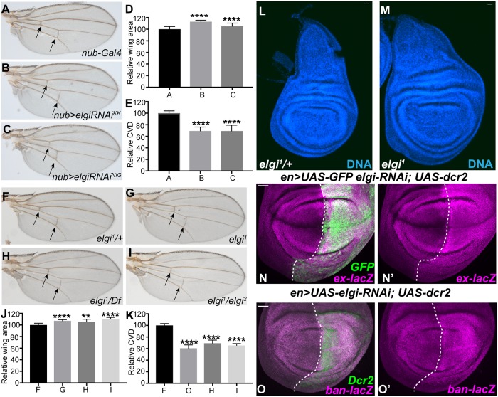
To identify contributions of protein stability to wing growth and patterning, we conducted a RNAi-based genetic screen, in which we depleted the RING domain E3 ubiquitin ligases encoded by the Drosophila genome within the wing pouch region of the developing wing imaginal disc using UAS RNAi lines and a nub-Gal4 driver. From this screen, we identified elgi, based on an increase in size of the adult wing, and a reduced spacing between the anterior and posterior cross veins when it is knocked down by either of two independent RNAi lines (Fig 1A–1E). These phenotypes are hallmark features of mutations in components of the Fat signaling pathway [8, 14, 15, 33, 35–39], suggesting that elgi could participate in Fat signaling.
Fig 1. elgi mutant phenotypes.
(A-C) Adult male wings from flies expressing nub-Gal4 UAS-Dcr2 control (A), nub-Gal4 UAS-Dcr2 UAS-elgi-RNAiKK (VDRC109617) (B), and nub-Gal4 UAS-Dcr2 UAS-elgi-RNAiNIG (17033R-3) (C). Arrows point to the crossveins. (D) Histogram of relative wing areas in flies of the genotypes in panels A-C, as indicated (normalized to the average area of control wings); mean of B is 112.9, and C is 105.2. (E) Histogram of the ratio of crossvein distance (CVD) to wing length, (normalized to the average CVD/wing length in control wings) in flies of the genotypes in panels A-C, as indicated. (F-I) Adult male wings from elgi1/+ (control) (F), homozygous elgi1 (G), hemizygous elgi1/Df(3L) BSC575 (H), trans-heterozygous elgi1 / elgi2 (I) flies. Arrows point to the crossveins. (J) Histogram of relative wing areas (normalized to the average area of control wings) in flies of the genotypes in panels F-I, as indicated. Mean of G is 107.1; H is 105.4 and I is 110.4. (K) Histogram of the ratios of crossvein distance (CVD) to wing length, (normalized to the average CVD/wing length ratio of control wings) in flies of the genotypes in panels F-I, as indicated. Data are shown as mean ± SD from measurements of 10–13 wings per genotype. **P<0.002 and ****P<0.001, (Student’s t test between control and the other genotypes); (L,M) Wing imaginal discs from age matched control elgi1/+ and homozygous elgi1 4 day old third instar larvae. (N-O’) Third instar wing imaginal discs expressing en-Gal4 UAS-dcr2 UAS-GFP UAS-elgi-RNAi and ex-lacZ (N, N’) or ban-lacZ (O, O’) stained for expression of ex-lacZ or ban-lacZ (magenta), with posterior cells marked by expression of GFP or Dcr2. Dashed white line marks the A-P compartment boundary. Scale bar is 33 μm.
elgi was previously characterized for a role in oogenesis, and named for a mutant phenotype including premature meiotic maturation [40]. Loss of function alleles that were isolated based on this oogenesis phenotype, elgi1 and elgi2, also increase the size of adult wings and reduce spacing between the anterior and posterior cross veins when homozygous, or when hemizygous with a chromosomal deficiency that deletes the elgi locus (Fig 1F–1K). elgi1 mutant wing discs are also larger than control wing discs (Fig 1L and 1M). These observations confirm that elgi regulates wing growth and patterning.
Fat signaling regulates growth by controlling Hippo signaling. To examine the possibility that elgi regulates wing size through Hippo signaling, we depleted elgi in the posterior compartment of wing imaginal discs by expressing UAS-RNAi elgi under the control of en-gal4, and then examined the effect of this Elgi depletion on the levels of the Yki target genes ex and ban. Using ex-lacZ and ban-lacZ reporter genes, we observed that loss of elgi increased Yki activity (Fig 1N and 1O), consistent with the inference that the increased wing size observed is due to impairment of Fat-Hippo signaling.
Elgi regulates Dachs and Vam levels
To further examine the possibility that Elgi could be involved in Ds-Fat signaling, and to begin to investigate how it might influence this pathway, we examined the effect of loss of elgi on the key Fat signaling components, Ds, Fat, Dachs and Vam. Depletion of elgi by RNAi did not result in any visible effect on the localization or levels of Ds (S2 Fig). Loss of elgi leads to a slight increase in Fat levels (Fig 2D), but this cannot explain the increased Yki activity and wing growth observed, because over-expression of Fat decreases Yki activity and wing growth [7, 41]. In contrast, in cells lacking elgi, both Dachs and Vam, examined using genomic GFP-tagged transgenes, accumulate at very high levels (Fig 2B and 2C, S4A Fig). Since over-expression of Dachs or Vam can increase wing size [8, 14, 15], this could potentially account for the increased wing growth associated with loss of elgi.
Fig 2. elgi regulates the levels of Dachs, Vam and Fat.
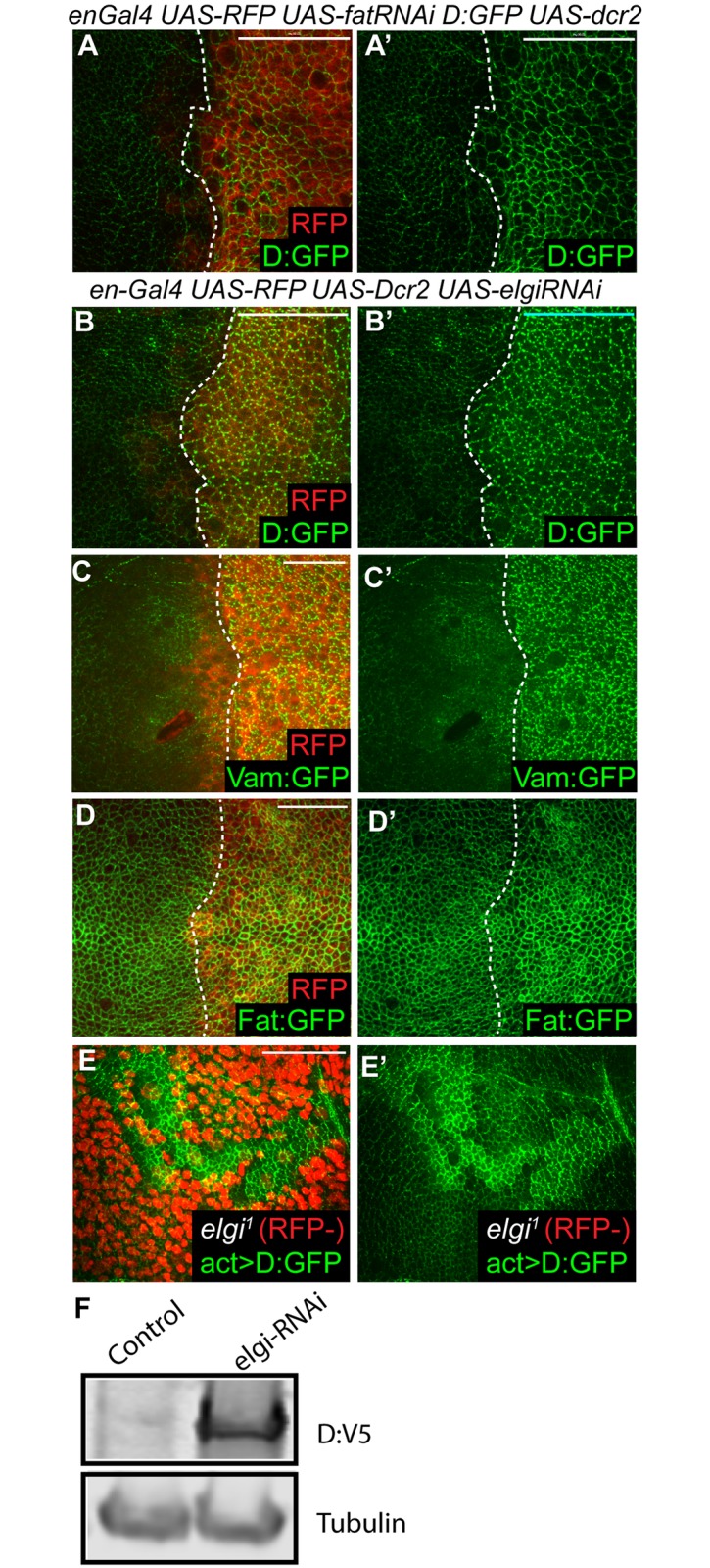
(A-D) Horizontal apical sections of wing imaginal discs expressing en-Gal4 UAS-Dcr2 UAS-RFP (red) with either UAS-fat-RNAi (A) or UAS-elgi-RNAi (B-D) and Dachs:GFP (D:GFP) (A, B), Vam:GFP (C) or Fat:GFP (D) showing increased levels of membrane localized Dachs:GFP, Vam:GFP and Fat:GFP in the posterior compartment. A and B were imaged using the same acquisition settings. (E) Horizontal apical sections of wing imaginal discs expressing Dachs:GFP under the control of the actin5C promoter (act>D:GFP) throughout, showing increased levels of membrane localized Dachs:GFP, in homozygous elgi1 mutant clones, marked by absence of RFP (red). (F) Western blot showing levels of Dachs:V5 (D:V5) from third instar wing disc lysate from flies expressing tub-Gal4 UAS-D:V5 without (control) or with elgi-RNAi. Tubulin was used as a control for loading and transfer. Scale bar is 33 μm (A,B,E), 19 μm (C) and 16.5μm (D).
Absence of fat also leads to increased Dachs levels. To compare the increases in Dachs generated by loss of elgi versus loss of fat, elgi or fat were knocked down by RNAi in the posterior compartment of wing discs expressing Dachs:GFP, which were examined using the same imaging settings for both genotypes. This revealed that apical Dachs levels appear higher in the absence of elgi than in the absence of fat (Fig 2A and 2B). However, in the absence of fat Dachs localizes evenly around the cell circumference at the sub-apical membrane, whereas in the absence of elgi Dachs localizes to the membrane in a punctate pattern, similar to its localization in wild-type cells.
To examine whether the increase in the levels of Dachs is transcriptional or post-transcriptional, we examined the influence of elgi-RNAi and elgi1 mutant clones on a transgene expressing Dachs:GFP under the control of the Act5c promoter. Act5c-Dachs:GFP accumulated at high levels in the apical cortex in the absence of elgi (Fig 2E, S4C and S4E Fig), just like Dachs:GFP expressed from its endogenous promoter, which indicates that elgi regulates Dachs at a post-transcriptional level. Consistent with this, western blotting of wing disc lysates revealed that loss of elgi leads to a significant increase in the levels of V5-tagged Dachs expressed ubiquitously under the control of a tub-Gal4 driver (Fig 2F).
Elgi acts genetically upstream of Dachs, and in parallel to Fat
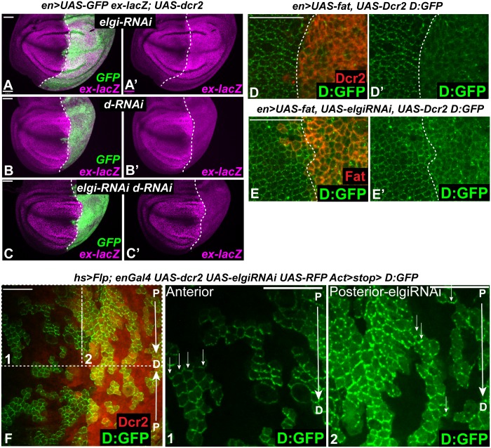
Mutation of upstream components of the Fat signaling pathway, including Fat, Ds, and Dco, increase Yki activity and wing growth by increasing levels of Dachs at the apical membrane [7, 8, 24]. To confirm that the increased Yki activity observed with elgi loss-of-function is due to increased Dachs, we investigated whether the elgi phenotype genetically depends upon dachs. Indeed, depletion of dachs by RNAi completely suppressed elevation of ex-lacZ expression by elgi RNAi, and wing disc cells with reduction of both proteins instead exhibit the loss of ex-lacZ expression characteristic of dachs RNAi (Fig 3A–3C).
Fig 3. elgi regulates Dachs in parallel to Fat and does not affect Dachs polarity.
(A-C) Third instar wing imaginal discs expressing en-Gal4 UAS-dcr2 UAS-GFP ex-lacZ and either UAS-elgi-RNAi (A, A’) or UAS-dachs-RNAi (UAS-d-RNAi) (B, B’) or UAS-elgi-RNAi and UAS-d-RNAi (C, C’), stained for expression of ex-lacZ (magenta), with posterior cells marked by expression of GFP (green). Dashed white line marks the A-P compartment boundary. Scale bar is 33 μm. (D, E) Horizontal apical sections of wing imaginal discs expressing en-Gal4 UAS-dcr2 D:GFP UAS-Fat without (D, D’) or with (E, E’) UAS-elgi-RNAi showing displacement of membrane localized Dachs:GFP (D:GFP) into the cytoplasm. Posterior cells are marked by expression of Dcr2 or Fat (red) Dashed white line marks the A-P compartment boundary. Scale bar is 16.5 μm. (F) Horizontal apical sections of wing imaginal discs expressing Dachs:GFP (D:GFP) in flipout clones and elgi-RNAi in the posterior compartment (marked by RFP, red). Arrows point in a proximal (P) to distal (D) direction. (1, 2) Magnified views of boxed areas in F with small arrows indicating preferential enrichment of D:GFP on the distal boundary of cells. Scale bar is 16.5 μm.
Overexpression of Fat can displace Dachs from the membrane into the cytoplasm [8, 16]. To examine the genetic relationship between elgi and fat, we investigated the influence of elgi on this regulation of Dachs by Fat. Fat was overexpressed in the absence or in the presence of elgi RNAi in the posterior compartment of wing discs expressing Dachs:GFP. Overexpression of Fat caused a displacement of Dachs from the plasma membrane even in the presence of elgi RNAi (Fig 3E). Thus, Fat over-expression is epistatic to elgi loss of function. Together with observations that elgi RNAi increases Fat levels, and that loss of Elgi or loss of Fat result in different patterns of increased Dachs, this suggests that fat and elgi function in parallel to regulate Dachs.
Normally Dachs localizes in a planar polarized manner, with a preferential localization to the distal side of wing disc cells [8]. To examine whether elgi affects Dachs polarity, we expressed Dachs:GFP in small clones in wing discs expressing elgi RNAi in the posterior compartment. While loss of elgi leads to an increase in the levels of Dachs:GFP, it is still mostly polarized to the distal side (Fig 3F). Fat signaling regulates PCP as well as Hippo signaling, and loss of Fat results in Dachs-dependent disruption of hair polarity in the proximal wing [13, 42]. However, wings from elgi1 mutant flies do not exhibit any defect in hair polarity (S1 Fig). These observations are consistent with the idea that the levels of membrane localized Dachs influence Hippo signaling, whereas the polarity of membrane Dachs influences PCP, and also further support the conclusion that Elgi and Fat act in parallel to regulate Dachs.
Elgi localizes to the cytoplasm and physically interacts with Dachs
To further investigate how Elgi regulates Dachs, we examined the localization of Elgi protein. We created a GFP-tagged genomic copy of Elgi, but the extremely weak signal from this construct was insufficient to clearly establish its localization. Therefore we expressed a HA-tagged Elgi in the wing pouch under nub-Gal4 control. Overexpression of Elgi-Myc-HA under UAS-Gal4 control results in a slightly smaller wing size (Fig 4D and 4F), opposite to the effect of elgi loss-of-function, while also causing a very mild decrease in cross vein spacing (Fig 4C–4G). This UAS-elgi-Myc-HA construct could rescue the reduced cross-vein spacing of an elgi mutant (S3 Fig). These observations indicate that it has Elgi activity. Anti-HA antibody staining revealed a dispersed cytoplasmic localization for Elgi (Fig 4A).
Fig 4. elgi localization and interaction with Dachs, and elgiCS phenotypes.
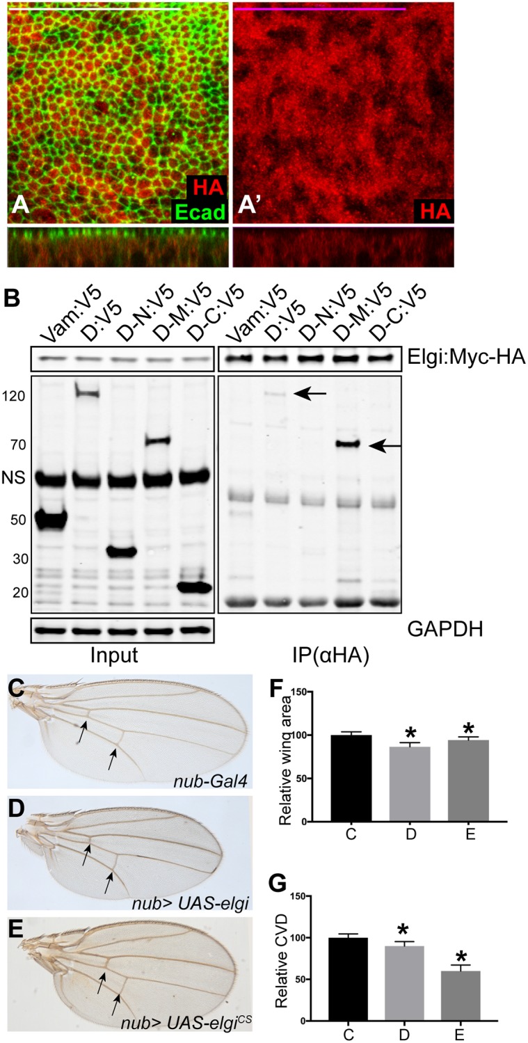
(A, A’) Horizontal apical sections (above) and vertical sections (below) of wing imaginal discs expressing nub-Gal4 UAS-elgi-MYC-HA and stained with anti E-cad (green) and anti HA (red) antibodies showing diffuse cytoplasmic localization of Elgi. Scale bar is 33.0 μm. (B) Western blot showing results of co-immunoprecipitation experiments on proteins co-expressed in S2 cells. V5-tagged Vam, Full length Dachs (D:V5), Dachs N terminus(D-N:V5), Dachs Middle domain (D-M:V5) or Dachs C terminus (D-C:V5) were co-expressed with full length Elgi-MYC-HA, and complexes were precipitated using Ezview red anti HA affinity resin and immunoblotted with Rabbit anti V5 and Rabbit anti HA antibodies. GAPDH was used as a control for loading and transfer in the input lanes. Arrows point to the co-immunoprecipitated bands. NS: non-specific (C-E) Adult male wings from flies expressing nub-Gal4 (control) (C), nub-Gal4 UAS-elgi (D) and nub-Gal4 UAS-elgiCS (E). Arrows point to the crossveins. (F-G) Histograms of relative wing areas (normalized to the average wing area of control wings) (F) and ratios of CVD to wing length, (normalized to the average CVD to wing length ratio of control wings) (G) in flies of the genotypes in panels C-E, as indicated. Data are shown as mean ± SD from measurements of 12 wings per genotype. *P<0.001, (Student’s t test between control and the other genotypes).
elgi encodes for a predicted RING domain E3 ubiquitin ligase, homologous to vertebrate RNF41 protein. Since we found that Elgi regulates the levels of Dachs and Vam, we examined whether Elgi physically interacts with them. To test this, we co-expressed Myc-HA-epitope-tagged Elgi along with V5-epitope-tagged full length Vam or Dachs, or N-terminal, middle or C-terminal fragments of Dachs, in S2 cells and conducted co-immunoprecipitation experiments. While Elgi does not interact with Vam, it can co-precipitate both full length Dachs, and the middle region of Dachs (Fig 4B). Since Dachs can influence levels of Vam [14, 15], this suggests that elgi could primarily affect Dachs, with the increase in Vam levels arising indirectly from the increase in Dachs levels. The middle region of Dachs is homologous to regions of myosin family proteins. Interestingly, a pulldown experiment identified Myo10A as an interactor of Elgi [43], suggesting that Elgi interacts with a subset of myosin domains.
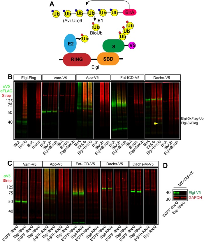
Since Elgi physically interacts with Dachs, influences Dachs levels, and encodes a predicted ubiquitin ligase, we examined whether it can ubiquitinate Dachs. We used a recently described method that employs biotinylated-Ubiquitin, as it is highly sensitive [44]. Briefly, when avi-tagged Ubiquitin fused to E.coli biotin ligase BirA (Bio-Ub) is expressed in cells, biotinylated Ubiquitin is produced, which is then incorporated by the ubiquitination machinery (Fig 5A). We expressed Dachs:V5 in cultured Drosophila S2 cells along with Bio-Ub with or without Elgi, immunoprecipitated Dachs:V5 using a mouse anti-V5 antibody, and then immunoblotted with rabbit anti-V5 to detect Dachs:V5 and with fluorescently-conjugated streptavidin to detect biotinylated-Ubiquitin. As a positive control, we examined Elgi auto-ubiquitination, which was readily detected (Figs 5B and 6H). When Dachs was expressed along with Bio-Ub we detected a smear of biotinylated bands, both at higher molecular weight (presumably corresponding to poly-ubiquitinated Dachs) and at lower molecular weight (presumably corresponding to ubiquitinylated degradation products). When Elgi was co-expressed along with Dachs:V5 and Bio-Ub, this signal was not altered, aside from the detection of the co-immunoprecipitated Elgi band. These results suggest that Dachs is ubiquitinated by an E3 ligase present in S2 cells. However, because this signal does not change when Elgi is over-expressed, it appears that Elgi does not ubiquitinate Dachs. We also examined if Dachs ubiquitination in S2 cells depends upon Elgi. Using dsRNA, Elgi could be efficiently depleted in S2 cells (Fig 5D). However, elgi RNAi did not decrease Dachs ubiquitination (Fig 5C).
Fig 5. elgi does not ubiquitinate Vam, App, Fat-ICD or Dachs.
(A) Schematic illustrating the ubiquitination assay used. BirA covalently conjugates Biotin to the fused Avi-tagged ubiquitin, which is processed by the E1 to produce biotinylated ubiquitin (BioUb), which is subsequently incorporated by the ubiquitination machinery onto the substrates. This can be detected by fluorescently conjugated streptavidin. (B) Western blot showing results of ubiquitination experiments on proteins expressed in S2 cells. For Elgi autoubiquitination, 3xFLAG tagged Elgi was co-expressed with either BirA or BirA-BioUb and immunoprecipitated using anti-Flag beads. Elgi was detected by anti-FLAG antibody. V5-tagged Full length Vam (Vam-V5), App (App-V5), Fat-ICD (Fat-ICD-V5) or Dachs (Dachs-V5) were co-expressed with either BirA alone or BirA-BioUb with or without Elgi-3xFLAG. V5-tagged proteins were immunoprecipitated from cell lysates, and detected with anti-V5 antibody. In all lanes, biotinylated Ubiquitin was detected with IRDYE680 conjugated streptavidin (Red). The yellow arrow points to a faint band of Elgi-3x-FLAG immunoprecipitated with Dachs:V5; the strong red band just above it is ubiquitinated Elgi-3x-FLAG, based on size and detection with anti-FLAG antibodies. Its appearance presumably reflects the strong association of Elgi with Dachs. (C) Western blot showing results of ubiquitination experiments on proteins expressed in S2 cells treated with dsRNA against either EGFP (control) or elgi. V5-tagged Full length Vam (Vam-V5), App (App-V5), Fat-ICD (Fat-ICD-V5), full length Dachs (Dachs-V5) or middle domain of Dachs (D-M-V5) were co-expressed with BirA-BioUb. V5-tagged proteins were immunoprecipitated from cell lysates and detected with anti-V5 antibody; Biotinylated Ubiquitin was detected with IRDYE680 conjugated streptavidin (Red). (D) Western blot showing efficiency of dsRNA-mediated depletion of elgi in S2 cells. V5-tagged Elgi (Elgi-V5) was expressed using MT-Gal4 with 0.5mM CuSO4 in presence of 10uM MG132 in S2 cells treated with dsRNA against either EGFP (control) or elgi, and whole cell lysate was examined for expression of Elgi-V5 with Rabbit anti-V5 antibody. GAPDH was used as a loading and transfer control.
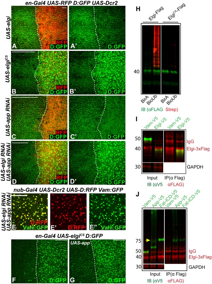
Fig 6. elgiCS has a neomorphic activity and interferes with app function.
(A-D) Horizontal apical sections of wing imaginal discs expressing en-Gal4 UAS-dcr2 D:GFP UAS-RFP along with UAS-elgi-Myc-HA (UAS-elgi) (A, A’), UAS-elgiCS-Myc-HA (UAS-elgiCS) (B, B’), UAS-app-RNAi (C, C’), or UAS-elgi-RNAi and UAS-app-RNAi (D, D’), showing the effect on Dachs:GFP (D:GFP, green) levels and localization in the posterior compartment (marked by RFP, red). Dashed white line marks the A-P compartment boundary. (E-E”) Horizontal apical sections of wing imaginal discs expressing nub-Gal4 UAS-dcr2 UAS-D:RFP UAS-Vam:GFP UAS-elgi-RNAi UAS-app-RNAi showing close up view of D:RFP (red) (E’) and Vam-GFP (green) (E’) colocalization. (F,G) Horizontal apical sections of wing imaginal discs expressing en-Gal4 UAS-elgiCS D:GFP (F) or along with UAS-app (G) showing the effect on localization of Dachs:GFP (D:GFP) in the posterior compartment (to the right). Dashed white line marks the A-P compartment boundary. Scale bar is 6 μm in A, 33 μm in B-D, 17 μm in E and 19 μm in F and G. (H) Western blot showing results of ubiquitination experiments on proteins co-expressed in S2 cells. 3xFLAG tagged Elgi or ElgiCS were co-expressed with either BirA or BirA-BioUb and immunoprecipitated using anti-Flag beads. Elgi was detected by anti-FLAG antibody and biotinylated Ubiquitin was detected with IRDYE680 conjugated streptavidin (Red). (I) Western blot showing results of co-immunoprecipitation experiments on proteins co-expressed in S2 cells. V5-tagged Vam or Elgi were co-expressed with full length Elgi-3x-FLAG, and complexes were precipitated using anti FLAG affinity resin and immunoblotted with anti-V5 and anti-FLAG antibodies. GAPDH was used as a control for loading and transfer in the input lanes. Elgi-3x-FLAG has a slower mobility compared to Elgi-V5 due to the larger epitope tag. (J) Western blot showing results of co-immunoprecipitation experiments on proteins co-expressed in S2 cells. V5-tagged Vam, App (App-V5) or Fat-ICD (Fat-ICD-V5) were co-expressed with Elgi-3x-FLAG, and complexes were precipitated using anti-FLAG affinity resin and then immunoblotted with anti-V5 and anti-FLAG antibodies. GAPDH was used as a control for loading and transfer in the input lanes. Arrow points to the full length App in the input lane.
A catalytic mutant form of Elgi has as a neomorphic activity and interferes with App function
To evaluate the potential contribution of Elgi catalytic activity to Dachs regulation, we created a mutant version of elgi, elgiCS, in which the two cysteines (C18 and C21) in the highly conserved RING domain were mutated to serines. In other E3 ubiquitin ligases, this type of mutation has been reported to abolish catalytic activity [45, 46]. Consistent with this, while wild type Elgi can auto-ubiquitinate, ElgiCS lacks this activity (Fig 6H). Over-expression of elgiCS results in a significant decrease in cross vein spacing in the adult wing, reminiscent of loss of elgi function (Fig 4E and 4G). However, unlike loss of elgi, it does not lead to an increase in wing size (Fig 4E and 4F). At the cellular level, expression of elgiCS resulted in a dramatic increase in the levels of Dachs (Fig 6B), reminiscent of elgi RNAi, and opposite to the decrease in levels of Dachs observed when wild-type Elgi is over-expressed (Fig 6A). These observations suggest that ElgiCS interferes with endogenous Elgi function. This could result from ElgiCS binding to its substrate and preventing access to wild type Elgi. Alternatively, Elgi could function as a homodimer or heterodimer with another E3 ligase, as many RING-domain ligases are known to function as dimers [47]. To test if Elgi forms homodimers, we coexpressed 3xFlag-tagged Elgi with either V5-tagged Vam or V5-tagged Elgi in S2 cells and immunoprecipitated 3x-Flag tagged Elgi and examined for their interaction. While Elgi:3xFlag did not interact with Vam:V5 it interacted with Elgi:V5 (Fig 6I), indicating that Elgi forms homodimers, which could explain why ElgiCS interferes with endogenous Elgi function.
ElgiCS also causes an additional effect on Dachs, as when it is expressed, Dachs localization to the subapical membrane is diminished, and instead it accumulates in bright punctae that are dispersed throughout the cytoplasm (Fig 6B, S4G and S4H Fig). This could explain why unlike elgi RNAi or mutants, animals expressing elgiCS do not display wing overgrowth (Fig 4E and 4F), as Dachs function depends upon its membrane localization. Expression of elgiCS also does not upregulate the Yki target gene, ex-lacZ (S5A Fig). The formation of cytoplasmic puncta is a Dachs-specific effect, as several other proteins including Fat, Ds, Crumbs, Armadillo and Jub that normally localize to the apical cortex are not affected by elgiCS (S5B–S5F Fig). Vam also gets mis-localized with Dachs when elgiCS is expressed (S5G Fig, Fig 8A), but this could be because Vam forms an obligate heterodimer with Dachs [14, 15].
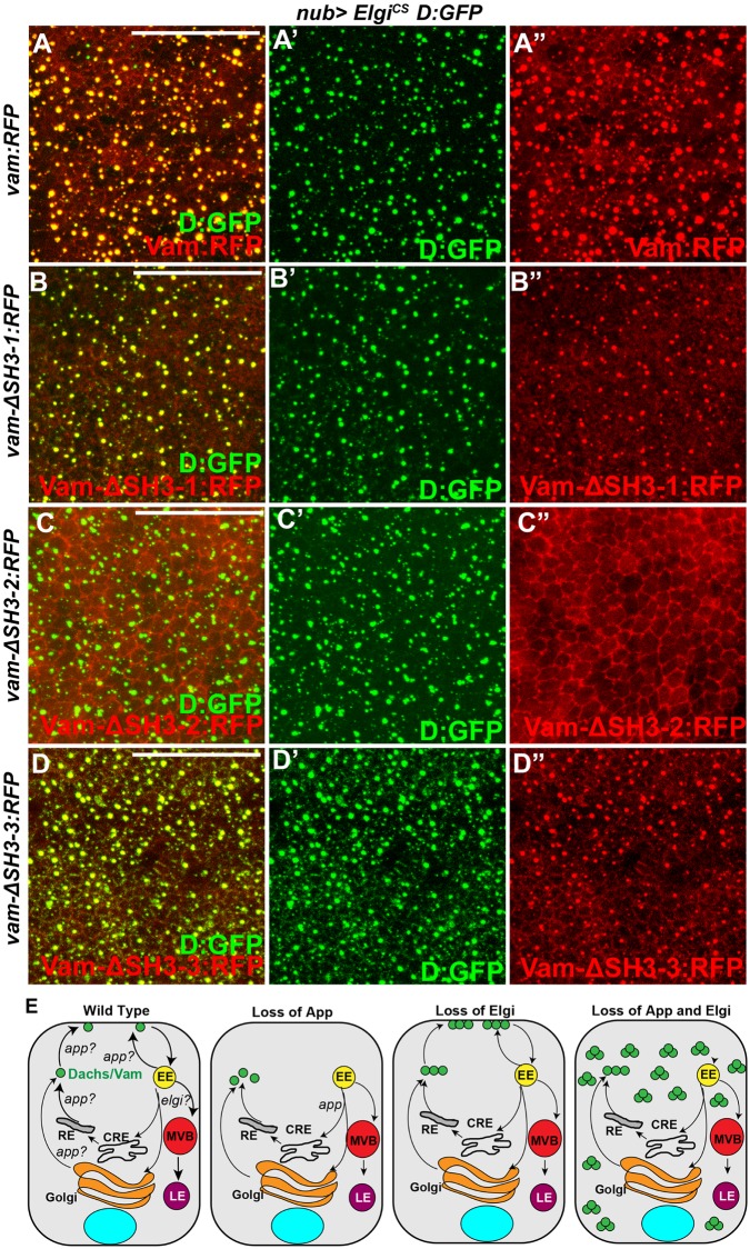
Fig 8. elgiCS affects Dachs rather than Vam.
(A-D) Horizontal apical sections of wing imaginal discs expressing nub-Gal4 UAS-elgiCS D:GFP along with either UAS-vam-RFP (A, A', A”), or UAS-vam-Δ-SH3-1-RFP (B, B', B”), UAS-vam-Δ-SH3-2-RFP (C, C', C”) or UAS-vam-Δ-SH3-3-RFP (D, D', D”) showing how the different SH3 domains of Vam (red) affect its co-localization with Dachs:GFP (D:GFP) (green) in the presence of elgiCS. (E) Schematic illustrating model showing that under normal conditions Elgi maintains limiting levels of Dachs (green circles) which localizes to the membrane in an App-dependent manner. In absence of App, Dachs is mostly found in the cytoplasm in a diffuse manner and in absence of Elgi there is higher amount of Dachs, but the endogenous App is sufficient to promote its membrane localization. However, in absence of both, there is excessive Dachs that fails to localize to the membrane and accumulates in punctae in the cytoplasm. Elgi might regulate lysosomal trafficking of a Dachs/Vam complex, or direct proteosomal degradation of a Dachs/Vam complex. EE: early endosome, MVB: multi vesicular body, CRE: common recycling endosome, RE: recycling endosome, LE: late endosome. Scale bar is 16.5 μm in A-D.
Since membrane localization of Dachs and Vam is promoted by App [14, 15, 33], the loss of Dachs and Vam from the apical membrane when elgiCS is expressed suggested that it might interfere with App function. We also note that expression of elgiCS causes rounder wings with a mild hair polarity defect in the proximal wing, similar to app loss-of-function (Fig 4E, S5I Fig) [33]. If ElgiCS interferes with App, concomitant depletion of elgi and app should cause similar localization defects of Dachs and Vam. To examine this, we compared loss of elgi or app alone with simultaneous knockdown of both genes. Loss of elgi and app together, but not individually, leads to accumulation of Dachs and Vam in cytoplasmic punctae similar to those observed when elgiCS is expressed, with Dachs and Vam co-localized (Fig 6B–6E). If elgiCS interferes with app function, we would also predict that overexpression of App could rescue the mis-localization of Dachs induced by expression of elgiCS. Indeed, over-expression of App restored Dachs membrane localization in cells expressing elgiCS (Fig 6G), indicating that elgiCS interferes with App function. The mechanism by which ElgiCS affects App is unclear. Elgi can bind to App and one of its identified substrates, the Fat-ICD (Fig 6J). However, Elgi does not detectably promote their ubiquitination (Fig 5B and 5C).
We were unable to determine the nature of the cytoplasmic accumulations of Dachs and Vam observed when elgi and app are knocked down or ElgiCS is expressed. They fail to colocalize with known markers of subcellular compartments including fast recycling endosomes (Rab4), early endosomes (Rab5), late endosomes (Rab7), recycling endosomes (Rab11), lysosomes (LAMP1), multi vesicular bodies (Hrs), mitochondria (Mito-GFP) and autophagosomes (Atg8a-GFP) (S6 Fig). It is possible that they correspond to unidentified vesicular compartments. Alternatively, they could represent a transient vesicular trafficking structure that accumulates in the absence of Elgi and App. They could arise from coacervation. It is also possible that in absence of App and Elgi, Dachs accumulates in misfolded aggregates. Interestingly, palmitoylation is known to be required for the proper function of a number of chaperones [48, 49], but it remains to be examined if App affects any chaperones.
Elgi and App primarily affect Dachs rather than Vam
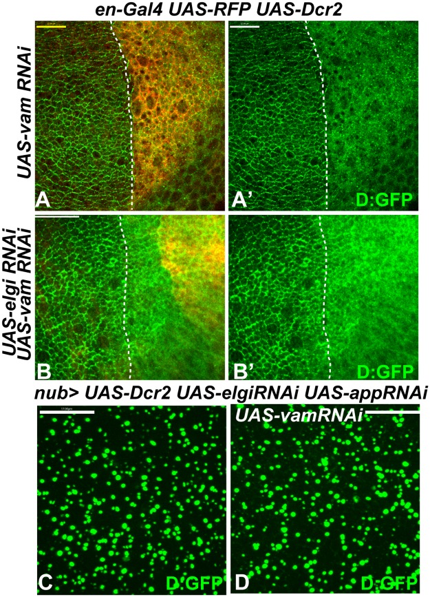
Expression of elgiCS or simultaneous depletion of elgi and app led to accumulation of both Dachs and Vam in cytoplasmic punctae (Figs 6B, 6D, 6E and 8A). App can palmitoylate Vam in S2 cells [15]. If App acts through Vam in vivo to regulate Dachs localization, then we would expect simultaneous depletion of elgi and vam to result in a similar mislocalization of Dachs. However, Dachs does not mislocalize to cytoplasmic punctae when elgi and vam are both knocked down (Fig 7B), suggesting that Vam may not be a bona fide substrate of App in vivo.
Fig 7. elgi and app affect Dachs in a Vam-independent manner.
(A-B) Horizontal apical sections of wing imaginal discs expressing en-Gal4 UAS-dcr2 UAS-RFP D:GFP along with UAS-vam-RNAi (A, A’), UAS-elgi-RNAi UAS-vam-RNAi (B, B’), showing their effects on D:GFP localization in the posterior compartment (marked by RFP, red). Dashed white line marks the A-P compartment boundary. (C, D) Horizontal apical sections of wing imaginal discs expressing nub-Gal4 UAS-dcr2 UAS-elgi-RNAi UAS-app-RNAi D:GFP (C) or along with UAS-vam-RNAi (D), showing that Vam is not required for Dachs:GFP (D:GFP) mislocalization in absence of app and elgi. Scale bar is 12 μm in A, 33 μm in B and 17μm in C and D.
The mislocalization of both Dachs and Vam in the absence of elgi and app could be explained either by Elgi and App directly affecting both Dachs and Vam, or alternatively by only directly affecting one of them, with the other mislocalized due to the physical interaction between Dachs and Vam. To distinguish between these possibilities, we examined whether Vam is required for Dachs mislocalization in absence of elgi and app. Depletion of Vam in cells lacking elgi and app did not affect the mislocalization of Dachs (Fig 7C and 7D), indicating that Vam is not required for Dachs mislocalization. To investigate the potential requirement for Dachs in Vam mislocalization, we took advantage of the observation that the second SH3 domain of Vam is specifically required for its interaction with Dachs [14, 15]. We examined how deletion of the different SH3 domains of Vam affected its localization in presence of elgiCS. Full length Vam-RFP as well as Vam-RFP lacking either the first (Vam-ΔSH3-1-RFP) or third (Vam-ΔSH3-3-RFP) SH3 domains were mislocalized along with Dachs:GFP (Fig 8A, 8B and 8D), but Vam-RFP lacking the second SH3 domain (Vam-ΔSH3-2-RFP), which fails to interact with Dachs, did not mislocalize with Dachs:GFP in presence of elgiCS (Fig 8C). Together, these experiments indicate that elgi and app primarily affect Dachs, and that in absence of both elgi and app, Vam gets mislocalized along with Dachs because it physically interacts with Dachs. In addition, they suggest that Vam does not seem to be the substrate of App that regulates Dachs localization.
If Vam is affected by Elgi solely through its interaction with Dachs, then Vam that cannot interact with Dachs could be more stable than wild-type Vam. To test this, we expressed either full length Vam-RFP or Vam-RFP lacking one of the SH3 domains, using a ubiquitously expressed tub-Gal4 and UAS-vam transgenes inserted at the same genomic locus. Western Blotting of wing disc lysates revealed that Vam-ΔSH3-2:RFP, which cannot interact with Dachs, is expressed at a higher level than the other Vam constructs (S7 Fig), suggesting that association with Dachs regulates Vam protein stability.
Discussion
Our results identify the E3 ubiquitin ligase Elgi as playing a crucial role in modulating Fat-Hippo signaling. Elgi regulates the stability of Dachs and, together with App, its membrane localization. Through its effects on Dachs, Elgi also indirectly regulates the levels and localization of Vam. Our observations suggest a model (Fig 8E) where, under normal conditions, Elgi maintains limiting levels of Dachs and Vam by facilitating their degradation, and controls their localization in coordination with App. In the absence of Elgi, Dachs and Vam levels increase, but App is still sufficient for their membrane localization. However, when elgiCS is expressed or when elgi and app are simultaneously depleted, the increased levels of Dachs and Vam are unable to localize to the membrane and instead accumulate in punctate structures in the cytoplasm. Due to the impact that membrane levels of Dachs have on Warts activity, Elgi influences the activity of Yki, the key transcription factor of the Hippo pathway.
Our observations indicate that Elgi regulates Dachs in parallel to Fat, and the distinct ways in which they regulate Dachs provide insight into how Dachs regulates Hippo signaling and PCP. The increase in Dachs at membranes appears greater when Elgi is knocked down than when Fat is knocked down but the increase in wing growth is greater when Fat is knocked down than when Elgi is knocked down. This might be explained by an influence of elgi on other proteins, which counteract the growth promoted by Dachs. Alternatively, it could be explained by the distinct patterns of Dachs membrane localization in fat versus elgi loss-of-function. In the absence of Fat, Dachs localizes to the membrane around the entire circumference of the cell, whereas in the absence of Elgi, Dachs still remains polarized. Investigations of Hippo signaling have revealed that Warts is activated in discrete complexes at the sub-apical membrane, where multiple upstream components co-localize [50, 51]. We suggest that when Dachs is membrane localized around the entire cell circumference, it is able to broadly disrupt Warts activation, whereas when Dachs is polarized, there could be regions of the sub-apical membrane where Warts activation can occur normally, resulting in a lower overall level of Yki activity than can be achieved with uniform Dachs membrane localization. The normal polarization of Dachs, together with the normal hair polarity in elgi mutants, is consistent with the conclusion that Fat signaling regulates PCP through the polarization of Dachs localization. Thus Elgi effectively separates the two distinct downstream branches of Fat signaling–it disrupts Hippo signaling, but leaves PCP unaffected.
Elgi is a homolog of mammalian RNF41/NRDP1, which regulates ErbB3 and ErbB4 receptors, BRUCE, BIRC6 and Parkin [52–54]. RNF41 also regulates trafficking of certain JAK2-associated type1 receptors [55]. Moreover, it can regulate PCP by ubiquitinating Dishevelled (DVL) [56]. Interestingly, RNF41 physically interacts with VANGL2 but does not ubiquitinate it [56]. Rather it ubiquitinates DVL, which is associated with VANGL2. RNF41 also stabilizes AP2S1 in an E3 ligase activity-independent manner [57].
Although Elgi can physically interact with Dachs, App and Fat-ICD, we were unable to detect direct ubiquitination of these proteins by Elgi. Thus Elgi might regulate Dachs stability through an unknown protein. Alternatively, it is possible that Elgi might function as an adapter to facilitate proteosomal or endolysosomal degradation of Dachs. For example, Elgi might directly link Dachs (or a Dachs-Vam complex) to the proteasome. Some E3 ligases, such as Stuxnet, have been reported to regulate protein degradation without ubiquitination by acting in this way [58]. If Elgi plays a role in vesicular trafficking, it might not only promote trafficking of Dachs (or a Dachs-Vam complex) to the lysosome, it could also affect the localization and levels of other proteins. The increase in Fat levels in elgi mutant cells is consistent with this possibility.
Dachs is also regulated by FbxL7 [16, 17], which like Elgi is predicted to be a ubiquitin ligase. It is also not clear if FbxL7 regulates Dachs by directly ubiquitinating it or instead by affecting vesicular trafficking. However, FbxL7 differs from Elgi in that FbxL7 regulates both the levels and the polarity of Dachs, and in that FbxL7 itself exhibits polarized membrane localization. The presence of multiple mechanisms to limit amounts of Dachs emphasize the importance of strict control over Dachs levels.
The palmitoyltransferase App is critical for membrane localization of Dachs [33]. Although App can palmitoylate Vam and the Fat ICD when overexpressed [15, 34], the mechanism by which it regulates Dachs localization in vivo is still not clear. Our studies revealed that concomitant loss of Elgi and App leads to accumulation of Dachs and Vam in cytoplasmic punctae. The detection of these puncta might reflect a role for App in trafficking of Dachs and Vam. In the presence of wild-type Elgi, the low levels of Dachs could preclude detection of this trafficking defect, but the dramatic increase in Dachs levels in the absence of elgi could exacerbate accumulation of Dachs/Vam in vesicles and make them easier to detect. Consistent with this possibility, basal puncta of Dachs were detected when Elgi was depleted and Dachs was expressed using the act5C promoter. Alternatively, Elgi and App might impair distinct activities that are redundantly required for trafficking of Dachs and Vam. Palmitoylation is well known to play a key role in vesicular trafficking [59]. Further characterization of the nature of these cytoplasmic accumulations of Dachs could help understand the exact mechanism by which App regulates Dachs localization.
Our genetic experiments also suggest that Vam may not be a relevant substrate of App in vivo, as concomitant loss of vam and elgi does not result in the accumulations of Dachs observed in the absence of elgi and app. This is consistent with the finding that mutating a predicted palmitoylation site within Vam does not abrogate its membrane localization [14]. Our experiments also revealed that elgi and app primarily affect Dachs, and only indirectly affect Vam through its physical association with Dachs. This is consistent with the observation that Elgi associates with Dachs but not with Vam. Interestingly, Vam lacking the second SH3 domain is expressed at a higher level compared to the full length Vam or Vam lacking the first or third SH3 domains. This suggests that Elgi-mediated degradation normally maintains Vam at low levels by acting on the Dachs-Vam heterodimer.
Materials and methods
Drosophila strains
The following previously described alleles and transgenes were used, elgi1, elgi2 [40], Df(3L)BSC575 (BL27587), en-Gal4 (BL30564), nub-Gal4 (BL25754), tub-Gal4, UAS-dcr2, ex-LacZ (BL44248), ban-lacZ (BL10154), UAS-mCD8:RFP (gift of G. Morata, Universidad Autónoma de Madrid, Madrid), Dachs:GFP [31], Ds:GFP [21], UAS-Dachs:V5 [8], UAS-Fat [41], UAS-vam-RNAi (BL38263), UAS-fat-RNAi (VDRC 9396), UAS-d-RNAi (VDRC 12555), UAS-app, UAS-app-RNAi [33], Vam:GFP, UAS-vam-3x-FLAG-RFP, UAS-vam-ΔSH3-1-3x-FLAG-RFP, UAS-vam-ΔSH3-2-3x-FLAG-RFP, UAS-vam-ΔSH3-3-3x-FLAG-RFP and ACT>Stop>Vam-3x-FLAG-GFP [14], Jub:GFP [60], UAS-D:RFP, UAS-elgi RNAi (VDRC109617, NIG 17033R-3) UAS-Mito-GFP (BL8442), UAS-atg8a-GFP (52005), LAMP1-YFP (DGGR 115517), Rab4-YFP (BL62542), Rab5-YFP (BL62543), Rab7-YFP (BL62545), Rab11-YFP (BL62549). The transgenes UAS-elgi-Myc-HA and UAS-elgiCS-Myc-HA were created in this study.
Immunohistochemistry
Dissected wing discs were fixed in 4% paraformaldehyde for 10 minutes at room temperature and stained with primary antibodies, rat anti-E-cad (1:400, DSHB DCAD2), rat-anti-Fat (1:4000) (Feng and Irvine, 2009), rat-anti-Ds (1: 5000) (Ma et al., 2003), mouse-anti-Crb (1:200, DSHB), mouse-anti-Arm (1:200, DSHB) mouse anti-β-gal (1:400, DSHB JIE7-c), Guinea pig anti-Hrs (gift from Hugo Bellen)(1:200); and secondary antibodies, donkey anti-rat-647 (1:100, Jackson, 712-605-150) and donkey anti-rat-Cy3 (3:400, Jackson, 712-165-150). GFP and RFP were detected by autofluorescence. Confocal images were captured using a Leica SP8 confocal microscope.
Molecular biology
To create pUAST-elgi-Myc-HA, elgi cDNA was PCR amplified with elgi-forward 5’TGAATAGGGAATTGGGAATTCATGGGCTACGATGTGAATCGCTT3’ and elgi-MYC-HA-rev CGCAAGATCTGTTAACGAATTCCTAAGCGTAATCTGGAACATCGTATGGGTACAGATCCTCTTCTGAGATGAGTTTTTGTTCCTCGATGCCATGGGCGAAG3’ primers and cloned into pUAST plasmid digested with EcoRI by Gibson assembly. To create pUAST-elgiCS-Myc-HA, elgi cDNA was PCR amplified with elgiCS forward primer, 5’TGAATAGGGAATTGGGAATTCATGGGCTACGATGTGAATCGCTTTCAGGGGGAGGTGGACGAGGAGCTCACCTCTCCCATCTCCTCCGGAGTGCTT 5’ and elgi-MYC-HA-rev primer and cloned into pUAST plasmid digested with EcoRI by Gibson assembly. All plasmids were sequence verified. pUAST-elgi-V5 and pUAST-elgi-3xFLAG were similarly created using the elgi-forward primer and elgi-V5 reverse 5’GCAAGATCTGTTAACGAATTCCTAGGTGCTGTCCAGGCCCAGCAGGGGGTTGGGGATGGGCTTGCCCTCGATGCCATGGGCGAAG 3’ and elgi-FLAG-reverse 5’ GCAAGATCTGTTAACAATTCTTACTTGTCATCGTCATCCTTGTAATCGATGTCATGATCTTTATAATCACCGTCATGGTCTTTGTAGTCCTCGATGCCATGGGCGAAG primers respectively. pACU2-app:V5 was created by amplifying the app CDNA using forward 5’gttcaattacagctcgaattcATGAATCTGCTGTGCTGCTGTTG 3’ and reverse primer 5’ cgcagatctgttaacgaattcTTAGGTGCTGTCCAGGCCCAGCAGGGGGTTGGGGATGGGCTTGCCGACTATGGCCACGTTTGTGGTC and and cloned into pACU2 plasmid digested with EcoRI, by Gibson assembly. To create pUAST-Vam-V5, Vam cDNA was amplified with Vam-for 5’TGAATAGGGAATTGGGAATTCATGGCATTTCTTTGCCCCGT3’ and Vam-V5-Rev 5’GCAAGATCTGTTAACGAATTCTTACGTAGAATCGAGACCGAGGAGAGGGTTAGGGATAGGCTTACCAAGGCTGGTCATCGCGGGTGGT3’primers and cloned into pUAST plasmid digested with EcoRI, by Gibson assembly. To create pUAST-D-RFP, Dachs cDNA was amplified with D-for 5’TGAATAGGGAATTGGGAATTCATGTTGACTACGACGATCTGGACAG 3’ and D-Rev 5’ GCTCTTCGCCCTTAGACACCATTTTACTGAGCGTCATGAACTGGAAGG 3’ primers. Tag-RFP was amplified with RFP-For 5’ ATGGTGTCTAAGGGCGAAGAG and RFP-Rev 5’ TCCTTCACAAAGATCCTCTAGATCAATTAAGTTTGTGCCCCAGTTTGC3 and cloned into pUAST plasmid digested with EcoRI and XbaI by Gibson assembly. All plasmids were verified by sequencing.
dsRNA production
Small fragments of elgi or EGFP coding sequences were selected using SnapDragon tool to avoid any off-target effect. For generating the template for dsRNA synthesis for elgi, elgi T7 forward 5’TAATACGACTCACTATAGGGGGATGCATTAACGAGTGGCTAACC 3’ and T7 Reverse 5’ TAATACGACTCACTATAGGGATAGTCAGCTGCTGGTCGGTG 3’ primers were used. For dsRNA synthesis for EGFP, a fragment was amplified with the EGFP-T7 forward 5’TAATACGACTCACTATAGGGACGTAAACGGCCACAAGTTC 3’ and EGFP-T7 reverse 5’ TAATACGACTCACTATAGGGTGTTCTGCTGGTAGTGGTCG 3’ primers. dsRNA synthesis was carried out using MEGAscript T7 transcription Kit (Invitrogen), following manufacturer’s instruction. After synthesis the dsRNA was purified using RNAeasy mini Kit (Quiagen).
Cell culture, immunoprecipitation and western blotting
S2 cells were grown in Schneider’s media and transfected with plasmids aw-GAL4, pUAST-D-:V5 [8] pUAST-D-N:V5, pUAST-D-M:V5, pUAST-D-C:V5 [10], pUAST-Vam-V5 [14] along with pUAST-elgi-Myc-HA, using effectene transfection reagent (Quiagen) following the manufacturer’s instruction. Cells were lysed in RIPA buffer supplemented with protease inhibitor cocktail and phosphatase inhibitor cocktail (Calbiochem). Cell lysates were incubated with affinity resin conjugated with anti HA antibody (Sigma) for two hours at 4 °C, following which they were washed four times. Proteins samples were boiled with Laemlii buffer and subjected to SDS-PAGE using 4–15% gradient gels (Bio Rad). Western transfer was carried out using Trans-Blot Turbo transfer system (Bio Rad) and immunoblotting was performed using the primary antibodies, rabbit-anti-V5 (Bethyl Laboratories, 1:5000), mouse anti-V5 (Invitrogen, 1:5000), mouse-anti-GAPDH (Covance, 1:1000), mouse anti α-Tubulin (1:20,000), mouse anti FLAG (Sigma, 1:5000) Rabbit anti FLAG (Sigma, 1:5000) and Guinea Pig anti App (gift from Seth Blair, 1:5000); and as secondary antibodies goat anti-mouse 680 (Li-Cor, 926–68,020), goat anti-rabbit-680 (Li-Cor, 926–68,021) and Donkey anti-Gunea Pig-780 (Li-Cor, 926–32411), all at a dilution of 1:10,000. Blots were scanned using an Odyssey Imaging System (Li-Cor). Co-immunoprecipitation experiments in Fig 6I and 6J were performed following similar protocols where S2 cells were transfected with aw-Gal4, pUAST-Elgi-3x-FLAG together with either pUAST-Vam-V5 or pUAST-Elgi-V5 (Fig 6I) or pACU2-App-V5 or pUAST-Fat-ICD-V5 (Fig 6J). Elgi-3x-Flag was immunoprecipitated using EZ view Red FLAG M2 affinity Gel and immunoblotted with Mouse anti-FLAG and Rabbit-anti-V5 primary antibodies and donkey anti-mouse 680 and donkey anti-rabbit 780 secondary antibodies.
Ubiquitination assay
Ubiquitination assays were carried out as previously described [44]. Briefly, S2 cells were transfected with either pUAST-Vam:V5, pACU2-App:V5, pUAST-Fat-ICD:V5 or pUAST-D:V5 with or without pUAST-BioUb-BirA and pUAST-elgi-3x-FLAG along with pmt-Gal4. After incubation for 72 hours, protein expression was induced by adding 0.5mM CuSO4. In all ubiquitination experiments, to prevent degradation of the ubiquitinated products, cells were treated with the proteasome inhibitor MG132 (10uM) at the same time as CuSO4 was added. After overnight incubation, cells were lysed in RIPA buffer supplemented with protease inhibitor cocktail (Roche) and phosphatase inhibitor cocktail (Calbiochem) and 10mM N-ethyl Maleimide (deubiquitinase inhibitor). Cell lysates were incubated with affinity resin conjugated with mouse anti-V5 antibody (Sigma) for two hours at 4 °C, following which they were washed four times. After washing, samples were boiled in Laemmli buffer and subjected to SDS-PAGE followed by Western transfer. Immunoblotting was performed using rabbit anti-FLAG and rabbit anti-V5 primary antibodies and goat anti-rabbit-800 (Licor) secondary antibody. IRDye-680RD-Streptavidin (Licor 925–68079) was used at 1:10000 dilution to detect biotin, and the blots were scanned using an Odyssey imaging system (LiCor). Elgi autoubiquitination was performed following a similar protocol, where S2 cells were transfected with pUAST-Elgi-3x-Flag or pUAST-ElgiCS-3x-Flag along with pUAST-BirA or pUAST-BioUb-BirA and Elgi-3x-Flag or ElgiCS-3x-Flag was immunoprecipitated using EZ view Red FLAG M2 affinity Gel (Sigma) and immunoblotted with Rabbit-anti-FLAG (Sigma) and above described secondary antibody and streptavidin.
For RNAi, cells were resuspended in serum free Schneider’s medium at a concentration of 1-4x10⁵ cells/ml and to 1ml of of cell suspension, 30μg of either EGFP-shRNA or elgi-shRNA was added and incubated for 30 minutes, following which 1ml of fresh medium containing 20% serum was added. Immediately, they were transfected with either pUAST-Vam:V5, pACU2-App:V5, pUAST-Dachs:V5, or pUAST-Fat-ICD:V5 plasmid along with the pMT-Gal4 and pUAST-BirA-bioUB plasmids, using Effectene transfection reagent (Quiagen) following manufacturer’s protocol. After incubation for 72 hours, protein expression was induced by adding 0.5mM CuSO4, in the presence of 10uM MG132. After overnight inductions the cells were collected and the proteins of interest were immunoprecipitated and processed as described above.
Statistical analyses
Statistical significance was calculated using Graphpad Prism Software. For pairwise comparisons we used t test.
Supporting information
Close up of proximal wing areas showing the normal orientation of hairs from control (A), homozygous elgi1 (B), and elgi1/Df(3L)BSC575 (C) flies.
(TIF)
Horizontal apical sections of wing imaginal discs expressing en-Gal4 UAS-dcr2 UAS-GFP (green) UAS-elgi-RNAi and stained with anti-Ds antibody (red) showing no effect on the levels or localization of Ds in the posterior compartment (marked by GFP, green). Dashed white line marks the A-P compartment boundary. Scale bar is 33.00μm.
(TIF)
(A-D) Adult male wings from UAS-elgi-Myc-HA/+; elgi1/+ (control) (A), UAS-elgi-Myc-HA/+; elgi1 (B), nub-Gal4 UAS-elgi-Myc-HA/+; elgi1/TM6b (C) and nub-Gal4 UAS-elgi-Myc-HA/+; elgi1/TM6b (D) flies. Arrows point to the crossveins. (E) Histogram of the ratio of crossvein distance (CVD) to wing span, (normalized to the average CVD/wing span in control wings in flies of the genotypes in panels A-D, as indicated. Error bars indicate Standard deviation. **** (p<0.0001), NS not significant.
(TIF)
(A-D) Horizontal apical (A, A’, C, C’), basal (B, B’, D, D’), and vertical (A1, A2, C1, C2) sections of wing imaginal discs expressing en-Gal4 UAS-dcr2 UAS-RFP (red) UAS-elgi-RNAi, with either Dachs:GFP (D:GFP) (A, B) or act5C promoter driven Dachs:GFP (act>D:GFP) (C, D) showing increased levels of membrane localized Dachs:GFP (green), in the posterior compartment (marked by red). In D and D’ some punctate cytoplasmic accumulations of Dachs:GFP is seen in the basal sections. (E,F) Horizontal apical (E,E’), basal (F, F’) and vertical (E1, E2) sections of wing imaginal discs expressing Dachs:GFP under the control of the actin5C promoter (act>D:GFP) throughout, showing increased levels of membrane localized Dachs:GFP (E, E’) as well as punctate cytoplasmic accumulations of Dachs:GFP in the basal sections (F, F’) in homozygous elgi1 mutant clones, marked by absence of RFP (red). E-cad is shown in blue. (G-H) Horizontal apical (G,G’), basal (H,H’) and vertical (G1,G2) sections of wing imaginal discs expressing en-Gal4 UAS-dcr2 D:GFP UAS-RFP along with UAS-elgiCS, showing the effect on Dachs:GFP (D:GFP)(green) levels and localization in the posterior compartment (marked by RFP, red). E-cad is shown in blue. Dashed white line marks the A-P compartment boundary. Scale bar is 16.5 μm in all panels.
(TIF)
(A, A’) Third instar wing imaginal discs expressing en-Gal4 UAS-GFP ex-lacZ UAS-elgiCS stained for expression of ex-lacZ (magenta), with posterior cells marked by expression of GFP (green). Dashed white line marks the A-P compartment boundary. (B-F) Horizontal apical sections of wing imaginal discs expressing en-Gal4 UAS-dcr2 UAS-elgiCS along with Fat:GFP (B, B’), Ds:GFP (C, C’), Jub:GFP (D, D’) or stained for Crumbs (E, E’) or armadillo (Arm) (F, F’) showing that it does not affect their localization in the posterior compartment (marked by Dcr2 staining, red). Dashed white line marks the A-P compartment boundary. (G-G”) Horizontal sections of wing imaginal discs expressing nub-Gal4 UAS-elgiCS along with D:GFP and Vam:RFP showing that Vam:RFP (red) (G”) gets mislocalized with Dachs:GFP (D:GFP, green) (G’) in presence of ElgiCS. Scale bar is 33μm in A-F and 16.5 μm in G. (H, I) Close up of proximal wing areas showing the orientation of hairs from flies carrying nub-Gal4 alone (H) or in combination with UAS-elgiCS (I). (J) Western blot showing levels of App from third instar wing disc lysate from flies expressing tub-Gal4 alone or with UAS-app or UAS-elgi-RNAi. Lysates from flies expressing tub-Gal4 UAS-app serves as positive control. Tubulin was used as a control for loading and transfer.
(TIF)
(A-E) Horizontal sections of wing imaginal discs expressing nub-Gal4 UAS-dcr2 UAS-elgi-RNAi UAS-app-RNAi UAS-D:RFP along with Rab4:YFP (green) (A, A’,A"), Rab5:YFP (green) (B, B’,B") Rab7:YFP (green) (C, C’,C"), Rab11:YFP (green) (D, D’,D") or LAMP1-YFP (green) (E,E’,E") showing that the cytoplasmic accumulations of Dachs:RFP (D:RFP, red) in absence of elgi and app do not colocalize with these markers. (F,G) Horizontal sections of wing imaginal discs expressing en-Gal4 UAS-elgiCS UAS-D:RFP (red) along with UAS-Mito-GFP (green) (F,F’,F") or UAS-atg8a-GFP (green) (G, G’,G"). (H, H’, H”) Horizontal sections of wing imaginal discs expressing en-Gal4 UAS-elgiCS, Dachs:GFP (green) stained with anti-Hrs antibody (red). Scale bar is 33 μm in all panels.
(TIF)
Western blot showing expression levels of full length Vam:RFP or Vam:RFP lacking the individual SH3 domains, from third instar wing disc lysate from flies expressing tub-Gal4 along with UAS-Vam:RFP, UAS-Vam-Δ-SH3-1-RFP, UAS-Vam-Δ-SH3-2-RFP or UAS-Vam-Δ-SH3-3-RFP, with all the Vam transgenes inserted at the same genomic location and detected by αRFP antibody. Tubulin was used as a control for loading and transfer.
(TIF)
Acknowledgments
We thank the Bloomington Drosophila stock center, Developmental Studies Hybridoma Bank, D. Strutt, D. Drumond-Barbosa, M. Simon, Y. Bellaiche, S. Blair and H. Bellen for providing Drosophila stocks and antibodies. We also thank Abhijit Ambegaonkar for making the pUAST-Dachs:RFP plasmid.
Data Availability
All relevant data are within the manuscript and its Supporting Information files.
Funding Statement
This research was supported by National Institute of Health (NIH) grants R01 GM78620 (KDI) and K99 HD092553 (JRM). The funders had no role in study design, data collection and analysis, decision to publish, or preparation of the manuscript.
References
- 1.Staley BK, Irvine KD. Hippo signaling in Drosophila: recent advances and insights. Developmental dynamics: an official publication of the American Association of Anatomists. 2012;241(1):3–15. 10.1002/dvdy.22723 . [DOI] [PMC free article] [PubMed] [Google Scholar]
- 2.Irvine KD, Harvey KF. Control of organ growth by patterning and hippo signaling in Drosophila. Cold Spring Harbor perspectives in biology. 2015;7(6). 10.1101/cshperspect.a019224 . [DOI] [PMC free article] [PubMed] [Google Scholar]
- 3.Matis M, Axelrod JD. Regulation of PCP by the Fat signaling pathway. Genes & development. 2013;27(20):2207–20. 10.1101/gad.228098.113 . [DOI] [PMC free article] [PubMed] [Google Scholar]
- 4.Sharma P, McNeill H. Regulation of long-range planar cell polarity by Fat-Dachsous signaling. Development. 2013;140(18):3869–81. 10.1242/dev.094730 . [DOI] [PubMed] [Google Scholar]
- 5.Thomas C, Strutt D. The roles of the cadherins Fat and Dachsous in planar polarity specification in Drosophila. Developmental dynamics: an official publication of the American Association of Anatomists. 2012;241(1):27–39. 10.1002/dvdy.22736 . [DOI] [PubMed] [Google Scholar]
- 6.Blair S, McNeill H. Big roles for Fat cadherins. Current opinion in cell biology. 2018;51:73–80. Epub 2017/12/20. 10.1016/j.ceb.2017.11.006 . [DOI] [PMC free article] [PubMed] [Google Scholar]
- 7.Cho E, Feng Y, Rauskolb C, Maitra S, Fehon R, Irvine KD. Delineation of a Fat tumor suppressor pathway. Nature genetics. 2006;38(10):1142–50. 10.1038/ng1887 . [DOI] [PubMed] [Google Scholar]
- 8.Mao Y, Rauskolb C, Cho E, Hu WL, Hayter H, Minihan G, et al. Dachs: an unconventional myosin that functions downstream of Fat to regulate growth, affinity and gene expression in Drosophila. Development. 2006;133(13):2539–51. 10.1242/dev.02427 . [DOI] [PubMed] [Google Scholar]
- 9.Mao Y, Tournier AL, Bates PA, Gale JE, Tapon N, Thompson BJ. Planar polarization of the atypical myosin Dachs orients cell divisions in Drosophila. Genes & development. 2011;25(2):131–6. 10.1101/gad.610511 . [DOI] [PMC free article] [PubMed] [Google Scholar]
- 10.Rauskolb C, Pan G, Reddy BV, Oh H, Irvine KD. Zyxin links fat signaling to the hippo pathway. PLoS biology. 2011;9(6):e1000624 10.1371/journal.pbio.1000624 . [DOI] [PMC free article] [PubMed] [Google Scholar]
- 11.Vrabioiu AM, Struhl G. Fat/Dachsous Signaling Promotes Drosophila Wing Growth by Regulating the Conformational State of the NDR Kinase Warts. Developmental cell. 2015;35(6):737–49. Epub 2015/12/26. 10.1016/j.devcel.2015.11.027 . [DOI] [PMC free article] [PubMed] [Google Scholar]
- 12.Ayukawa T, Akiyama M, Mummery-Widmer JL, Stoeger T, Sasaki J, Knoblich JA, et al. Dachsous-dependent asymmetric localization of spiny-legs determines planar cell polarity orientation in Drosophila. Cell reports. 2014;8(2):610–21. 10.1016/j.celrep.2014.06.009 . [DOI] [PubMed] [Google Scholar]
- 13.Ambegaonkar AA, Irvine KD. Coordination of planar cell polarity pathways through Spiny-legs. eLife. 2015;4 Epub 2015/10/28. 10.7554/eLife.09946 . [DOI] [PMC free article] [PubMed] [Google Scholar]
- 14.Misra JR, Irvine KD. Vamana Couples Fat Signaling to the Hippo Pathway. Developmental cell. 2016;39(2):254–66. Epub 2016/10/26. 10.1016/j.devcel.2016.09.017 . [DOI] [PMC free article] [PubMed] [Google Scholar]
- 15.Zhang Y, Wang X, Matakatsu H, Fehon R, Blair SS. The novel SH3 domain protein Dlish/CG10933 mediates fat signaling in Drosophila by binding and regulating Dachs. eLife. 2016;5 Epub 2016/10/04. 10.7554/eLife.16624 . [DOI] [PMC free article] [PubMed] [Google Scholar]
- 16.Rodrigues-Campos M, Thompson BJ. The ubiquitin ligase FbxL7 regulates the Dachsous-Fat-Dachs system in Drosophila. Development. 2014;141(21):4098–103. Epub 2014/09/27. 10.1242/dev.113498 . [DOI] [PMC free article] [PubMed] [Google Scholar]
- 17.Bosch JA, Sumabat TM, Hafezi Y, Pellock BJ, Gandhi KD, Hariharan IK. The Drosophila F-box protein Fbxl7 binds to the protocadherin fat and regulates Dachs localization and Hippo signaling. eLife. 2014;3:e03383 Epub 2014/08/12. 10.7554/eLife.03383 . [DOI] [PMC free article] [PubMed] [Google Scholar]
- 18.Pan G, Feng Y, Ambegaonkar AA, Sun G, Huff M, Rauskolb C, et al. Signal transduction by the Fat cytoplasmic domain. Development. 2013;140(4):831–42. 10.1242/dev.088534 . [DOI] [PMC free article] [PubMed] [Google Scholar]
- 19.Brittle AL, Repiso A, Casal J, Lawrence PA, Strutt D. Four-jointed modulates growth and planar polarity by reducing the affinity of dachsous for fat. Current biology: CB. 2010;20(9):803–10. 10.1016/j.cub.2010.03.056 . [DOI] [PMC free article] [PubMed] [Google Scholar]
- 20.Ishikawa HO, Takeuchi H, Haltiwanger RS, Irvine KD. Four-jointed is a Golgi kinase that phosphorylates a subset of cadherin domains. Science. 2008;321(5887):401–4. 10.1126/science.1158159 . [DOI] [PMC free article] [PubMed] [Google Scholar]
- 21.Simon MA, Xu A, Ishikawa HO, Irvine KD. Modulation of fat:dachsous binding by the cadherin domain kinase four-jointed. Current biology: CB. 2010;20(9):811–7. 10.1016/j.cub.2010.04.016 . [DOI] [PMC free article] [PubMed] [Google Scholar]
- 22.Matakatsu H, Blair SS. Interactions between Fat and Dachsous and the regulation of planar cell polarity in the Drosophila wing. Development. 2004;131(15):3785–94. 10.1242/dev.01254 . [DOI] [PubMed] [Google Scholar]
- 23.Ambegaonkar AA, Pan G, Mani M, Feng Y, Irvine KD. Propagation of Dachsous-Fat planar cell polarity. Current biology: CB. 2012;22(14):1302–8. 10.1016/j.cub.2012.05.049 . [DOI] [PMC free article] [PubMed] [Google Scholar]
- 24.Brittle A, Thomas C, Strutt D. Planar polarity specification through asymmetric subcellular localization of Fat and Dachsous. Current biology: CB. 2012;22(10):907–14. 10.1016/j.cub.2012.03.053 . [DOI] [PMC free article] [PubMed] [Google Scholar]
- 25.Hale R, Brittle AL, Fisher KH, Monk NA, Strutt D. Cellular interpretation of the long-range gradient of Four-jointed activity in the Drosophila wing. eLife. 2015;4 Epub 2015/02/25. 10.7554/eLife.05789 . [DOI] [PMC free article] [PubMed] [Google Scholar]
- 26.Misra JR, Irvine KD. The Hippo signaling network and its biological functions. Annual review of genetics. 2018;52:65–87. 10.1146/annurev-genet-120417-031621 . [DOI] [PMC free article] [PubMed] [Google Scholar]
- 27.Willecke M, Hamaratoglu F, Sansores-Garcia L, Tao C, Halder G. Boundaries of Dachsous Cadherin activity modulate the Hippo signaling pathway to induce cell proliferation. Proceedings of the National Academy of Sciences of the United States of America. 2008;105(39):14897–902. 10.1073/pnas.0805201105 . [DOI] [PMC free article] [PubMed] [Google Scholar]
- 28.Feng Y, Irvine KD. Fat and expanded act in parallel to regulate growth through warts. Proceedings of the National Academy of Sciences of the United States of America. 2007;104(51):20362–7. 10.1073/pnas.0706722105 . [DOI] [PMC free article] [PubMed] [Google Scholar]
- 29.Silva E, Tsatskis Y, Gardano L, Tapon N, McNeill H. The tumor-suppressor gene fat controls tissue growth upstream of expanded in the hippo signaling pathway. Current biology: CB. 2006;16(21):2081–9. Epub 2006/09/26. 10.1016/j.cub.2006.09.004 . [DOI] [PubMed] [Google Scholar]
- 30.Bennett FC, Harvey KF. Fat cadherin modulates organ size in Drosophila via the Salvador/Warts/Hippo signaling pathway. Current biology: CB. 2006;16(21):2101–10. Epub 2006/10/19. 10.1016/j.cub.2006.09.045 . [DOI] [PubMed] [Google Scholar]
- 31.Bosveld F, Bonnet I, Guirao B, Tlili S, Wang Z, Petitalot A, et al. Mechanical control of morphogenesis by Fat/Dachsous/Four-jointed planar cell polarity pathway. Science. 2012;336(6082):724–7. 10.1126/science.1221071 . [DOI] [PubMed] [Google Scholar]
- 32.Rogulja D, Rauskolb C, Irvine KD. Morphogen control of wing growth through the Fat signaling pathway. Developmental cell. 2008;15(2):309–21. 10.1016/j.devcel.2008.06.003 . [DOI] [PMC free article] [PubMed] [Google Scholar]
- 33.Matakatsu H, Blair SS. The DHHC palmitoyltransferase approximated regulates Fat signaling and Dachs localization and activity. Current biology: CB. 2008;18(18):1390–5. 10.1016/j.cub.2008.07.067 . [DOI] [PMC free article] [PubMed] [Google Scholar]
- 34.Matakatsu H, Blair SS, Fehon RG. The palmitoyltransferase Approximated promotes growth via the Hippo pathway by palmitoylation of Fat. The Journal of cell biology. 2017;216(1):265–77. Epub 2016/12/30. 10.1083/jcb.201609094 . [DOI] [PMC free article] [PubMed] [Google Scholar]
- 35.Mahoney PA, Weber U, Onofrechuk P, Biessmann H, Bryant PJ, Goodman CS. The fat tumor suppressor gene in Drosophila encodes a novel member of the cadherin gene superfamily. Cell. 1991;67(5):853–68. . [DOI] [PubMed] [Google Scholar]
- 36.Bryant PJ, Huettner B, Held LI Jr., Ryerse J, Szidonya J. Mutations at the fat locus interfere with cell proliferation control and epithelial morphogenesis in Drosophila. Developmental biology. 1988;129(2):541–54. Epub 1988/10/01. . [DOI] [PubMed] [Google Scholar]
- 37.Clark HF, Brentrup D, Schneitz K, Bieber A, Goodman C, Noll M. Dachsous encodes a member of the cadherin superfamily that controls imaginal disc morphogenesis in Drosophila. Genes & development. 1995;9(12):1530–42. 10.1101/gad.9.12.1530 [DOI] [PubMed] [Google Scholar]
- 38.Mao Y, Kucuk B, Irvine KD. Drosophila lowfat, a novel modulator of Fat signaling. Development. 2009;136(19):3223–33. 10.1242/dev.036152 . [DOI] [PMC free article] [PubMed] [Google Scholar]
- 39.Villano JL, Katz FN. four-jointed is required for intermediate growth in the proximal-distal axis in Drosophila. Development. 1995;121(9):2767–77. Epub 1995/09/01. . [DOI] [PubMed] [Google Scholar]
- 40.Von Stetina JR, Tranguch S, Dey SK, Lee LA, Cha B, Drummond-Barbosa D. alpha-Endosulfine is a conserved protein required for oocyte meiotic maturation in Drosophila. Development. 2008;135(22):3697–706. Epub 2008/10/18. 10.1242/dev.025114 . [DOI] [PMC free article] [PubMed] [Google Scholar]
- 41.Matakatsu H, Blair SS. Separating the adhesive and signaling functions of the Fat and Dachsous protocadherins. Development. 2006;133(12):2315–24. 10.1242/dev.02401 . [DOI] [PubMed] [Google Scholar]
- 42.Ma D, Yang CH, McNeill H, Simon MA, Axelrod JD. Fidelity in planar cell polarity signalling. Nature. 2003;421(6922):543–7. 10.1038/nature01366 . [DOI] [PubMed] [Google Scholar]
- 43.Liu R, Woolner S, Johndrow JE, Metzger D, Flores A, Parkhurst SM. Sisyphus, the Drosophila myosin XV homolog, traffics within filopodia transporting key sensory and adhesion cargos. Development. 2008;135(1):53–63. Epub 2007/11/30. 10.1242/dev.011437 . [DOI] [PubMed] [Google Scholar]
- 44.Franco M, Seyfried NT, Brand AH, Peng J, Mayor U. A novel strategy to isolate ubiquitin conjugates reveals wide role for ubiquitination during neural development. Mol Cell Proteomics. 2011;10(5):M110 002188. Epub 2010/09/24. 10.1074/mcp.M110.002188 . [DOI] [PMC free article] [PubMed] [Google Scholar]
- 45.Yeh E, Zhou L, Rudzik N, Boulianne GL. Neuralized functions cell autonomously to regulate Drosophila sense organ development. The EMBO journal. 2000;19(17):4827–37. Epub 2000/09/06. 10.1093/emboj/19.17.4827 . [DOI] [PMC free article] [PubMed] [Google Scholar]
- 46.Riley BE, Lougheed JC, Callaway K, Velasquez M, Brecht E, Nguyen L, et al. Structure and function of Parkin E3 ubiquitin ligase reveals aspects of RING and HECT ligases. Nat Commun. 2013;4:1982 Epub 2013/06/19. 10.1038/ncomms2982 . [DOI] [PMC free article] [PubMed] [Google Scholar]
- 47.Buetow L, Huang DT. Structural insights into the catalysis and regulation of E3 ubiquitin ligases. Nature reviews Molecular cell biology. 2016;17(10):626–42. Epub 2016/08/04. 10.1038/nrm.2016.91 . [DOI] [PMC free article] [PubMed] [Google Scholar]
- 48.Greaves J, Salaun C, Fukata Y, Fukata M, Chamberlain LH. Palmitoylation and membrane interactions of the neuroprotective chaperone cysteine-string protein. The Journal of biological chemistry. 2008;283(36):25014–26. Epub 2008/07/04. 10.1074/jbc.M802140200 . [DOI] [PMC free article] [PubMed] [Google Scholar]
- 49.Lynes EM, Raturi A, Shenkman M, Ortiz Sandoval C, Yap MC, Wu J, et al. Palmitoylation is the switch that assigns calnexin to quality control or ER Ca2+ signaling. Journal of cell science. 2013;126(Pt 17):3893–903. Epub 2013/07/12. 10.1242/jcs.125856 . [DOI] [PubMed] [Google Scholar]
- 50.Sun S, Reddy BV, Irvine KD. Localization of Hippo signalling complexes and Warts activation in vivo. Nat Commun. 2015;6:8402 Epub 2015/10/01. 10.1038/ncomms9402 . [DOI] [PMC free article] [PubMed] [Google Scholar]
- 51.Yin F, Yu J, Zheng Y, Chen Q, Zhang N, Pan D. Spatial organization of Hippo signaling at the plasma membrane mediated by the tumor suppressor Merlin/NF2. Cell. 2013;154(6):1342–55. 10.1016/j.cell.2013.08.025 . [DOI] [PMC free article] [PubMed] [Google Scholar]
- 52.Diamonti AJ, Guy PM, Ivanof C, Wong K, Sweeney C, Carraway KL 3rd. An RBCC protein implicated in maintenance of steady-state neuregulin receptor levels. Proceedings of the National Academy of Sciences of the United States of America. 2002;99(5):2866–71. Epub 2002/02/28. 10.1073/pnas.052709799 . [DOI] [PMC free article] [PubMed] [Google Scholar]
- 53.Qiu XB, Markant SL, Yuan J, Goldberg AL. Nrdp1-mediated degradation of the gigantic IAP, BRUCE, is a novel pathway for triggering apoptosis. The EMBO journal. 2004;23(4):800–10. Epub 2004/02/07. 10.1038/sj.emboj.7600075 . [DOI] [PMC free article] [PubMed] [Google Scholar]
- 54.Zhong L, Tan Y, Zhou A, Yu Q, Zhou J. RING finger ubiquitin-protein isopeptide ligase Nrdp1/FLRF regulates parkin stability and activity. The Journal of biological chemistry. 2005;280(10):9425–30. Epub 2005/01/06. 10.1074/jbc.M408955200 . [DOI] [PubMed] [Google Scholar]
- 55.Wauman J, De Ceuninck L, Vanderroost N, Lievens S, Tavernier J. RNF41 (Nrdp1) controls type 1 cytokine receptor degradation and ectodomain shedding. Journal of cell science. 2011;124(Pt 6):921–32. Epub 2011/03/08. 10.1242/jcs.078055 . [DOI] [PMC free article] [PubMed] [Google Scholar]
- 56.Wald JH, Hatakeyama J, Printsev I, Cuevas A, Fry WHD, Saldana MJ, et al. Suppression of planar cell polarity signaling and migration in glioblastoma by Nrdp1-mediated Dvl polyubiquitination. Oncogene. 2017;36(36):5158–67. Epub 2017/05/10. 10.1038/onc.2017.126 . [DOI] [PMC free article] [PubMed] [Google Scholar]
- 57.Masschaele D, Wauman J, Vandemoortele G, De Sutter D, De Ceuninck L, Eyckerman S, et al. High-Confidence Interactome for RNF41 Built on Multiple Orthogonal Assays. Journal of proteome research. 2018;17(4):1348–60. Epub 2018/03/22. 10.1021/acs.jproteome.7b00704 . [DOI] [PubMed] [Google Scholar]
- 58.Du J, Zhang J, He T, Li Y, Su Y, Tie F, et al. Stuxnet Facilitates the Degradation of Polycomb Protein during Development. Developmental cell. 2016;37(6):507–19. Epub 2016/06/22. 10.1016/j.devcel.2016.05.013 . [DOI] [PMC free article] [PubMed] [Google Scholar]
- 59.Aicart-Ramos C, Valero RA, Rodriguez-Crespo I. Protein palmitoylation and subcellular trafficking. Biochimica et biophysica acta. 2011;1808(12):2981–94. Epub 2011/08/09. 10.1016/j.bbamem.2011.07.009 . [DOI] [PubMed] [Google Scholar]
- 60.Das Thakur M, Feng Y, Jagannathan R, Seppa MJ, Skeath JB, Longmore GD. Ajuba LIM proteins are negative regulators of the Hippo signaling pathway. Current biology: CB. 2010;20(7):657–62. Epub 2010/03/23. 10.1016/j.cub.2010.02.035 . [DOI] [PMC free article] [PubMed] [Google Scholar]
Associated Data
This section collects any data citations, data availability statements, or supplementary materials included in this article.
Supplementary Materials
Close up of proximal wing areas showing the normal orientation of hairs from control (A), homozygous elgi1 (B), and elgi1/Df(3L)BSC575 (C) flies.
(TIF)
Horizontal apical sections of wing imaginal discs expressing en-Gal4 UAS-dcr2 UAS-GFP (green) UAS-elgi-RNAi and stained with anti-Ds antibody (red) showing no effect on the levels or localization of Ds in the posterior compartment (marked by GFP, green). Dashed white line marks the A-P compartment boundary. Scale bar is 33.00μm.
(TIF)
(A-D) Adult male wings from UAS-elgi-Myc-HA/+; elgi1/+ (control) (A), UAS-elgi-Myc-HA/+; elgi1 (B), nub-Gal4 UAS-elgi-Myc-HA/+; elgi1/TM6b (C) and nub-Gal4 UAS-elgi-Myc-HA/+; elgi1/TM6b (D) flies. Arrows point to the crossveins. (E) Histogram of the ratio of crossvein distance (CVD) to wing span, (normalized to the average CVD/wing span in control wings in flies of the genotypes in panels A-D, as indicated. Error bars indicate Standard deviation. **** (p<0.0001), NS not significant.
(TIF)
(A-D) Horizontal apical (A, A’, C, C’), basal (B, B’, D, D’), and vertical (A1, A2, C1, C2) sections of wing imaginal discs expressing en-Gal4 UAS-dcr2 UAS-RFP (red) UAS-elgi-RNAi, with either Dachs:GFP (D:GFP) (A, B) or act5C promoter driven Dachs:GFP (act>D:GFP) (C, D) showing increased levels of membrane localized Dachs:GFP (green), in the posterior compartment (marked by red). In D and D’ some punctate cytoplasmic accumulations of Dachs:GFP is seen in the basal sections. (E,F) Horizontal apical (E,E’), basal (F, F’) and vertical (E1, E2) sections of wing imaginal discs expressing Dachs:GFP under the control of the actin5C promoter (act>D:GFP) throughout, showing increased levels of membrane localized Dachs:GFP (E, E’) as well as punctate cytoplasmic accumulations of Dachs:GFP in the basal sections (F, F’) in homozygous elgi1 mutant clones, marked by absence of RFP (red). E-cad is shown in blue. (G-H) Horizontal apical (G,G’), basal (H,H’) and vertical (G1,G2) sections of wing imaginal discs expressing en-Gal4 UAS-dcr2 D:GFP UAS-RFP along with UAS-elgiCS, showing the effect on Dachs:GFP (D:GFP)(green) levels and localization in the posterior compartment (marked by RFP, red). E-cad is shown in blue. Dashed white line marks the A-P compartment boundary. Scale bar is 16.5 μm in all panels.
(TIF)
(A, A’) Third instar wing imaginal discs expressing en-Gal4 UAS-GFP ex-lacZ UAS-elgiCS stained for expression of ex-lacZ (magenta), with posterior cells marked by expression of GFP (green). Dashed white line marks the A-P compartment boundary. (B-F) Horizontal apical sections of wing imaginal discs expressing en-Gal4 UAS-dcr2 UAS-elgiCS along with Fat:GFP (B, B’), Ds:GFP (C, C’), Jub:GFP (D, D’) or stained for Crumbs (E, E’) or armadillo (Arm) (F, F’) showing that it does not affect their localization in the posterior compartment (marked by Dcr2 staining, red). Dashed white line marks the A-P compartment boundary. (G-G”) Horizontal sections of wing imaginal discs expressing nub-Gal4 UAS-elgiCS along with D:GFP and Vam:RFP showing that Vam:RFP (red) (G”) gets mislocalized with Dachs:GFP (D:GFP, green) (G’) in presence of ElgiCS. Scale bar is 33μm in A-F and 16.5 μm in G. (H, I) Close up of proximal wing areas showing the orientation of hairs from flies carrying nub-Gal4 alone (H) or in combination with UAS-elgiCS (I). (J) Western blot showing levels of App from third instar wing disc lysate from flies expressing tub-Gal4 alone or with UAS-app or UAS-elgi-RNAi. Lysates from flies expressing tub-Gal4 UAS-app serves as positive control. Tubulin was used as a control for loading and transfer.
(TIF)
(A-E) Horizontal sections of wing imaginal discs expressing nub-Gal4 UAS-dcr2 UAS-elgi-RNAi UAS-app-RNAi UAS-D:RFP along with Rab4:YFP (green) (A, A’,A"), Rab5:YFP (green) (B, B’,B") Rab7:YFP (green) (C, C’,C"), Rab11:YFP (green) (D, D’,D") or LAMP1-YFP (green) (E,E’,E") showing that the cytoplasmic accumulations of Dachs:RFP (D:RFP, red) in absence of elgi and app do not colocalize with these markers. (F,G) Horizontal sections of wing imaginal discs expressing en-Gal4 UAS-elgiCS UAS-D:RFP (red) along with UAS-Mito-GFP (green) (F,F’,F") or UAS-atg8a-GFP (green) (G, G’,G"). (H, H’, H”) Horizontal sections of wing imaginal discs expressing en-Gal4 UAS-elgiCS, Dachs:GFP (green) stained with anti-Hrs antibody (red). Scale bar is 33 μm in all panels.
(TIF)
Western blot showing expression levels of full length Vam:RFP or Vam:RFP lacking the individual SH3 domains, from third instar wing disc lysate from flies expressing tub-Gal4 along with UAS-Vam:RFP, UAS-Vam-Δ-SH3-1-RFP, UAS-Vam-Δ-SH3-2-RFP or UAS-Vam-Δ-SH3-3-RFP, with all the Vam transgenes inserted at the same genomic location and detected by αRFP antibody. Tubulin was used as a control for loading and transfer.
(TIF)
Data Availability Statement
All relevant data are within the manuscript and its Supporting Information files.


