Abstract
Myeloproliferative neoplasms (MPNs) driver mutations are usually found in JAK2, MPL and CALR genes, however, 10–15% of cases are triple negative (TN). A previous study showed lower rate of JAK2 V617F in Primary Myelofibrosis (PMF) patients exposed to low doses of ionizing radiation (IR) from Chernobyl accident. To examine distinct driver mutations, we enrolled 281 Ukrainian IR-exposed and unexposed MPN patients. Genomic DNA was obtained from peripheral blood leukocytes. JAK2 V617F, MPL W515, type 1- and 2-like CALR mutations were identified by Sanger Sequencing and RT-PCR. Chromosomal alterations were assessed by oligo-SNP microarray platform. Additional genetic variants were identified by whole exome and targeted sequencing. Statistical significance was evaluated by Fisher’s exact test and Wilcoxon’s rank sum test (R, version 3.4.2). IR-exposed MPN patients exhibited a different genetic profile versus unexposed: lower rate of JAK2 V617F (58.4% vs 75.4%, P = 0.0077), higher rate of type 1-like CALR mutation (12.2% vs 3.1%, P = 0.0056), higher rate of TN cases (27.8% vs 16.2%, P = 0.0366), higher rate of potentially pathogenic sequence variants (mean numbers: 4.8 vs 3.1, P = 0.0242). Furthermore, we identified several potential drivers specific to IR-exposed TN MPN patients: ATM p.S1691R with copy-neutral loss of heterozygosity at 11q; EZH2 p.D659G at 7q and SUZ12 p.V71M at 17q with copy number loss. Thus, IR-exposed MPN patients represent a group with distinct genomic characteristics worthy of further study.
Keywords: Myeloproliferative Neoplasms, Ionizing Radiation, Triple Negative, Sequence Variant
Introduction
Ionizing radiation (IR) is a well-established human carcinogen that can cause genetic mutations or copy number alterations. Exposure to IR is associated with the induction of solid cancers and hematological malignancies including myeloproliferative neoplasms (MPNs).1,2 On April 25–26, 1986, the Chernobyl nuclear power plant accident at reactor 4 occurred in Ukraine, classified as a level 7 event by the International Nuclear Event Scale. Over 500,000 people were involved in clean-up work following the accident during 1986–1991. Nearly 400 million people resided in territories that were contaminated with dangerous levels of radioactivity from April to July 1986. About 5 million people still live with dangerous levels of radioactive contamination in Ukraine, Belarus, and European Russia, thus the hematologic effects of ionizing radiation are of ongoing concern.
Philadelphia-chromosome negative chronic myeloproliferative neoplasms (MPNs) are clonal diseases of hematopoietic stem cells that encompass Polycythemia Vera (PV), Essential Thrombocythemia (ET) and Primary Myelofibrosis (PMF). Today the cause of MPNs can be identified on the molecular level in the majority of cases.3,4 Acquired somatic mutations that drive the myeloproliferative phenotype usually arise in a mutually exclusive manner in three known genes: Janus Kinase 2 (JAK2), Thrombopoietin Receptor (MPL) and Calreticulin (CALR), all of which activate JAK/STAT signaling.4–6 The most frequent mutation of the JAK2 gene, JAK2 V617F, is associated with > 95% of PV cases and with 50 – 60% of ET and PMF cases.5,7,8 The frequencies of the CALR gene mutations in patients with ET and PMF vary from 14% to 31% and from 12% to 35% of cases, respectively, as shown in meta-analysis of 19 studies.9 A small proportion, 5 – 10%, of MPN patients have activating mutations in the MPL gene.5 However, somatic mutations of JAK2, MPL, or CALR gene are absent in 10 – 15% of MPN patients, and this group is in a molecular diagnostic gap.4,5,10,11 Thus, other molecular mechanisms might be involved in disease development in this group of patients. Numerous additional mutations have been described in MPNs, which can be mutually exclusive or co-exist with one of the three usual driving mutations. These include mutations affecting genes involved in cell signaling (c-CBL, SOCS, LNK, PTPN11, FLT3, KIT, KRAS, NF1), tumor suppression (CEBPA, NPM1, RUNX1), RNA maturation and slicing (SF3B1, SRSF2, U2AF1), epigenetic regulation (ATRX, DAXX, TET2, DNMT3A, EZH2, ASXL1) or leukemic transformation (IDH1/2, NRAS/KRAS, TP53, RUNX1).12–16
Rapidly proliferating hematopoietic tissue is the most radiosensitive in a human body. When exposed to IR at doses in excess of 1 – 2 Gy, the sequence of predictable events appears within hours or days and is referred to as acute radiation syndrome which includes hematopoietic, gastrointestinal and neurovascular subsyndromes. The severity and duration of bone marrow depression is dose dependent at doses >1 Gy (deterministic or nonstochastic effects). Late IR effects caused by lower doses can manifest months or years after irradiation and do not exhibit a dose threshold (non-deterministic or stochastic effects). This study is focused on these late stochastic IR effects. The fundamental alterations (DNA base damages and changes, DNA-DNA and DNA-protein cross linking, single-strand breaks, and double-strand breaks) that lead to late effects occur immediately after exposure. Radiogenic acute leukemia is one of the best studied neoplasms with the shortest minimal latent period and peak-incidence after IR exposure (2 and 5 −7 years, respectively). In contrast, the minimal latent period for IR-induced multiple myeloma is approximately 20 years with unknown peak-incidence time after exposure. For IR-induced MPNs the latent period and peak-incidence after exposure are unknown.17,18
In this study, we provide a molecular and cytogenetic characterization of patients who developed MPN’s following exposure to IR during the Chernobyl nuclear accident, in comparison with other Ukrainian MPN patients who did not report significant exposure to IR. A preliminary study of the same cohort of patients showed that the JAK2 V617F mutation was less common in IR-exposed PMF patients than in unexposed PMF patients.1,19 Thus, we hypothesized that IR-exposed MPN patients would exhibit a lower rate of the usual driver mutations, but would exhibit novel mutations and genomic alterations which could contribute to MPN development and evolution. In this study, we confirmed this hypothesis and identified combinations of sequence variants and copy number variants that may serve as drivers of MPN in IR-exposed patients.
Methods
Patients and controls
The study included 281 MPN patients diagnosed in different clinics of Ukraine between 2009 and 2016 and referred to the National Research Center for Radiation Medicine of Ukraine (Kyiv, Ukraine). The group of patients included 90 MPN patients exposed to IR during the Chernobyl nuclear accident in Ukraine and 191 IR-unexposed MPN patients. In the majority of cases, the cleanup workers received 20 – 500 mSv, while permanent residents in the radionuclide contaminated territories received average total dose 5.9 – 31 mSv (≤ 0.5 – 5 mSv/year).20 The controls included 96 female patients with average age 64 years (range: 49 – 79) and 25 years after IR exposure (dose range: 8 – 650 mSv), who were not diagnosed with any oncological condition at the moment of DNA sampling; and 89 IR-unexposed Ukrainians: 34 males and 55 females with average age 45 years (range: 19 – 77) who were not diagnosed with oncological conditions or other severe diseases and considered as healthy at the moment of DNA sampling. The patients and controls provided written informed consent in accordance with the Declaration of Helsinki. The study was approved by the local National Research Center for Radiation Medicine (Kyiv, Ukraine) and Dana-Farber/Harvard Cancer Center (Boston, USA) ethics committees. World Health Organization (WHO) 2016 MPN diagnostic criteria were used to classify the types of MPN. Clinical data (complete blood count, spleen size, history of thrombosis, transfusion dependence, transformation to acute leukemia) were also analyzed.
Samples
Blood samples were obtained from MPN patients and controls and processed by density gradient centrifugation to obtain the peripheral blood mononuclear cells (PBMCs). PBMCs were used immediately for genomic DNA extraction with Quiamp DNA extraction kit (Qiagen, Hilgen, Germany) or innuPREP Mini DNA extraction kit (Analytik Jena, Jena, Germany). Remaining PBMCs were stored at −20°C. DNA samples for 26 MPN patients were extracted from frozen PBMCs.
Detection of usual driver mutations
Real Time polymerase chain reaction (RT-PCR) TaqMan Assay was used for JAK2 V617F mutation detection according to the method described21 (Supplementary Material Table 1, Methods). Patients negative for JAK2 V617F mutation were tested for MPL and CALR gene mutations. Sanger Sequencing was used to detect MPL gene mutations. A 212-bp fragment containing MPL exon 10 sequence was amplified. Purified products were bidirectionally sequenced using forward and reverse nested primers as described20 at the Boston Children’s Hospital IDDRC Molecular Genetics Core Facility (Supplementary Material Table 1, Methods). The chromatograms of MPL exon 10 were analyzed using SeqMan Pro 14 software (DNASTAR, Madison, Wisconsin USA). Sequence NM_005373.2 was used as reference. Type 1-like CALR gene mutation was detected by RT-PCR and Melting Point Analysis. A 134-bp fragment containing CALR exon 9 was amplified and the melting point analysis was performed using Applied Biosystems 7500 Fast Real-Time PCR System. The result was analyzed using 7500 Software version 2.0.4. RT-PCR TaqMan Assay was used to detect type 2-like CALR gene mutation. Specific oligonucleotide probes were designed using NM_004343.3 sequence in Primer3Plus software (Supplementary Material Table 1). PCR was performed as described in Supplementary Material Methods.
High density array assay
Copy-number alterations and copy-neutral loss of heterozygosity (cnLOH) were assessed on the PBMC DNA from 30 PMF patients using the high-density CytoScan HD microarray platform (Affymetrix, Santa Clara, CA, US), which includes 2.67 million probes. DNA digestion, labeling, and hybridization were performed following the manufacturer’s recommendations. The Chromosome Analysis Suite (ChAS) software version 3.1 (Affymetrix) based on the genome assembly version GRCh37/hg19 was used to analyze the data considering at least 50 markers over 200 kb (50 markers over 100 kb for oncology regions) for gains, 30 markers over 50 kb (15 markers over 20 kb for oncology regions) for losses, and cnLOH with a minimum length of 5 Mb (3 Mb for oncology regions). All genomic alterations were visually inspected and confirmed, and regions with poor quality were excluded.
Whole-exome sequencing
Whole-exome sequencing (WES) was performed at the Broad Institute on the PBMC DNA from the same 30 PMF patients. Somatic variants from 30 samples were called using MuTect 2 (M2) over the ICE intervals. The germline DNA for these tumor samples were not available, thus germline events and sequencing noise were filtered using a panel of normals (PoN) gathered from 406 samples and the HapMap 20-plex as an unmatched normal. Called variants that existed in 5% or more of ~60,000 samples in ExAC were filtered out (Supplementary Material Methods). As a reference, GRCh37/hg19 human genome assembly was used. The sequence variants within coding regions were analyzed. To avoid sequence variants that were detected as a result of unfaithful PCR amplification or sequencing, a sequence variant was only further analyzed when the following conditions were met: (1) the variant was detected in at least 5 (absolute) reads; and (2) the variant was detected in > 5% of all reads at the variant site.
Targeted DNA sequencing
Targeted sequencing of suspected driver genes was performed on PBMC DNA from 28 MPN patients negative for usual driver mutations in three genes (JAK2, MPL and CALR), 96 IR-exposed Ukrainian subjects without oncologic diseases, and 89 unexposed healthy Ukrainian controls. The group of unexposed healthy individuals was analyzed to identify germline single nucleotide polymorphisms (SNPs) or sequence variants that may be unique to the Ukrainian population. 309 cancer-related genes were chosen for targeted sequencing, including genes with sequence variants detected by WES and genes located in the regions of genomic alterations identified by high-density CytoScan HD microarray assay and classified as cancer-related according to Catalog of Somatic Mutations in Cancer (COSMIC) database. Genomic DNA libraries were generated from PBMC DNA using SureSelectXT2 Target Enrichment System for Illumina Paired-End Multiplexed Sequencing (Agilent Technologies, Santa Clara, CA, USA) according to the manufacturer’s instructions, using the SureSelect design wizard. DNA from the 89 healthy IR-unexposed controls were pooled and indexed as a single sample, while the other samples were processed individually. The libraries were sequenced with the Illumina HiSeq platform, 150-bp paired-end configuration using Illumina HiSeq 4000 System. The raw sequencing data was processed to remove any adaptor, PCR primers and low-quality reads using trimmomatic program.22 The quality of the reads was checked using FastQC program.23 High quality reads were aligned against human genome using Burrows-Wheeler Alignment algorithm in paired manner.24 As a reference, GRCh38 human genome assembly was used. Variant calling was performed using Haplotype caller function in GATK workflow.25 Further annotation of variants was performed using the ANNOVAR tool.26 Nonsynonymous nucleotide variants with adequate read depth (at least 5 variant reads and variant read count >5% of reads) were compared between MPN patients and controls (healthy IR-exposed and unexposed groups).
Statistical Analysis
Statistical significance for categorical variables and continuous variables were evaluated by Fisher’s exact test and Wilcoxon’s rank sum test using R, version 3.4.2.
Results
Demographic and clinical features of MPN patients
We studied 281 patients (106 PV patients, 58 ET patients, 108 PMF patients and 9 MPN, unclassifiable). Supplementary Material Table 2 and 3 report the demographic, clinical, and hematologic features of IR-exposed and unexposed MPN patients at the time of diagnosis, classified by disease subtype according to the World Health Organization (WHO) classification of 201627. Overall, 151 males and 128 females were enrolled in the study. There were more males than females among IR-exposed MPN patients (P < 0.0001), the same pattern of sex distribution was observed in disease subtypes of IR-exposed MPN patients including PV, ET and PMF patients. This is consistent with more males being involved in the cleanup work of the Chernobyl nuclear accident consequences. IR-exposed MPN patients were older (P < 0.0001) at the time of diagnosis with median age 60 years (range 11 – 79), than IR-unexposed MPN patients with median age 53 years (range 19 – 87). The median age at the time of exposure to low doses of IR was 35 years (range 0 – 57). After IR-exposure, the median time to MPN diagnosis was 25 years (range 2 – 30 years).
The clinical features of both groups, IR-exposed and unexposed MPN patients (cases of thrombosis, splenomegaly, transfusion dependence, transformation to acute leukemia), were similar and comparable to the reported data in retrospective studies.10,28–31 However, more PMF IR-exposed patients were transfusion dependent (32.4%), than PMF unexposed patients (14.1%) (P = 0.042), indicating enhanced severity of radiation-associated PMF. No significant difference in the rate of transformation to acute leukemia was observed (Supplementary Material Table 2). However, for most MPN patients the follow up period was <10 years from diagnosis, which may not be sufficient time to observe leukemia transformation.
Frequencies of the usual driver mutations in JAK2, MPL and CALR genes in IR-exposed and IR-unexposed MPN patients
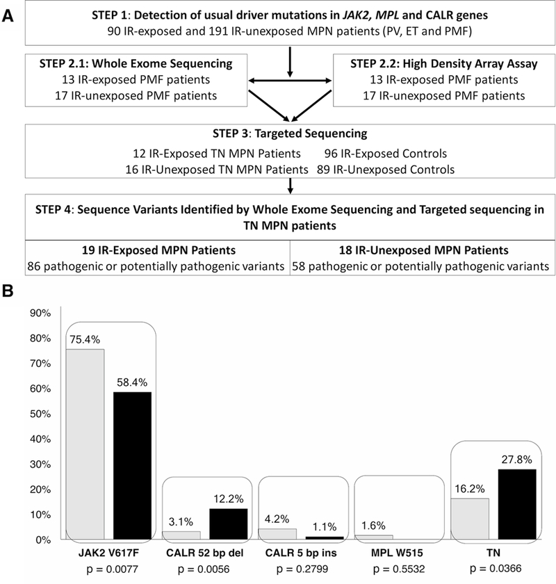
We next carried out a series of studies to identify the spectrum of genomic alterations in IR-exposed versus unexposed MPN patients (Figure 1A). We first tested for the presence of the JAK2 V617F mutation. There were significantly more JAK2 V617F-positive cases (75.4%) among IR-unexposed MPN patients, compared to IR-exposed patients (58.4%) (P = 0.0077) (Figure 1B, Supplementary Material Table 4). This did not appear to reflect a higher prevalence of PV among IR-unexposed MPN patients as there was also a trend toward higher JAK2 mutation frequency in IR-unexposed versus exposed patients with PV (92.9% vs 65%, P = 0.1014). MPN patients who were JAK2 V617F-negative were tested for MPL and CALR gene mutations (Figure 1B, Supplementary Material Table 4). We did not observe a significant difference in the rate of MPL or CALR type 2-like mutations in IR-exposed vs unexposed MPN patients, but the rate of CALR type 1-like mutation was significantly increased in IR-exposed MPN patients compared to unexposed MPN patients (12.2% vs 3.1%, P = 0.0056). This increase in CALR type 1-like mutation rate held true for PMF and ET subtypes: (18.9% vs 4.2% and 33.3% vs 6.5%, P = 0.0279 and P = 0.0298, respectively). Consistent with our prior hypothesis, we observed a higher rate of MPNs that are triple negative for JAK2, MPL and CALR mutations (TN MPN) in the IR-exposed vs unexposed patients (27.8% vs 16.2%, P = 0.0366). The JAK2 V617F and CALR mutation frequencies in unexposed to IR MPN patients in this study were similar to the reported data.9,32–37 The frequency of MPL W515 gene mutation was lower in MPN patients in our study, in comparison to published data.5,38–40.
Figure 1.

(A) Study flow chart. (B) Frequencies of JAK2 V617F, CALR type 1-like (52 bp del) and 2-like (5 bp ins), MPL W515 and negative cases for these mutations (TN) in IR-exposed (black) and IR-unexposed (white) MPN patients. IR: Ionizing Radiation; MPN: Myeloproliferative neoplasms.
Genetic variants identified in IR-exposed and IR-unexposed PMF patients
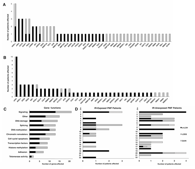
Whole Exome Sequencing was performed on the PBMC DNA from 30 PMF patients (13 IR-exposed and 17 unexposed PMF patients). WES identified previously reported and unreported nonsynonymous nucleotide variants considered as pathogenic or potentially pathogenic based on COSMIC, dbSNP, 1000Genoms, ClinVar, Varsome and UniProt databases. Overall, excluding usual mutations in JAK2, MPL and CALR genes, there were more pathogenic or potentially pathogenic sequence variants identified in IR-exposed than unexposed PMF patients. The mean number of pathogenic or potentially pathogenic nonsynonymous variants detected in IR-exposed PMF patients was 4.8 (range 1–9), while the mean number of nonsynonymous nucleotide variants detected in unexposed PMF patients was 3.1 (1–8), (P = 0.0242). Among recurrently affected genes in IR-exposed PMF patients were ASXL1, U2AF1, PEG3, LAMB4, NF1, JARID2, EZH2, DNMT3A, TET2 and ATM, in addition to JAK2 and CALR (Figure 2, Supplementary Material Table 5, 6). In IR-unexposed patients recurrently affected were genes ASXL1, EZH2, PEG3, RTEL1, SUZ12, RBBP8, BRCA2, FLT3LG and ATM, in addition to JAK2, MPL and CALR. Interestingly, ASXL1 gene was affected more frequently in IR-exposed (five cases, 38.5%) versus unexposed (three cases, 17.6%) PMF patients. However, this difference does not reach statistical significance (P = 0.242) (Figure 2A, B, Supplementary Material Table 5, 6).
Figure 2.

Frequencies of genes with variants considered as pathogenic or potentially pathogenic in (A) IR-exposed and (B) IR-unexposed PMF patients with known driver mutation (black) and in TN patients (white) as identified by whole exome sequencing of PBMC. (C) Pathways potentially affected in IR-exposed (black) and unexposed (white) PMF patients. (D) Copy-number alterations and copy-neutral loss of heterozygosity in 13 PMF patients previously exposed to ionizing radiation and in 17 unexposed PMF patients. IR: Ionizing Radiation; PMF: Primary Myelofibrosis; TN: patients negative for usual driver mutations in JAK2, MPL and CALR genes; Chr: chromosome; cnLOH: copy-nutral loss of heterozygosity; LOSS: copy-number loss; GAIN: copy-number gain.
Among the subset of IR-exposed patients negative for mutations in JAK2, MPL and CALR (triple negative, TN), recurrently affected genes were EZH2, DNMT3A, TET2 and ATM. Genes affected in single cases in this IR-exposed TN subset were KIT, SUZ12, CUX1, KRAS, UMODL1, CBL and SF3B1 (Figure 2A). Overall, the gene functions most frequently affected in these 30 PMF patients are involved in signal transduction, DNA damage, splicing, and epigenetic regulation (Figure 2C).
Copy number alterations and cnLOH in IR-exposed and IR-unexposed PMF patients
Analysis of copy-number alterations and copy-neutral loss of heterozygosity (cnLOH) for 30 PMF patients was assessed by High Density Array Assay in addition to WES. This revealed frequent alterations, but no significant difference in the rates of copy-number loss, copy-number gain, cnLOH, or multiple chromosomal alterations, respectively in the IR-exposed versus unexposed groups (30.8% vs 47.1%, P = 0.4651; 15.4% vs 17.6%, P = 1; 38.5% vs 70.6%, P = 0.1376; and 30.8% vs 47.1%, P = 0.4651) (Figure 2D, Table 1). The most common chromosomal abnormalities were cnLOH at 1p, 9p, 11q and copy-number loss at 7q, 13q. These findings are consistent with published data indicating that the most frequent chromosomal abnormalities in MPNs are LOH at 1p, 9p, 14q, gain of chromosome 8, 9, 14, gain at 1q, and loss at 11q, 13q, 18p, 20q, while loss at 5q, 7p, 7q and LOH at 17q have been observed in single cases.41,42
Table 1.
Copy-number alterations and copy-neutral loss of heterozygosity in PMF patients
| Chr | Patients’s ID | Exposure to IR |
Region altered | Type of alteration |
Size, Mb |
Genes within the regions | Pathogenic or potentially pathogenic variants* |
|---|---|---|---|---|---|---|---|
| 1 | 740 | Exp | 1p36.32p36.22, 1q32.3q41 | cnLOH | 12.8 | RBP7 | |
| 818 | Exp | 1p36.33p34.3 | cnLOH | 34.8 | RAP1GAP, RBP7 | ||
| 615 | Exp | 1q21.1q44 | GAIN | 103.8 | RBBP5, RBM34, TP53BP2 | ||
| 842 | Un | 1p34 | cnLOH | 1 | MPL | MPL c.1543T>A p.W515R | |
| 848 | Un | 1p36.33p33 | cnLOH | 48.2 | CSF3R, MPL, RAP1GAP, RBP7 | MPL c.1543T>A p.W515R | |
| 983 | Un | 1p33p32.3 | cnLOH | 3 | |||
| 702 | Un | 1p36.36p22.1 | cnLOH | 93.6 | CSF3R, JAK1, MPL, RAP1GAP, RBMXL1, RBP7, RPL5 | MPL c.664C>T p.P222S | |
| 2 | 638 | Un | 2p22.2, 2p23.3, 2p25.3, 2q35 | LOSS | 3.3 | DNMT3A, TP53I3 | |
| 702 | Un | 2q22.3q23.3 | LOSS | 3.9 | |||
| 3 | 852 | Un | 3p14.2 | LOSS | 0.3 | ||
| 4 | 740 | Exp | 4q31.23q31.3 | cnLOH | 4.9 | FBXW7 | |
| 1131 | Un | 4p16.3 | LOSS | 1.8 | |||
| 926 | Un | 4q31.3 | LOSS | 0.364 | |||
| 724 | Un | 4q22.3q23 | cnLOH | 3.5 | RAP1GDS1 | ||
| 5 | 724 | Un | 5p13.2q11.2 | cnLOH | 9.7 | ||
| 638 | Un | 5p, 5q multiple alterations1 | LOSS | 134.4 | IRF1, RBM22, NPM1, DDX41 | ||
| 852 | Un | 5p15.2p14.3 | cnLOH | 6.1 | |||
| 6 | 740 | Exp | 6q21q22.31 | cnLOH | 15.2 | ||
| 7 | 740 | Exp | 7q21.3 | cnLOH | 4.3 | ||
| 818 | Exp | 7q22.3q36.2 | LOSS | 47.9 | BRAF, EZH2, LAMB4, POT1 | ||
| 846 | Exp | 7q35q36.2 | LOSS | 6.8 | EZH2 | EZH2 c.1976A>G p.D659G | |
| 638 | Un | 7q11.23, 7q11.23q21.11, 7q21.2q21.3 | GAIN | 12.9 | RBM48 | ||
| 638 | Un | 7q21.11q21.2, 7q21.3q36.3 | LOSS | 71.7 | BRAF, CUX1, EZH2, LAMB4, POT1, RBM33 | ||
| 702 | Un | 7q21.3q31.31 | LOSS | 25.1 | CUX1, EZH2, LAMB4 | ||
| 842 | Un | 7q36.1q36.2 | cnLOH | 3.3 | |||
| 8 | 846 | Exp | 8p23.3q24.3 | GAIN | 146.4 | CSMD1, RAD21-AS1, RBM12B, RUNX1T1, TP53INP1 | |
| 9 | 615 | Exp | 9p24.3p13.3 | cnLOH | 35.9 | FANCG, JAK2 | JAK2 c.1849G>T p.V617F |
| 818 | Exp | 9q32q33.1 | LOSS | 5.5 | |||
| 724 | Un | 9p24.3p13.1, 9q34.2q34.3 | cnLOH | 42.4 | FANCG, JAK2 | JAK2 c.1849G>T p.V617F | |
| 539 | Un | 9p24.3p23 | cnLOH | 13.7 | JAK2 | JAK2 c.1849G>T p.V617F | |
| 702 | Un | 9q21.11q21.13 | cnLOH | 6.5 | |||
| 11 | 740 | Exp | 11p15.5p15.4 | LOSS | 1.7 | ||
| 740 | Exp | 11q13.2q25 | cnLOH | 67.7 | ATM, CBL, | ATM c.5071A>C p.S1691R, CBL c.1258C>G p.R420G | |
| 1131 | Un | 11q12.3q13.2, 11q13.3q25 | cnLOH | 72.1 | ATM, CBL, RBM7, TP53AIP1, RBM14, RBM4B | CBL c.1139T>C p.L380P | |
| 1008 | Un | 11q23.3q24.1 | cnLOH | 6.4 | CBL | ||
| 12 | 615 | Exp | 12p13.33p11.1 | LOSS | 34.7 | AEBP2, GPRC5A, KRAS | AEBP2 c.198_199insG p.G66fs |
| 638 | Un | 12q multiple alterations2 | LOSS | 12.5 | SH2B3, NCOR2 | ||
| 926 | Un | 12q21.2q21.31 | cnLOH | 4.1 | |||
| 13 | 702 | Un | 13q12.3q14.3 | LOSS | 19.8 | BRCA2, RB1 | |
| 638 | Un | 13q14.13q14.3 | LOSS | 4.8 | RB1 | ||
| 15 | 740 | Exp | 15q23q24.2 | cnLOH | 7.7 | ||
| 743 | Un | 15q13.3 | GAIN | 0.433 | |||
| 1014 | Un | 15q24.1q24.2 | LOSS | 1.4 | |||
| 16 | 743 | Un | 16q23.1 | LOSS | 0.174 | ||
| 17 | 818 | Exp | 17p13.3q21.2 | LOSS | 39.6 | PRPF8, RAP1GAP2, SUZ12, TP53 | SUZ12 c.211G>A |
| 638 | Un | 17p13.1p11.2 | cnLOH | 9.1 | |||
| 638 | Un | 17p13.3p13.1, 17q21.31q21.32, 17q21.33 | LOSS | 13.3 | PRPF8, RAP1GAP2, TP53 | TP53 c.527G>A p.C176Y | |
| 638 | Un | 17q23.2q25.3 | GAIN | 22.5 | RBFOX3, SRSF2 | ||
| 18 | 702 | Un | 18q12.2q21.1 | cnLOH | 12.4 | SETBP1 | |
| 19 | 538 | Exp | 19p13.3p12 | cnLOH | 22.9 | CALR, CALR3, ELANE, JAK3, ZSWIM4 | CALR c.1154_1155insTTGTC p.K385fs |
| 740 | Exp | 19q12q13.12 | cnLOH | 3.5 | CEBPA | ||
| 20 | 904 | Un | 20p13 | GAIN | 0.226 | ||
| 842 | Un | 20p13p12.3 | cnLOH | 7 | RAD21L1, RBCK1 | ||
| 702 | Un | 20q11.21q13.13 | LOSS | 18.8 | ASXL1, RBL1, RBM12, RBM39, RBPJL, TP53INP2, TP53RK | ||
| 743 | Un | 20q13.13q13.33 | cnLOH | 15.7 | CBLN4, CTCFL, DIDO1, GNAS, RTEL1, TP53RK | ||
| 21 | 724 | Un | 21q11.2 | LOSS | 0.335 | ||
| 22 | 703 | Exp | 22q12.1q12.3 | cnLOH | 4.4 |
PMF: Primary Myelofibrosis; Chr: Chromosome; ID: Identification; IR: Ionizing Radiation; Exp: Exposed to IR PMF patients; Un: Unexposed to IR PMF patients; cnLOH: copy-neutral loss of heterozygosity; GAIN: copy-number gain; LOSS: copy-number loss; Mb: megabase.
Pathogenic or potentially pathogenic variants identified by Whole Exome Sequencing.
5p15.32p15.31, 5q11.1q11.2, 5q11.2, 5q11.2q12.1, 5q12.1q12.3, 5q12.3, 5q12.3q21.3, 5q21.3q22.1, 5q22.2q23.1, 5q23.1q31.3, 5q31.3, 5q31.3q33.1, 5q33.2q33.3, 5q33.3q34, 5q34, 5q34q35.1, 5q35.1q35.3, 5q35.3, 5p14.2p14.1, 5p14.3
12q12q13.11, 12q13.11q13.12, 12q13.3q14.1, 12q21.2, 12q22, 12q24.11q24.12, 12q24.23q24.31, 12q24.33
The cnLOH we found at 1p in IR-unexposed PMF patients duplicated the pathogenic MPL W515R mutation in two out of four cases and also a novel MPL P222S variant in one case. In contrast, the 1p alterations did not involve the MPL gene in IR-exposed cases. Copy-number LOH at 19p duplicated the pathogenic CALR K385fs mutation in one out of two cases in IR-exposed PMF patients and did not affect chromosome 19 in IR-unexposed PMF patients. This supports our results suggesting that CALR gene alterations contribute more and MPL gene alterations less to the disease development in IR-exposed MPN patients.
Copy-number losses of EZH2 at 7q (ID 846) and SUZ12 at 17q (ID 818), both components of Polycomb Repressive Complex 2 (PRC2), were identified in conjunction with nonsynonymous missense variants in IR-exposed PMF patients who were negative for usual driver mutations in the JAK2, MPL and CALR genes. This may suggest the contribution of impaired PRC2 (by loss-of-function alterations) to MPN development. Copy-neutral LOH involving ATM gene at 11q with identified nonsynonymous missense variant in another TN IR-exposed patient (ID 740) suggests homozygous loss of ATM function and subsequent DNA damage repair defects. In the same patient (ID 740) cnLOH at 11q duplicated an additional missense mutation CBL R420G (89% allele frequency) with enhancing cell survival capacity, but without confirmed influence on cell proliferation.43 Finally, copy-number loss of TP53 at 17p was identified in one JAK2-positive (24% allele frequency) unexposed PMF patient in conjunction with a likely pathogenic missense variant p.C176Y (c.527G>A) (23% allele frequency), which suggests its contribution to the disease evolution due to loss of DNA repair function.
Targeted sequencing of candidate driver genes in TN MPN patients
Based on the Whole Exome Sequencing and High Density Array Assay data we identified 309 genes that were altered in at least one case and may be pathogenic. To identify novel sequence variants that may drive MPN development in TN patients, we performed targeted sequencing of the 309 genes on an additional cohort of IR-exposed (12) and unexposed (16) TN MPN patients’ DNA. In parallel we similarly examined 89 IR-unexposed healthy controls to identify germline SNPs or sequence variants that may be unique to the Ukrainian population. We also examined 96 IR-exposed healthy controls to further identify novel SNPs or sequence variants and potentially find evidence of subclinical MPN (Figure 1A).
Overall, there were 133 nonsynonymous sequence variants (Supplementary Material Table 7–9) in the 28 TN MPN patients with a range of 1 – 11 sequence variants per sample. It should be noted that despite filtering sequence variants, many variants had allele frequencies around 50% that could be clonal mutations, but we are not able to exclude that these are rare germline sequence variants.
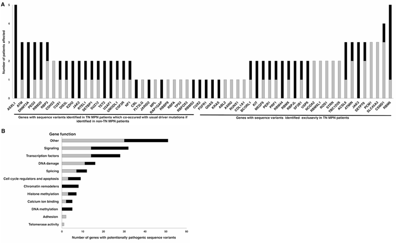
IR-exposed and unexposed TN MPN patients did not differ regarding mean number of sequence variants per patient, 4.5 (range 1–9) and 4.9 (1–11), respectively (P = 0.3208). In contrast, TN IR-unexposed patients displayed a higher mean number of sequence variants per patient (4.9, range 1–11) versus non-TN IR-exposed patients (3, range 1–8) (P = 0.0112), suggesting a greater role for DNA damage in the group of IR-unexposed TN MPN patients. Overall, combining the targeted sequencing data from the IR-exposed and unexposed patients, genes affected in TN MPN patients were involved in signal transduction, transcriptional regulation, DNA damage and splicing (Figure 3B).
Figure 3.

(A) Frequencies of genes with sequence variants identified by whole exome sequencing and targeted sequencing in IR-exposed (black) and unexposed (white) MPN patients negative for usual driver mutations in JAK2, MPL and CALR genes. (B) Pathways potentially affected in 12 IR-exposed (black) and 16 unexposed (white) MPN patients negative for usual driver mutations in JAK2, MPL and CALR genes. IR: Ionizing Radiation; MPN: myeloproliferative neoplasms.
Recurrently affected genes in TN IR-exposed MPN patients
We next combined WES and Targeted Sequencing data to focus on TN cases (negative for usual mutations in JAK2, MPL and CALR genes) (Figure 3A). First, we asked whether there are recurrently affected genes that are specific to TN IR-exposed MPN patients. Several potentially pathogenic sequence variants were detected exclusively in TN IR-exposed MPN patients in the following genes: KIT, JAK3, USP6, RBPJL and RBM44 (Figure 3A). One PMF patient (ID 547) harbored KIT D816V missense mutation with 16% allele frequency reported in patients with systemic mastocytosis.44
Recurrently affected genes in TN IR-exposed and IR-unexposed MPN patients
We next identified recurrently affected genes that were exclusive to TN cases (IR-exposed or unexposed), which included SF3B1, ATMIN, BCORL1, CSMD1, SEPT9, RBM6 and MEGF6. One of the identified SF3B1 variants was a missense mutation K700E with 32% allele frequency in an unexposed TN PMF patient (ID 1007). K700E, the most frequently reported mutation in the SF3B1 gene, has been identified in chronic lymphocytic leukemia, myelodysplastic syndrome, breast and pancreatic cancers.45–48
Recurrently affected genes in TN IR-exposed and IR-unexposed MPN patients also identified in non-TN cases
Several recurrently affected genes in TN MPN patients (IR-exposed or unexposed) were also involved in MPN patients with usual driver mutations in JAK2, MPL or CALR genes. Among them were ASXL1, ATM, DNMT3A, EZH2, SUZ12, TET2, RTEL1, SETBP1, U2AF1, CSF3R and UMOD1 (Figure 3A). Although mutations of ASXL1 and DNMT3A were found in cases with usual driver mutations, they were more frequent in TN MPN patients, and only in the IR-exposed TN MPN patients. ASXL1 variants were found in five (29.4%) IR-exposed TN cases with allele frequencies varying from 27 to 56%, and DNMT3A variants were found in three (17.6%) IR-exposed TN cases with allele frequencies varying from 26 to 48%.
Two sequence variants of the recurrently affected CSF3R gene were identified in IR-exposed (ID 1360) and unexposed (ID 879) TN ET patients. CSF3R T618I (observed in ID 1360 with 37% allele frequency) is a highly prevalent and well-studied specific mutation in chronic neutrophilic leukemia.49,50 The other patient (ID 879) had a previously undescribed missense variant CSF3R G415R (46% allele frequency) and in addition TP53 V31I variant (45% allele frequency) which has been previously identified in patients with acute myeloid leukemia.51
Finally, non-canonical mutations in JAK2 gene were identified in two TN cases. JAK2 R938Q missense variant with 45% allele frequency was detected in a IR-exposed TN PMF patient (ID 1283) The JAK2 R938Q somatic mutation was reported previously in a hereditary thrombocythemia case and B-cell acute lymphoblastic leukemia case.52,53 An IR-exposed PV patient (ID 1887) exhibited a frameshift variant JAK2 K539fs of uncertain biological significance with 23% allele frequency.
We also identified potentially pathogenic variants in a healthy 68 years old female who was exposed to IR (documented dose 50mSv). These were missense U2AF1 p.Q157P (c.470A>C) with 23% allele frequency and nonsense DNMT3A p.C368X (c.C1104A) mutations with 21% allele frequency. U2AF1 p.Q157P (c.470A>C) was previously reported in patients with MDS54,55 and PMF56. Interestingly, a daughter of this woman was diagnosed with acute leukemia. We did not identify any potential pathogenic variants in healthy IR-unexposed controls.
Discussion
In this study, we evaluated the effect of prior exposure to IR during the Chernobyl nuclear accident on the clinical characteristics and genomic profiles of MPN patients in Ukraine. IR-exposed MPN patients exhibited a higher mean age and were predominantly men, which likely reflects the time elapsed from the Chernobyl disaster and the male sex-bias of cleanup workers. The clinical profiles of the IR-exposed and unexposed patients were similar, although the rate of transfusion-dependence was slightly higher among IR-exposed MPN patients. We found that IR-exposed MPN patients exhibited a different genetic profile from that of unexposed MPN patients: a lower rate of JAK2 V617F mutation, a higher rate of type 1-like CALR mutation, a higher rate of TN cases, and a higher rate of potentially pathogenic sequence variants. No difference in the rates of chromosomal alterations in the IR-exposed and unexposed groups may be related to the fact that some of the patients who developed chromosomal alterations immediately following IR exposure >30 years ago developed leukemia and did not survive to be included in this cohort.
Using Next Generation Sequencing we identified a spectrum of affected genes with sequence variants in both IR-exposed and unexposed MPN patients. TN IR-exposed and unexposed MPN patients did not differ regarding median numbers of sequence variants per patient and exhibited mutations in several of the same genes. This may suggest a common molecular basis of TN MPNs in Ukrainian IR-exposed patients with a documented history of irradiation and unexposed MPN patients without such history, but for whom we could not exclude the possibility of exposure to IR due to external (visiting contaminated with radionuclides territories, travelling by flight or undergoing specific medical examinations) or internal irradiation (contaminated food, using radioactive substances for medical purposes).
Nevertheless, in IR-exposed TN MPN patients, in comparison to unexposed TN MPN cases and healthy controls, we identified several nonsynonymous sequence variants in conjunction with copy number alteration of the corresponding chromosomal regions that we propose as potential drivers of MPN development. Among these genetic variants, ATM p.S1691R (c.5071 A>C) variant (91% allele frequency) with cnLOH at 11q22.3 was identified in one TN IR-exposed PMF patient (ID 740) diagnosed 13 years after IR-exposure at the age of 47. Patient’s DNA was collected at the age of 58 when he presented with anemia (HGB 88 g/l), thrombocytopenia (80 × 109/l), leukocytosis (18.7 × 10⁹/l), increased level of LDH (589 U/l), palpable splenomegaly (218 mm by ultrasound) and hepatomegaly (191 mm by ultrasound) and was erythrocyte-transfusion dependent. Previously the same ATM S1691R variant was reported in an Ataxia-Telangiectasia family57 and in patients with chronic lymphocytic leukemia, including one case with LOH at D11S217958,59, breast cancer60–62, and melanoma63, but not in MPN patients.
Missense mutations in EZH2 at 7q36.1 and SUZ12 at 17q11.2 with copy number loss were also identified separately in TN IR-exposed PMF patients (ID 846 and 818, respectively). Loss-of-function mutations and cytogenetic alterations of EZH2 and SUZ12 impair function of PRC2 and are frequently reported in MPNs/MDS.64–66 Missense mutation EZH2 p.D659G (c.1976 A>G) at 7q36.1 with copy number loss (66% allele frequency) was identified in a 69 years old IR-exposed TN PMF (ID 846) patient. The patient was diagnosed 26 years after IR-exposure and presented with mild anemia (HGB 125 g/l), leukocytosis (14.8 × 10⁹/l), increased level of LDH (907 U/l), palpable splenomegaly (171 mm by ultrasound) and later became RBC-transfusion dependent. Frequently described in MPNs, loss-of-function mutations of EZH2 gene (~10%) are mostly early events in leukemogenesis and associated with a poor prognosis and MPN phenotype modifications.65,67
Missense mutation SUZ12 p.V71M (c.211G>A) at 17q11.2 with copy number loss and 50% allele frequency was identified in a 58 years old TN PMF patient 25 years after IR-exposure (ID 818). The patient presented with mild anemia (HGB 125 g/l), leukopenia (1.9 × 10⁹/l), palpable splenomegaly (249 mm by ultrasound) and hepatomegaly (187 mm by ultrasound). SUZ12 gene mutations in MPNs are relatively rare (1.4%66; 1.6%68), although in the range of MPL gene mutation frequencies.
This study provides the first extensive chromosomal and genomic characterization of patients who developed MPN’s following exposure to IR and the Chernobyl nuclear disaster in particular. The limitations of this study include the retrospective nature and the relatively small number of samples evaluated by WES and HD Array Assay, which limited the statistical power to discern the effect of IR on the incidence of specific subgroups of MPN, specific nonsynonymous nucleotide variants, or specific copy number alterations. Another limitation was that the only DNA obtained was from PBMC’s, which likely included both somatic and germline contributions, as a germline source of DNA was not available. To overcome this limitation, we performed targeted sequencing of DNA from healthy Ukrainian controls, to filter out germline nonsynonymous variants that occur frequently in the general Ukrainian population. Supporting the argument that the nonsynonymous sequence variants in potential MPN driver genes that we identified in MPN patients were somatic, rather than germline, none of these sequence variants were identified in unexposed healthy controls. Despite these limitations, we have identified a wide spectrum of nonsynonymous sequence variants among IR-exposed and unexposed MPN patients. We found that while TN IR-exposed and unexposed MPN patients often exhibited similar DNA alterations suggesting a common molecular basis for MPN development, there were unusual genetic variants specific to some of the IR-exposed TN MPN patients and that TN MPN is more common among IR-exposed MPN patients. These findings indicate that IR exposure contributes particularly to the development of TN MPN, which in turn is driven by a diversity of oncogenic genomic alterations.
Supplementary Material
Acknowledgements.
We would like to thank Takuto Sato, Broad Institute, for performing the WES data analyses.
Funding source.
This research was funded by DOD Peer Reviewed Cancer Research Program, Project #CA150529 (PGF and SB). Grant W9111NF-09–001 from the Institute for Collaborative Biotechnologies of the Army Research Office (EF).
REFERENCES:
- 1.Mishcheniuk OY, Kostukevich OM, Dmytrenko I V., et al. Molecular characterization of ph-negative myeloproliferative neoplasms in ukraine. Exp Oncol. 2013. [PubMed] [Google Scholar]
- 2.Klymenko SV, Smida J, Atkinson MJ, Bebeshko VG, Nathrath M, Rosemann M. Allelic imbalances in radiation-associated acute myeloid leukemia. Genes (Basel). 2011. doi: 10.3390/genes2020384 [DOI] [PMC free article] [PubMed] [Google Scholar]
- 3.Murati A, Brecqueville M, Devillier R, Mozziconacci MJ, Gelsi-Boyer V, Birnbaum D. Myeloid malignancies: Mutations, models and management. BMC Cancer. 2012. doi: 10.1186/1471-2407-12-304 [DOI] [PMC free article] [PubMed] [Google Scholar]
- 4.Mead AJ, Mullally A. Myeloproliferative neoplasm stem cells. Blood. 2017. doi: 10.1182/blood-2016-10-696005 [DOI] [PMC free article] [PubMed] [Google Scholar]
- 5.Milosevic Feenstra JD, Nivarthi H, Gisslinger H, et al. Whole-exome sequencing identifies novel MPL and JAK2 mutations in triple-negative myeloproliferative neoplasms. Blood. 2016. doi: 10.1182/blood-2015-07-661835 [DOI] [PMC free article] [PubMed] [Google Scholar]
- 6.Lim K-H, Lin H-C, Chen CG-S, et al. Rapid and sensitive detection of CALR exon 9 mutations using high-resolution melting analysis. Clin Chim Acta. 2015. doi: 10.1016/j.cca.2014.11.011 [DOI] [PubMed] [Google Scholar]
- 7.Tefferi A, Thiele J, Vardiman JW. The 2008 World Health Organization classification system for myeloproliferative neoplasms: order out of chaos. Cancer. 2009. doi: 10.1002/cncr.24440 [DOI] [PubMed] [Google Scholar]
- 8.Constantinescu SN, Leroy E, Gryshkova V, Pecquet C, Dusa A. Activating Janus kinase pseudokinase domain mutations in myeloproliferative and other blood cancers. Biochem Soc Trans. 2013. doi: 10.1042/BST20130084 [DOI] [PubMed] [Google Scholar]
- 9.Kong H, Liu Y, Luo S, Li Q, Wang Q. Frequency of Calreticulin (CALR) Mutation and Its Clinical Prognostic Significance in Essential Thrombocythemia and Primary Myelofibrosis: A Meta-analysis. Intern Med. 2016. doi: 10.2169/internalmedicine.55.6214 [DOI] [PubMed] [Google Scholar]
- 10.Guglielmelli P, Pacilli A, Rotunno G, et al. Presentation and outcome of patients with 2016 WHO diagnosis of prefibrotic and overt primary myelofibrosis. Blood. 2017. doi: 10.1182/blood-2017-01-761999 [DOI] [PubMed] [Google Scholar]
- 11.Rumi E, Cazzola M. Diagnosis, risk stratification, and response evaluation in classical myeloproliferative neoplasms. Blood. 2017. doi: 10.1182/blood-2016-10-695957 [DOI] [PMC free article] [PubMed] [Google Scholar]
- 12.Martínez-Avilés L, Besses C, Álvarez-Larrán A, Torres E, Serrano S, Bellosillo B. TET2, ASXL1, IDH1, IDH2, and c-CBL genes in JAK2- and MPL-negative myeloproliferative neoplasms. Ann Hematol. 2012. doi: 10.1007/s00277-011-1330-0 [DOI] [PubMed] [Google Scholar]
- 13.McCabe MT, Ott HM, Ganji G, et al. EZH2 inhibition as a therapeutic strategy for lymphoma with EZH2-activating mutations. Nature. 2012. doi: 10.1038/nature11606 [DOI] [PubMed] [Google Scholar]
- 14.Bartels S, Lehmann U, Büsche G, et al. SRSF2 and U2AF1 mutations in primary myelofibrosis are associated with JAK2 and MPL but not calreticulin mutation and may independently reoccur after allogeneic stem cell transplantation. Leukemia. 2015. doi: 10.1038/leu.2014.277 [DOI] [PMC free article] [PubMed] [Google Scholar]
- 15.Lundberg P, Karow A, Nienhold R, et al. Clonal evolution and clinical correlates of somatic mutations in myeloproliferative neoplasms. Blood. 2014. doi: 10.1182/blood-2013-11-537167 [DOI] [PubMed] [Google Scholar]
- 16.Yogarajah M, Tefferi A. Leukemic Transformation in Myeloproliferative Neoplasms: A Literature Review on Risk, Characteristics, and Outcome. Mayo Clin Proc. 2017. doi: 10.1016/j.mayocp.2017.05.010 [DOI] [PubMed] [Google Scholar]
- 17.Shao L, Luo Y, Zhou D. Hematopoietic Stem Cell Injury Induced by Ionizing Radiation. Antioxid Redox Signal. 2014. doi: 10.1089/ars.2013.5635 [DOI] [PMC free article] [PubMed] [Google Scholar]
- 18.Fajardo L-GLF, Berthrong M, Anderson RE. Radiation Pathology. Oxford University Press, Inc.; 2001. [Google Scholar]
- 19.Klymenko S, Trott K, Atkinson M, et al. AML1 gene rearrangements and mutations in radiation-associated acute myeloid leukemia and myelodysplastic syndromes. J Radiat Res. 2005. doi: 10.1269/jrr.46.249 [DOI] [PubMed] [Google Scholar]
- 20.Likhtarev A, Kovgan L, Ivanova O, Masiuk S, Chepurny M, Boiko Z. Integrated dosimetric passportization of settlements of Ukraine and reconstruction of individualized doses of the Ukrainian State Register of persons affected by Chernobyl accident. J NAMS Ukr. 2016;22(2):208–21. [Google Scholar]
- 21.Furtado LV, Weigelin HC Elenitoba-Johnson KSJ, Betz BL. Detection of MPL mutations by a novel allele-specific PCR-based strategy. J Mol Diagnostics. 2013. doi: 10.1016/j.jmoldx.2013.07.006 [DOI] [PubMed] [Google Scholar]
- 22.Bolger AM, Lohse M, Usadel B. Trimmomatic: A flexible trimmer for Illumina sequence data. Bioinformatics. 2014. doi: 10.1093/bioinformatics/btu170 [DOI] [PMC free article] [PubMed] [Google Scholar]
- 23.Andrews S FastQC: A quality control tool for high throughput sequence data. Http://Www.Bioinformatics.Babraham.Ac.Uk/Projects/Fastqc/. doi:citeulike-article-id:11583827 [Google Scholar]
- 24.Li H, Durbin R. Fast and accurate short read alignment with Burrows-Wheeler transform. Bioinformatics. 2009. doi: 10.1093/bioinformatics/btp324 [DOI] [PMC free article] [PubMed] [Google Scholar]
- 25.McKenna A, Hanna M, Banks E, et al. The Genome Analysis Toolkit: a MapReduce framework for analyzing next-generation DNA sequencing data. Genome Res. 2010. doi: 10.1101/gr.107524.110 [DOI] [PMC free article] [PubMed] [Google Scholar]
- 26.Wang K, Li M, Hakonarson H. ANNOVAR: Functional annotation of genetic variants from high-throughput sequencing data. Nucleic Acids Res. 2010. doi: 10.1093/nar/gkq603 [DOI] [PMC free article] [PubMed] [Google Scholar]
- 27.Barbui T, Thiele J, Gisslinger H, Finazzi G, Vannucchi AM, Tefferi A. The 2016 revision of WHO classification of myeloproliferative neoplasms: Clinical and molecular advances. Blood Rev. 2016. doi: 10.1016/j.blre.2016.06.001 [DOI] [PubMed] [Google Scholar]
- 28.Casini A, Fontana P, Lecompte TP. Thrombotic complications of myeloproliferative neoplasms: Risk assessment and risk-guided management. J Thromb Haemost. 2013. doi: 10.1111/jth.12265 [DOI] [PubMed] [Google Scholar]
- 29.Mitra D, Kaye JA, Piecoro LT, et al. Symptom burden and splenomegaly in patients with myelofibrosis in the United States: A retrospective medical record review. Cancer Med. 2013. doi: 10.1002/cam4.136 [DOI] [PMC free article] [PubMed] [Google Scholar]
- 30.Andriani A, Latagliata R, Anaclerico B, et al. Spleen enlargement is a risk factor for thrombosis in essential thrombocythemia: Evaluation on 1,297 patients. Am J Hematol. 2016. doi: 10.1002/ajh.24269 [DOI] [PubMed] [Google Scholar]
- 31.Tefferi A, Rumi E, Finazzi G, et al. Survival and prognosis among 1545 patients with contemporary polycythemia vera: An international study. Leukemia. 2013. doi: 10.1038/leu.2013.163 [DOI] [PMC free article] [PubMed] [Google Scholar]
- 32.Baxter EJ, Scott LM, Campbell PJ, et al. Acquired mutation of the tyrosine kinase JAK2 in human myeloproliferative disorders. Lancet. 2005. doi: 10.1016/S0140-6736(05)74230-6 [DOI] [PubMed] [Google Scholar]
- 33.Levine RL, Wadleigh M, Cools J, et al. Activating mutation in the tyrosine kinase JAK2 in polycythemia vera, essential thrombocythemia, and myeloid metaplasia with myelofibrosis. Cancer Cell. 2005. doi: 10.1016/j.ccr.2005.03.023 [DOI] [PubMed] [Google Scholar]
- 34.Iványi J, Marton E, Plander M. Significance of the JAK2V617F mutation in patients with chronic myeloproliferative neoplasia. Orv Hetil. 2011;152(45):1975–803. [DOI] [PubMed] [Google Scholar]
- 35.Duletić AN, Dekanić A, Hadzisejdić I, et al. JAK2-v617F mutation is associated with clinical and laboratory features of myeloproliferative neoplasms. Coll Antropol. 2012;36:859–65. [PubMed] [Google Scholar]
- 36.Takata Y, Seki R, Kanajii T, et al. Association between thromboembolic events and the JAK2 V617F mutation in myeloproliferative neoplasms. Kurume Med J. 2014. doi: 10.2739/kurumemedj.MS63001 [DOI] [PubMed] [Google Scholar]
- 37.Azevedo AP, Silva S, Reichert A, Lima F, J�nior E, Rueff J. Prevalence of the Janus kinase 2 V617F mutation in Philadelphia‑negative myeloproliferative neoplasms in a Portuguese population. Biomed Reports. 2017. doi: 10.3892/br.2017.977 [DOI] [PMC free article] [PubMed] [Google Scholar]
- 38.Ghotaslou A, Nadali F, Chahardouli B, et al. Low frequency of c-MPL gene mutations in Iranian patients with Philadelphia-negative myeloproliferative disorders. Iran J Ped Hematol Oncol. 2015;5(1):43–49. [PMC free article] [PubMed] [Google Scholar]
- 39.Shirane S, Araki M, Morishita S, et al. JAK2, CALR, and MPL mutation spectrum in japanese patients with myeloproliferative neoplasms. Haematologica. 2015. doi: 10.3324/haematol.2014.115113 [DOI] [PMC free article] [PubMed] [Google Scholar]
- 40.Chaligné R, Tonetti C, Besancenot R, et al. New mutations of MPL in primitive myelofibrosis: Only the MPL W515 mutations promote a G1/S-phase transition. Leukemia. 2008. doi: 10.1038/leu.2008.137 [DOI] [PubMed] [Google Scholar]
- 41.Rice KL, Lin X, Wolniak K, et al. Analysis of genomic aberrations and gene expression profiling identifies novel lesions and pathways in myeloproliferative neoplasms. Blood Cancer J. 2011. doi: 10.1038/bcj.2011.39 [DOI] [PMC free article] [PubMed] [Google Scholar]
- 42.Klampfl T, Harutyunyan A, Berg T, et al. Genome integrity of myeloproliferative neoplasms in chronic phase and during disease progression. Blood. 2011. doi: 10.1182/blood-2011-01-331678 [DOI] [PubMed] [Google Scholar]
- 43.Sargin B, Choudhary C, Crosetto N, et al. Flt3-dependent transformation by inactivating c-Cbl mutations in AML. Blood. 2007. doi: 10.1182/blood-2007-01-066076 [DOI] [PubMed] [Google Scholar]
- 44.Jara-Acevedo M, Teodosio C, Sanchez-Muñoz L, et al. Detection of the KIT D816V mutation in peripheral blood of systemic mastocytosis: Diagnostic implications. Mod Pathol. 2015. doi: 10.1038/modpathol.2015.72 [DOI] [PubMed] [Google Scholar]
- 45.Wan Y, Wu CJ. SF3B1 mutations in chronic lymphocytic leukemia. Blood. 2013. doi: 10.1182/blood-2013-02-427641 [DOI] [PMC free article] [PubMed] [Google Scholar]
- 46.Graubert TA, Shen D, Ding L, et al. Recurrent mutations in the U2AF1 splicing factor in myelodysplastic syndromes. Nat Genet. 2012. doi: 10.1038/ng.1031 [DOI] [PMC free article] [PubMed] [Google Scholar]
- 47.Cancer Genome Atlas Network. Comprehensive molecular portraits of human breast tumours. Nature. 2012. doi: 10.1038/nature11412 [DOI] [PMC free article] [PubMed] [Google Scholar]
- 48.Biankin AV, Waddell N, Kassahn KS, et al. Pancreatic cancer genomes reveal aberrations in axon guidance pathway genes. Nature. 2012. doi: 10.1038/nature11547 [DOI] [PMC free article] [PubMed] [Google Scholar]
- 49.Fleischman AG, Maxson JE, Luty SB, et al. The CSF3R T618I mutation causes a lethal neutrophilic neoplasia in Mice that is responsive to therapeutic JAK inhibition. Blood. 2013. doi: 10.1182/blood-2013-06-509976 [DOI] [PMC free article] [PubMed] [Google Scholar]
- 50.Pardanani A, Lasho TL, Laborde RR, et al. CSF3R T618I is a highly prevalent and specific mutation in chronic neutrophilic leukemia. Leukemia. 2013. doi: 10.1038/leu.2013.122 [DOI] [PMC free article] [PubMed] [Google Scholar]
- 51.Hou H-A, Chou W-C, Kuo Y-Y, et al. TP53 mutations in de novo acute myeloid leukemia patients: longitudinal follow-ups show the mutation is stable during disease evolution. Blood Cancer J. 2015. doi: 10.1038/bcj.2015.59 [DOI] [PMC free article] [PubMed] [Google Scholar]
- 52.Sadras T, Heatley SL, Kok CH, et al. A novel somatic JAK2 kinase-domain mutation in pediatric acute lymphoblastic leukemia with rapid on-treatment development of LOH. Cancer Genet. 2017. doi: 10.1016/j.cancergen.2017.07.008 [DOI] [PubMed] [Google Scholar]
- 53.Langabeer SE. JAK2 mutations to the fore in hereditary thrombocythemia. JAKSTAT. 2014;3(3):e957618. [DOI] [PMC free article] [PubMed] [Google Scholar]
- 54.Li B, Liu J, Jia Y, et al. Clinical features and biological implications of different U2AF1 mutation types in myelodysplastic syndromes. Genes Chromosom Cancer. 2018. doi: 10.1002/gcc.22510 [DOI] [PMC free article] [PubMed] [Google Scholar]
- 55.Wu S-J, Tang J-L, Lin C-T, et al. Clinical implications of U2AF1 mutation in patients with myelodysplastic syndrome and its stability during disease progression. Am J Hematol. 2013. doi: 10.1002/ajh.23541 [DOI] [PubMed] [Google Scholar]
- 56.Tefferi A, Barraco D, Lasho TL, et al. U2AF1 mutation variants and their phenotypic and prognostic relevance in primary myelofibrosis. Blood. 2016;128:42–48. [Google Scholar]
- 57.Stankovic T, Kidd AM, Sutcliffe A, et al. ATM mutations and phenotypes in ataxia-telangiectasia families in the British Isles: expression of mutant ATM and the risk of leukemia, lymphoma, and breast cancer. Am J Hum Genet. 1998. doi: 10.1086/301706 [DOI] [PMC free article] [PubMed] [Google Scholar]
- 58.Nadeu F, Delgado J, Royo C, et al. Clinical impact of clonal and subclonal TP53, SF3B1, BIRC3, NOTCH1, and ATM mutations in chronic lymphocytic leukemia. Blood. 2016. doi: 10.1182/blood-2015-07-659144 [DOI] [PMC free article] [PubMed] [Google Scholar]
- 59.Bullrich F, Rasio D, Kitada S, et al. ATM mutations in B-cell chronic lymphocytic leukemia. Cancer Res. 1999. [PubMed] [Google Scholar]
- 60.Stredrick DL, Garcia-Closas M, Pineda MA, et al. The ATM missense mutation p.Ser49Cys (c.146C>G) and the risk of breast cancer. Hum Mutat. 2006. doi: 10.1002/humu.20323 [DOI] [PMC free article] [PubMed] [Google Scholar]
- 61.Teraoka SN, Malone KE, Doody DR, et al. Increased frequency of ATM mutations in breast carcinoma patients with early onset disease and positive family history. Cancer. 2001. doi: [pii] [DOI] [PubMed] [Google Scholar]
- 62.Broeks A, Braaf LM, Huseinovic A, et al. The spectrum of ATM missense variants and their contribution to contralateral breast cancer. Breast Cancer Res Treat. 2008. doi: 10.1007/s10549-007-9543-6 [DOI] [PMC free article] [PubMed] [Google Scholar]
- 63.Hamblin A, Wordsworth S, Fermont JM, et al. Clinical applicability and cost of a 46-gene panel for genomic analysis of solid tumours: Retrospective validation and prospective audit in the UK National Health Service. PLoS Med. 2017. doi: 10.1371/journal.pmed.1002230 [DOI] [PMC free article] [PubMed] [Google Scholar]
- 64.Vainchenker W, Isabelle P. EZH2: A molecular switch of the MPN phenotype. Blood. 2016. doi: 10.1182/blood-2016-04-711770 [DOI] [PubMed] [Google Scholar]
- 65.Sashida G, Wang C, Tomioka T, et al. The loss of Ezh2 drives the pathogenesis of myelofibrosis and sensitizes tumor-initiating cells to bromodomain inhibition. J Exp Med. 2016. doi: 10.1084/jem.20151121 [DOI] [PMC free article] [PubMed] [Google Scholar]
- 66.Score J, Hidalgo-Curtis C, Jones AV., et al. Inactivation of polycomb repressive complex 2 components in myeloproliferative and myelodysplastic/myeloproliferative neoplasms. Blood. 2012. doi: 10.1182/blood-2011-07-367243 [DOI] [PubMed] [Google Scholar]
- 67.Rinke J, Müller JP, Blaess MF, et al. Molecular characterization of EZH2 mutant patients with myelodysplastic/myeloproliferative neoplasms. Leukemia. 2017. doi: 10.1038/leu.2017.190 [DOI] [PubMed] [Google Scholar]
- 68.Brecqueville M, Rey J, Bertucci F, et al. Mutation analysis of ASXL1, CBL, DNMT3A, IDH1, IDH2, JAK2, MPL, NF1, SF3B1, SUZ12, and TET2 in myeloproliferative neoplasms. Genes Chromosom Cancer. 2012. doi: 10.1002/gcc.21960 [DOI] [PubMed] [Google Scholar]
Associated Data
This section collects any data citations, data availability statements, or supplementary materials included in this article.


