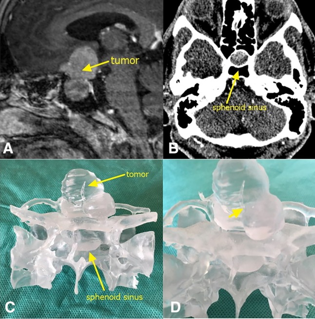
Fig. 2.

a MRI of the sella turcica region showing tumor lesions. The yellow arrow points to the tumor. b CT of the sphenoidal sinus. The yellow arrow points to the sphenoidal sinus. c Frontal view of the 3D-printed tumor model. d Enlarged image of a part of the tumor. The yellow arrow shows that the tumor wraps around the left side of the anterior communicating artery
