Abstract
We present a case of redo atrial fibrillation (AF) ablation for pulmonary vein reconnection. In mapping of posterior wall of left upper pulmonary vein (LUPV) with HD grid, a new and unique multipolar mapping catheter, it demonstrated presence of local electrocardiogram signals (EGM). But during mapping with the Tacticath ablation catheter, these signals were not visible. Nevertheless, ablation at this point resulted in isolation of LUPV. The unique mapping technology offered by HD grid mapping catheter may enable us to discover local EGM not otherwise visible in conventional bipole parallel recordings, to have more accurate maps and deliver for effective therapies.
Keywords: AF ablation, HD grid, high density non contact mapping, pulmonary vein isolation
1. INTRODUCTION
We present a 58‐year‐old lady who underwent a repeat atrial fibrillation (AF) ablation for persistent AF. She had a previous pulmonary vein isolation and roof line linear ablation for her index AF ablation procedure 1 year ago.
2. CASE REPORT
For the current AF ablation procedure, we used Advisor HD Grid mapping catheter which has 16 electrodes, 3 mm equidistant electrode spacing (Abbott, St Paul, MN, USA) to create a high density contact voltage map via the Ensite Velocity 3D mapping system (Abbott). The ablation catheter was the 3.5 mm irrigated tip Tacticath ablation catheter (Abbott) with 2‐2‐2mm interelectrode distance.
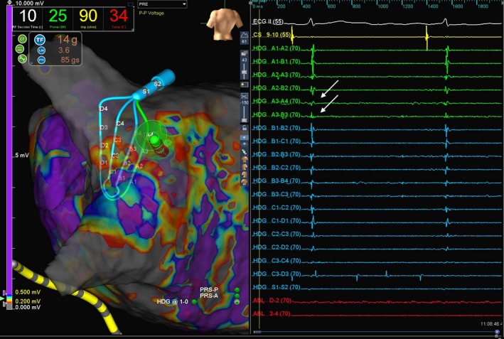
During mapping of the posterior wall of the left upper pulmonary vein (LUPV), we discovered a localized region of reconnection illustrated in arrows on Figure 1. As we were about to use ablation catheter for ablation, we noticed that the local EGM of reconnection was not visible on the ablation bipole (corresponding arrows on ablation bipoles on Figure 1). Signal amplitude/magnification for both HD Grid and ablation catheter were set at 60. Bipolar high pass filter 30 Hz, low pass 300 Hz, noise filter switched on. Unipolar high pass filter 0.5 Hz, low pass 100 Hz, noise filter switched on. Ablation catheter contact was 8‐10 g of force.
Figure 1.

Top panel shows geometry and voltage map of left atrium in posterior anterior view. The HD Grid multipolar mapping catheter and the ablation catheter are lying on top on the posterior antrum of the left upper pulmonary vein (LUPV). Bottom panel shows the electrogram (EGM) recording via Ensite Velocity 3D mapping system. Solid arrows show local EGM from the LUPV recorded by HD Grid catheter. However, these were not picked up on the ablation bipole
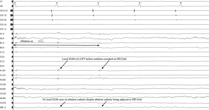
On the electrophysiology (EP) recording system, we were also able to see local EGM on the HD Grid catheter, albeit only on some bipoles (A1A2, B1B2, A1B1, A2B2) as illustrated on Figure 2. For the EP recording system, high pass filter 30Hz, low pass filter 500 Hz with notch filter switched on. Ablation with 30W power at this spot resulted in isolation of LUPV (Figure 2) and loss of these local EGM on the HD Grid catheter.
Figure 2.

Electrogram (EGM) recorded by the recording system showing local EGM from the left upper pulmonary vein (LUPV) as depicted by the arrows, but these were not recorded on the ablation bipole. Ablation at this spot, however, led to isolation of LUPV in 2.2 seconds after switching on power. This goes against the conventional wisdom of needing to see electrogram on ablation catheter prior to ablation
3. CASE DISCUSSION
The HD grid multipolar mapping catheter is a new and unique multipolar mapping catheter in that it allows bipole recording parallel and perpendicular to the splines via 16 electrodes, compared to conventional multipolar mapping catheters and ablation catheter which only allows bipole recording parallel to the splines. Recent multipolar mapping catheters such as Pentaray® (Biosense Webster, Irvine, CA, USA) and Orion® (Boston Scientific, Marlborough, MA, USA) have unique features when combined with their respective mapping system to create high density maps rapidy.1 However, these catheters essentially record local EGM parallel to their bipoles.
When HD Grid is paired with the Ensite mapping system, the algorithm displays signals amalgamated from orthogonal recordings of each bipole and displaying the highest amplitude signal. This would explain why only certain bipoles display the local EGM of reconnection on the EP recording system. This is despite the HD Grid having a greater interelectrode distance (3 mm) compared to the Tacticath (2 mm). With greater sensitivity to record electrical signals regardless of direction of activation, HD grid offers the capability to perform high density mapping to define the anatomical substrate and identify the low voltage isthmus. In this case, we were able to define the pulmonary vein connection displayed on the voltage map. By targeting this low voltage isthmus, despite not recording any EGM on the ablation catheter even with adequate contact of 14 g, we were able to achieve pulmonary vein isolation within a few seconds of applying radiofrequency ablation. This goes against the conventional wisdom of needing to see EGM on ablation catheter prior to ablation.
Being able to display only the highest amplitude signals from orthogonal recordings, HD grid may be able to detect local EGM signals which previously would have been missed and thus may offer more effective therapeutics.
CONFLICT OF INTERESTS
Authors declare no conflict of interests for this article.
Supporting information
Yeo C, Tan VH, Wong KCK. Pulmonary vein reconnection mapping with Advisor HD Grid demonstrating local EGM which were not visible on Tacticath ablation catheter. J Arrhythmia. 2019;35:152–154. 10.1002/joa3.12144
REFERENCE
- 1. Lin CY, Te ALD, Lin YJ, et al. High‐resolution mapping of pulmonary vein potentials improved the successful pulmonary vein isolation using small electrodes and inter‐electrode spacing catheter. Int J Cardiol. 2018;272:90–6. [DOI] [PubMed] [Google Scholar]
Associated Data
This section collects any data citations, data availability statements, or supplementary materials included in this article.
Supplementary Materials


