Abstract
Cecropia species are traditionally used in Latin American folk medicine and are available as food supplements with little information warranting their quality. The optimum conditions for the extraction of chlorogenic acid (CA), total flavonoids (TF) and flavonolignans (FL) from leaves of Cecropia species were determined using a fractional factorial design (FFD) and a central composite design (CCD). A reversed-phase high-performance liquid chromatographic method coupled to a diode array detector (HPLC-DAD) was validated for the quantification of CA, TF and FL, following the ICH guidelines. Quantitative and Principal Component Analysis (PCA) was also performed. The extraction-optimization methodology enabled us developing an appropriate extraction process with a time-efficient execution of experiments. The experimental values agreed with those predicted, thus indicating suitability of the proposed model. The validation parameters for all chemical markers of the quantification method were satisfactory. The results revealed that the method had excellent selectivity, linearity, precision (repeatability and intermediate precision were below than 2 and 5%, respectively) and accuracy (98–102%). The limits of detection and quantification were at nanogram per milliliter (ng/mL) level. In conclusion, the simultaneous quantification of chemical markers using the proposed method is an appropriate approach for species discrimination and quality evaluation of Cecropia sp.
Introduction
Plant species, popularly known as ‘yarumo’, ‘guarumo’, ‘guarumbo’, ‘embauba’, ‘ambay’ and ‘trumped tree’, belong to the genus Cecropia Loefl. (Urticaceae), which comprises 61 species and are distributed across the tropical and subtropical rainforest from Mexico over Central to South America below an altitude of 2600 m1,2.
Some Cecropia species, such as C. obtusifolia Bertol., C. peltata L., C. pachystachya Trécul, C. hololeuca Miq. and C. glaziovii Snethl. are traditionally used in Latin American folk medicine for the treatment of a variety of diseases, like diabetes3, hypertension4 and inflammation5. Additionally, they have been reported for their wound healing6 and antimalarial7 activities. These pharmacological properties have been correlated to their content of phenolic compounds, such as phenolic acids, flavonoids and terpenoids5,8,9.
A number of studies on Cecropia species suggested the relationship between the major compounds of the leaf extracts (relative concentration above 5 µg/g), chlorogenic acid and glycosyl flavonoids (orientin, isoorientin, vitexin and rutin) and the anti-diabetic10, anti-inflammatory11 and anti-hypertensive12 activities. Recently, a detailed phytochemical study on Cecropia species collected in Panama, also revealed the presence of flavonolignans in C. obtusifolia, C. peltata, C. insignis for the first time (Rivera-Mondragón et al.2 Submitted).
Taking this into consideration, chlorogenic acid, total flavonoids and flavonolignans have been selected as suitable chemical markers for the qualitative evaluation of leaves of plants of the genus Cecropia2. The relationship between these phenolic compounds and their pharmacological properties produces a big interest in the development and validation of an adequate analytical method, which will be useful for the quantification of herbal products containing these compounds. Additionally, the optimization of their extraction is of interest, since the chromatographic analysis only allows observing compounds properly extracted from the plant material.
A literature survey revealed that very little was published on developing and validating analytical methods for the quantitative determination of the main phenolic compounds in Cecropia species. For instance, a spectrophotometric colorimetric method (aluminium chloride) and HPLC-DAD methods have been developed for the determination of the seasonal variation of total flavonoids, isoorientin and isovitexin contents in dry leaves of C. glaziovii leaves8,13. In other investigations, LC methods have been described for the chemical standardization of C. glaziovii14–16, to isolate C-glycosyl flavones in C. lyratiloba17, to chemically fingerprint and quantify aqueous, methanolic and ethyl-acetate extracts of C. pacystachya11,18,19, and to evaluate the phytochemical composition of ethanolic extracts of C. obtusifolia and C. peltata20. Furthermore, two validated HPLC-DAD and UPLC-DAD methods for the quantification of chlorogenic acid and main flavone C-glycosides in leaves of C. glaziovii, C. pachystachya and C. hololeuca have been reported21,22.
However, to our knowledge, optimal extraction conditions and a complete validated method for the simultaneous identification and quantification of chlorogenic acid, total flavonoids and flavonolignans in Cecropia leaves have not been reported yet in peer-reviewed literature.
In this paper, we report a fully validated, sensitive and specific HPLC-DAD method for the quantification of CA, TF and FL in different authentic and commercial products of leaves of Cecropia species, collected in different regions of the Republic of Panama and obtained from the market. Furthermore, the extraction of the compounds of interest was optimized.
Results and Discussion
HPLC-DAD analysis of CA, TF and FL
Liquid chromatography coupled with mass spectrometry (LC-MS) offers better sensitivity and selectivity for compound identification in comparison with UV detection. However, the high cost of this equipment together with the absence of primary standards may be a limiting factor for low-budget analytical laboratories. On the other hand, DAD is inexpensive, broadly applied to natural product analysis, has a reliable and reproducible performance, and provides the possibility of online collection of UV spectra, inducing a viable added value for quantitative analysis23. Hence, we present an HPLC-DAD method for the routine analysis of CA, TF and FL in samples Cecropia species.
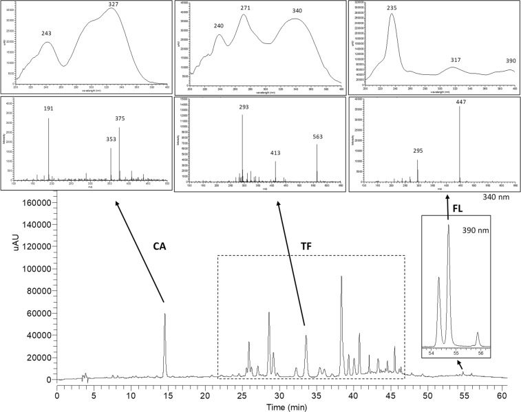
A representative HPLC-DAD profile of a leaf extract from a Cecropia species mixture is given in Fig. 1. Analysis of online UV spectra, obtained from peaks between 25–46 min, let us recognize typical UV absorption bands for flavonoids (Band I, λ max around 300–354 nm and Band II, λ max around 240–285 nm)24. As previously reported by our research group, flavone C-glycosides were predominately detected in C. obtusifolia, C. peltata and C. insignis; while quercetin O-glycosides were the main flavonoids described in C. hispidissima (Rivera-Mondragón et al., 2018. Submitted). Considering this, we decided to express O-glycosyl flavonols and C-glycosyl flavones as rutin (RU) and vitexin (VX) equivalents, respectively. FL such as mururin A and vaccinin A are not available as reference standard on the market. Therefore, VX was chosen as a secondary analytical standard for the quantification of these compounds.
Figure 1.
HPLC chromatogram of leaf extracts from a Cecropia species mixture measured on DA detector at 340 and 390 nm, and the observed characteristic UV and MS spectra of chlorogenic acid (CA), total flavonoids (TF) and flavonolignans (FL).
Extraction optimization
Variables with major effects over the extraction yields were optimized. First, ultrasound assisted extraction (UAE) was selected as the most appropriate extraction method due to its efficacy offering a great reduction of extraction time and low environmental impact25–27. Following, mixtures of water: methanol and acetone were chosen as extraction solvents, since these solvents have widely described as the most suitable systems for polyphenol extraction from plant material28,29.
The screening and optimization steps of TF, CA and FL from Cecropia species leaves were conducted by fractional factorial (FFD) and central composite (CCD) designs, respectively.
Screening (FFD)
Seven factors were chosen to be screened: methanol fraction (%, v/v) (A), extraction time (min) (B), number of extractions with methanol (C), extraction temperature (°C) (D), mass/solvent ratio (w/v) (E), number of extractions with acetone (F) and particle size of the plant material (µm) (G). The FFD, including the fraction levels and the experimental values of TF, CA and FL, is presented in the supplementary material (Table S1). The estimated effect of each factor on the responses and the corresponding critical effects are shown in Table 1. Those factors with effects below the critical effect (Ecritical) were considered statistically not significant factors affecting the yield of phenolic extraction.
Table 1.
Factor effects from screening the Cecropia species leaves extraction process by FFD.
| Factor | Effects | ||
|---|---|---|---|
| TF | CA | FL | |
| A | −92.88* | −19.99* | 1.12* |
| B | 38.54 | 9.66 | −0.05 |
| C | 76.98 | 24.70* | 0.94* |
| D | 149.19* | 31.99* | 1.55* |
| E | 39.50 | 18.37* | 0.40 |
| F | 12.65 | 0.58 | 0.85 |
| G | −73.20 | −13.01* | −0.03 |
| A × B | −22.59 | −6.84 | −0.08 |
| A × C | −14.37 | 1.38 | −0.20 |
| A × D | 50.12 | 4.40 | −0.44 |
| A × E | −8.85 | −3.20 | −0.31 |
| A × F | −24.34 | −5.63 | −0.08 |
| A × G | −47.06 | −3.64 | −0.02 |
| B × D | −27.05 | 1.27 | −0.45 |
| Ecritical | 84.54 | 10.97 | 0.47 |
Factors: (A) methanol fraction (%, v/v), (B) extraction time (min), (C) number of methanol extractions, (D) temperature (°C), (E) mass:solvent ratio (w/v), (F) number of acetone extractions, (G) particle size of the plant material (µm). Responses: sum of peak areas of total flavonoids (TF), chlorogenic acid (CA) and flavonolignans (FL). The responses are represented as mean; SD are not given. *Significant effects.
TF, CA and FL extraction yields were significantly affected by the methanol fraction (A) and the extraction temperature (D). CA and FL contents were also significantly affected by the number of extractions (C). Additionally, CA content was significantly affected by mass/solvent ratio (E) and particle size (G). For the optimization step, we selected the three factors with the major effects on TF, CA and FL yields: methanol fraction (A), number of extractions with methanol (C) and extraction temperature (D). The number of extractions (C), a discrete variable, was fixed at three (3) extractions. The less significant factors were mass/solvent ratio (E) and particle size (G), and were established as 1:50 (m/v) and ≤125 µm, respectively, since those are the best tested to obtain the highest CA yield. In addition, one (1) acetone extraction (F) was set, because it has a significant positive effect on FL peak area. Extraction time (B) was set at 30 min, since it has no significant effect neither on CA, TF or FL.
Optimization (CCD)
A response surface methodology (RSM), using a CCD, was employed to optimize the effects of the methanol fraction (X1) and extraction temperature (X2) on the extraction yields of TF (Y1), CA (Y2) and FL (Y3). The other variables were set as mentioned in the previous section (Screening FFD). Supplementary Tables S2 and S3 show the independent variables and their levels used for this experiment, and the actual design and the experimental data for the response variables, respectively. Second-order polynomial equations were built to describe the relationship between X1 and X2 (Table 2).
Table 2.
Quadratic polynomial equations for the three responses.
| Responses | Equations |
|---|---|
| TF | Y1 = 1737.8 − 106.1X1 + 43.8X2–86.9X12 − 6.8X22 + 47.3X1X2 |
| CA | Y2 = 340.7 − 22.0X1 + 14.6X2 − 23.2X12 − 2.4X22 + 1.8X1X2 |
| FL | Y3 = 14.0 + 0.35X1 + 0.33X2 − 0.63X12 − 0.27X22 − 0.16X1X2 |
Y1, Y2 and Y3 are the responses. X1 and X2 are the independent variables, methanol fraction (%, v/v) and temperature extraction (°C), respectively.
The analysis of variance (ANOVA) for the quadratic polynomial models developed for the response variables indicated that the linear effects of X1 and X2 were found to be significant (p < 0.05) for TF, CA and FL extraction (Table 3). Besides, X12 is significant (p < 0.05) for all response variables, while X22 was not significant (p > 0.05) for any response. On the other hand, the interaction effect of X1X2 is only significant (p < 0.05) for TF.
Table 3.
A t-test for the quadratic polynomial models developed for the response variables: total flavonoids (TF), chlorogenic acid (CA) and flavonolignans (FL) of Cecropia sp. mixture.
| Term | TF (Y1) | CA (Y2) | FL (Y3) | ||||||
|---|---|---|---|---|---|---|---|---|---|
| Coefficient | Standard error | p-Value | Coefficient | Standard error | p-Value | Coefficient | Standard error | p-Value | |
| Model | <0.0001 | <0.0001 | 0.0028 | ||||||
| Intercept | 1737.8 | 29.3 | 3.48E-16 | 340.7 | 3.64 | 1.50E-18 | 13.96 | 0.302 | 6.82E-15 |
| Linear terms | |||||||||
| X 1 | −106.1 | 10.4 | 2.75E-07 | −22.0 | 1.29 | 8.59E-10 | 0.350 | 0.107 | 0.0066 |
| X 2 | 43.8 | 10.4 | 0.0012 | 14.6 | 1.29 | 8.92E-08 | 0.329 | 0.107 | 0.0094 |
| Quadratic terms | |||||||||
| X 1 2 | −86.9 | 17.2 | 0.0003 | −23.2 | 2.14 | 1.47E-07 | −0.635 | 0.177 | 0.0038 |
| X 2 2 | −6.8 | 17.2 | 0.6980 | −2.40 | 2.14 | 0.2830 | −0.273 | 0.177 | 0.1484 |
| Interaction | |||||||||
| X 1 X 2 | 47.3 | 14.6 | 0.0072 | 1.81 | 1.82 | 0.3400 | −0.161 | 0.151 | 0.3060 |
In order to visualize the factor and interaction effects on the extraction efficiency, the three-dimensional response surface plots were generated as a function of X1 and X2 as shown in Fig. 2. In general, the peak areas of TF, CA and FL increased within a range of methanol fraction of 70–75% (v/v), 55–72% (v/v) and 70–80% (v/v), respectively. However, a methanol fraction below or above this ranges appeared to decrease the extraction yields of these compounds. Furthermore, the highest TF, CA and FL extraction yields were observed with extraction temperature ranges from 70–75 °C, 65–75 °C and 55–65 °C, respectively. FL extraction efficiency was negatively affected when the extraction temperature was above 65 °C. For TF and CA the extraction yields were only affected to a limited extend by the temperature.
Figure 2.
Response surface plots demonstrating the influence of the methanol fraction (%, v/v) and the extraction temperature (°C) on the peak areas of TF (A), CA (B) and FL (C).
Verification of the predicted optimal extraction conditions
The optimal X1 and X2 variables were determined by maximizing the responses using MODDE Pro Software. In order to confirm the reliability of the mathematical model, experimental extractions were performed under conditions selected as optimal: a methanol fraction of 70% (v/v), a extraction temperature of 64 °C, 30 min of extraction time, three (3) repeated extractions with methanol and one (1) with acetone, a mass/solvent ratio of 1:50, w/v, and particle size of ≤125 µm. The predicted and experimental responses demonstrated no significant differences (t-test, p > 0.05) and the difference between the predicted and experimental values were less than 2.0%, indicating an appropriate fitness of the predicted model (See Table 4).
Table 4.
Experimental and predicted values of TF, CA and FL at optimal conditions.
| Responses | Optimal extraction conditions | Maximun value | |||
|---|---|---|---|---|---|
| X1 (%, v/v) | X2 (°C) | Predicted | Experimental (n = 3) | % Difference (CV) | |
| TF | 70 | 64 | 1786.0 ± 29.3a | 1736.3 ± 7.9a | 0.94 |
| CA | 356.6 ± 3.6b | 354.4 ± 3.2b | 0.85 | ||
| FL | 13.9 ± 0.3c | 14.1 ± 0.3c | 1.79 | ||
All the values are means ± standard deviations and those sharing the same superscript letter in the same row are not significantly different from each other (p > 0.05).
In order to develop a less time- and solvent-consuming method, the robustness of the yield, was determined from different combinations of mass/solvent ratio (1:30, 1:15 and 1:10) and number of acetone extractions (1 and 0) (Fig. 3). One-way ANOVA followed by a Bonferroni post hoc test revealed not significant differences in CA, TF and FL contents (p >0.05) in comparison with the yield at the optimized conditions. Results show that mass/solvent ratio and number of acetone extractions may be changed (reduced) without compromising the analytes extraction. During the validation of the method, the mass/solvent ratio was set at 1:30, while the extraction with acetone-extraction was eliminated from the procedure.
Figure 3.
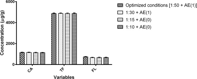
Evaluation of simultaneous variation of mass solvent ratio (1:30, 1:15 and 1:10) and number of acetone extractions (AE) compared to the optimized method conditions situation [1:50 + AE(1)].
Validation of the HPLC-DAD method
Specificity
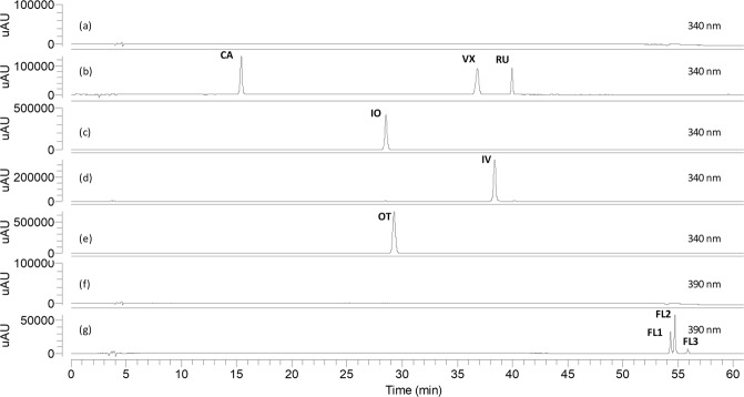
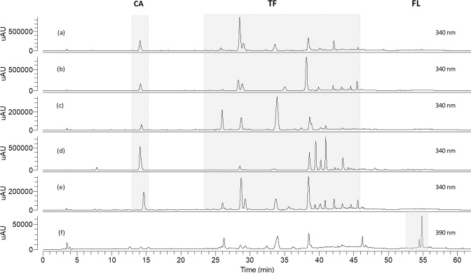
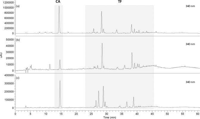
Blank solution, authentic standards and isolated compounds from Cecropia obtusifolia (FL1, FL2 and FL3) were analyzed under the analytical method conditions established here (Fig. 4). Authentic and commercial sample extracts from different species were analyzed as well in order to verify for possible interferences (Figs 5 and 6). Representative UV and MS spectra inspection of each analyte (Fig. 1), shows no relevant interferences at the regions (retention times) of interest. Despite some overlapping peaks were found (Fig. 5c), identified as flavonoids, the quantification of the total flavonoids is not interfered.
Figure 4.
HPLC chromatograms of blank solution (a,f), chlorogenic acid (CA), vitexin (VX) and rutin (RU) (b), isoorientin (IO) (c), isovitexin (IV) (d), orientin (OT) (e), flavonolignan 1 (FL1), flavonolignan 2 (FL2) and flavonolignan 3 (FL3) (g). Chromatograms (a–e) were obtained at 340 nm, (f,g) at 390 nm.
Figure 5.
HPLC chromatograms of authentic samples: C. obtusifolia CO-5 (a), C. peltata CP-1 (b), C. insingnis CI-1 (c), C. hispidissima CH-1 (d), mixture of Cecropia species (e), and C. obtusifolia CO-6 (f).
Figure 6.
HPLC chromatograms of commercial samples: C. obtusifolia CO-C (a), C. peltata CP-C (b) and C. hololeuca CHO-C (c).
Linearity
Four curves were plotted at different levels (n ≥ 5) within an appropriate concentration range of CA, VX and RU, and the residuals were observed to be homoscedastic and with % RSD values below than 5% (Supplementary Fig. S2). As shown in Table 5, all curves have a linear response with r2 > 0.999, t-tests (N-2, p = 95%) demonstrated that intercepts were not significantly different from zero, while slopes were significantly different from zero. Additionally, 95% CI of intercepts include zero, which means that quantification analysis can be performed based on a single-point calibration approach. Moreover, Mandel’s fitting tests indicate that first-order calibration function (linear equation) provides a significantly (α = 0.01) better fit than second-order calibration (quadratic equation).
Table 5.
Parameters of the calibration model for CA, VX and RU. Confidence interval (CI). Vitexin detected at 340 nm is represented as VX-1.
| Analyte | Concentration range (µg/mL) | Equation | r2 | 95% CI of the intercept | LoD (ng/mL) | LoQ (ng/mL) |
|---|---|---|---|---|---|---|
| CA | 0.16–50.30 | y = 67.6x − 15.2 | >0.99 | −32.62–2.27 | 160.7 | 401.9 |
| VX-1 | 0.16–211.70 | y = 74.4x − 12.4 | >0.99 | −42.64–17.92 | 131.2 | 328.0 |
| VX-2 | 1.63–352.83 | y = 5.2x − 3.4 | >0.99 | −7.16–0.46 | 423.6 | 903.8 |
| RU | 1.49–194.64 | y = 41.4x − 11.4 | >0.99 | −23.31–0.57 | 131.0 | 388.6 |
Vitexin detected at 390 nm as VX-2.
Precision
Repeatability (intra-day and intra-concentration level, n = 6) and intermediate precision (between 4 days, and three concentration levels, n = 6) were evaluated in order to assess the precision of the method. Overall, Cochran’s C test for homogeneity of variances (95% confidence level) indicated that variances between days and concentration levels are homogeneous (Supplementary Table S4). Values of % RSD for all parameters were below than 2.0% and 5.0% for repeatability and intermediate precision, respectively. In addition, all values did not exceed the levels calculated by the Horwitz equation and thus confirmed the expected performance of the method in terms of precision. Results are summarized in Table 6.
Table 6.
Precision of the method for CA, TF and FL during 4 days and at three concentration levels.
| Analyte | Relative standard deviation (% RSD) | |||||||||
|---|---|---|---|---|---|---|---|---|---|---|
| Days (100%) | Concentration levels | Overall repeatability | Intermediate precision | Horwitz equation | ||||||
| 1 | 2 | 3 | 4 | 50% | 150% | RSDr | RSDR | |||
| CA | 0.91 | 1.30 | 1.43 | 0.83 | 0.96 | 1.14 | 1.10 | 4.34 | 3.79 | 5.68 |
| TF-1a | 0.70 | 0.36 | 0.58 | 0.68 | 0.88 | 0.70 | 0.67 | 3.54 | 2.92 | 4.37 |
| TF-2b | 0.24 | 0.25 | 0.32 | 0.26 | 0.25 | 0.23 | 0.26 | 2.92 | 2.58 | 3.86 |
| FL | 1.19 | 1.39 | 1.86 | 0.99 | 0.91 | 1.46 | 1.32 | 4.77 | 4.23 | 6.34 |
n = 6, overall repeatability: intra days and levels, intermediate precision: inter days and levels. Overall variances were homogenous according Cochran’s C test (95% confidence level). aTF expressed as VX equivalent (C. species mixture). bTF expressed as rutin equivalent (C. hispidissima).
Accuracy
The accuracy was determined by means of a recovery experiment, adding known quantities of CA, VX and RU in three concentration levels (75, 100 and 125%). Fortified solutions were analyzed and the results were reported as percent recovery (%). Table 7 shows that accuracy data are in agreement with the acceptance criteria at all three concentrations levels since recovery values varied between 98 and 102%, 95% CI of the mean include 100%, and % RSD for each level concentration were lower or equal to % RSDr30.
Table 7.
Accuracy of the method for CA, VX-1, VX-2 and RU determined by recovery (%). Confidence interval (CI). n = 3.
| Analyte | Recovery (%) | |||||
|---|---|---|---|---|---|---|
| 75% | 100% | 125% | Mean | % RSD | 95% CI | |
| CA | 101.41 | 100.10 | 99.96 | 100.49 | 0.99 | 99.73–101.25 |
| VX-1a | 99.77 | 99.55 | 100.21 | 99.84 | 0.57 | 99.40–100.28 |
| VX-2b | 100.40 | 99.02 | 100.06 | 99.83 | 0.90 | 99.13–100.52 |
| RU | 100.09 | 100.40 | 100.22 | 100.24 | 0.21 | 100.08–100.40 |
aVitexin detected at 340 nm (VX-1). bVitexin detected at 390 nm (VX-2).
Limit of detection (LoD) and limit of quantification (LoQ)
LoD and LoQ were estimated based on the calibration curves of CA, VX and RU, constructed in a 1.5–50 µg/mL range (data not shown). Additionally, these estimates were subsequently validated by the independent analysis of six quantified samples (prepared by serial dilutions) in order to find concentration levels around LoD and LoQ. All compounds showed LoD and LoQ of the order of magnitude of ng/mL. CA, VX and RU showed lower concentrations of LoD and LoQ than those calculated by the mathematical method, except for VX detected at 390 nm, which displayed a higher LoD and a similar LoQ to those determined by its calibration curve (See supplementary material Table S5). This method is able to detect and quantify CA, TF (expressed as vitexin or rutin equivalent) and FL (expressed as vitexin equivalent) of Cecropia species in concentrations below than 0.5 µg/mL and 1.0 µg/mL, respectively (Table 5).
Quantitative and multivariate analysis
Subsequent to the optimization of the extraction and the validation of the proposed analytical method, the contents of CA, TF and FL from fourteen authentic Cecropia leaves and three commercial products were assessed. The concentrations of phenolics found in Cecropia are shown in Table 8. It was observed that CA was present in all samples ranging from 78.7 ± 4.3 (CP-C) to 14724.5 (CO-7) (µg/g). Quantitative analysis of samples from Cerro Azul, Camino de Cruces and Cerro Campana showed higher CA concentrations collected during the rainy season (October, 2015) compared to those collected in the second dry period during the rainy season (known as First Canicula, July 2016); except for CO-7, which presented the highest CA content and was collected in Chiriqui (July, 2017) (See Supplemenatary Fig. S3).
Table 8.
Concentrations of CA, TF and FL (µg/g) in authentic and commercial Cecropia leaf samples.
| Compounds | CO-1 | CO-2 | CO-3 | CO-4 | CO-5 | CO-6 | CO-7 | CO-C | CP-1 | CP-2 | |
|---|---|---|---|---|---|---|---|---|---|---|---|
| Phenolic acids | |||||||||||
| 1 | Chlorogenic acid | 1933.2 ± 36.9 | 1634.0 ± 40.7 | 243.6 ± 2.8 | 4238.1 ± 33.9 | 1393.3 ± 56.4 | 1091.5 ± 17.9 | 14724.5 ± 102.7 | 5612.6 ± 228.5 | 2217.8 ± 59.6 | 949.0 ± 35.0 |
| Flavonoids C-glycosides and di-C,O-glycosides | |||||||||||
| 2 | Luteolin C-hexoside-O-hexoside (1) | 181.4 ± 4.0 | 277.6 ± 14.5 | 33.3 ± 1.7 | <LOQ | <LOQ | 40.2 ± 1.5 | <LOQ | 83.2 ± 0.4 | <LOQ | <LOQ |
| 3 | Isoorientin-2′′-O-xyloside | 455.7 ± 9.8 | 884.0 ± 11.1 | 196.9 ± 3.0 | <LOQ | 284.5 ± 6.3 | 69.7 ± 1.3 | <LOQ | 612.2 ± 9.7 | <LOQ | <LOQ |
| 4 | Luteolin C-hexoside-O-hexoside (2) | 65.7 ± 1.8 | 233.8 ± 3.3 | <LOQ | <LOQ | <LOQ | 366.8 ± 8.1 | <LOQ | 85.8 ± 3.2 | <LOQ | <LOQ |
| 5 | Isoorientin-4′′-O-xyloside | 105.1 ± 10.5 | 370.2 ± 11.5 | <LOQ | <LOQ | <LOQ | <LOQ | <LOQ | 204.2 ± 12.2 | <LOQ | <LOQ |
| 6 | Isoorientin | 384.6 ± 8.3 | 1077.7 ± 10.2 | 40.2 ± 3.3 | 6399.4 ± 25.5 | 4187.9 ± 119.8 | 160.9 ± 1.5 | 5177.7 ± 54.5 | 3661.7 ± 74.0 | 1796.2 ± 39.2 | 2127.9 ± 54.4 |
| 7 | Orientin | <LOQ | <LOQ | <LOQ | 1117.6 ± 9.7 | 1167.4 ± 35.5 | <LOQ | 4523.8 ± 44.9 | <LOQ | 1076.9 ± 13.6 | 749.3 ± 18.2 |
| 8 | Isoorientin-2′′-O-rhamnoside | 612.0 ± 17.0 | 241.2 ± 5.7 | 164.0 ± 5.0 | <LOQ | <LOQ | 129.8 ± 1.6 | <LOQ | 823.0 ± 20.1 | <LOQ | <LOQ |
| 9 | Luteolin C-hexoside-O-pentoside | 84.8 ± 3.7 | 145.1 ± 4.6 | 23.9 ± 4.0 | <LOQ | <LOQ | <LOQ | <LOQ | 119.7 ± 7.7 | <LOQ | <LOQ |
| 10 | Isovitexin-2′′-O-glucoside | 240.4 ± 6.7 | 465.3 ± 8.4 | 269.6 ± 2.4 | <LOQ | 112.1 ± 7.4 | 315.3 ± 8.4 | <LOQ | 118.3 ± 5.9 | <LOQ | <LOQ |
| 11 | Isovitexin-2′′-O-xyloside | 761.4 ± 19.7 | 1671.4 ± 26.2 | 1936.3 ± 11.4 | <LOQ | 1044.7 ± 36.3 | 2142.9 ± 71.9 | <LOQ | 787.1 ± 16.8 | 266.5 ± 3.1 | 410.0 ± 19.6 |
| 12 | Vitexin | 170.1 ± 5.9 | <LOD | <LOQ | <LOQ | 98.0 ± 1.3 | <LOQ | 295.2 ± 3.6 | <LOQ | 198.2 ± 2.1 | 457.4 ± 37.2 |
| 13 | Apigenin C-hexoside-O-pentoside | 156.2 ± 4.1 | 511.8 ± 6.5 | 80.6 ± 1.5 | <LOQ | <LOQ | 233.4 ± 3.0 | <LOQ | 138.8 ± 4.3 | <LOQ | <LOQ |
| 14 | Diosmetin C-hexoside-O-pentoside | 58.0 ± 1.0 | 134.1 ± 1.9 | <LOQ | <LOQ | <LOQ | <LOQ | <LOQ | <LOQ | <LOQ | <LOQ |
| 15 | Isovitexin 2′′-O-rhamnoside | 364.4 ± 8.5 | 424.8 ± 7.8 | 1057.6 ± 5.2 | <LOQ | 1376.9 ± 36.8 | 1356.7 ± 30.8 | <LOQ | <LOQ | 1107.2 ± 4.7 | 2338.1 ± 103.2 |
| 16 | Apigenin C-hexoside-O-xyloside | 177.7 ± 5.1 | 436.9 ± 5.2 | 287.5 ± 2.5 | <LOQ | <LOQ | 179.1 ± 4.7 | <LOQ | <LOQ | <LOQ | <LOQ |
| 17 | Isovitexin | <LOQ | <LOQ | <LOQ | 502.8 ± 2.3 | <LOQ | <LOQ | 991.2 ± 12.1 | <LOQ | <LOQ | <LOQ |
| 18 | Diosmetin-C-hexoside-O-deoxyhexoside | <LOQ | <LOQ | 38.1 ± 0.7 | <LOQ | <LOQ | <LOQ | <LOQ | <LOQ | <LOQ | <LOQ |
| 19 | Diosmetin-C-hexoside | <LOQ | <LOQ | <LOQ | <LOQ | <LOQ | <LOQ | <LOQ | <LOQ | <LOQ | <LOQ |
| 20 | Luteolin-O-malonyl-C-hexoside (1) | <LOQ | <LOQ | <LOQ | 318.8 ± 1.4 | 251.0 ± 59.2 | <LOQ | <LOQ | <LOQ | 457.8 ± 11.1 | 444.2 ± 4.5 |
| 21 | Luteolin-O-malonyl-C-hexoside (2) | <LOQ | <LOQ | <LOQ | 1486.3 ± 14.2 | 651.1 ± 17.0 | <LOQ | 1622.9 ± 16.7 | <LOQ | 426.8 ± 3.4 | 631.3 ± 24.1 |
| 22 | Apigenin-O-malonyl-C-hexoside (1) | <LOQ | <LOQ | <LOQ | <LOQ | <LOQ | <LOQ | <LOQ | <LOQ | <LOQ | <LOQ |
| 23 | Apigenin-O-deoxyhexoside-O-malonyl-C-hexoside (1) | <LOQ | <LOQ | <LOQ | <LOQ | <LOQ | <LOQ | <LOQ | <LOQ | <LOQ | 172.1 ± 12.0 |
| 24 | Apigenin-O-malonyl-C-hexoside (2) | <LOQ | <LOQ | <LOQ | <LOQ | <LOQ | <LOQ | <LOQ | 124.9 ± 1.7 | <LOQ | <LOQ |
| 25 | Apigenin-O-deoxyhexoside-O-malonyl-C-hexoside (2) | <LOQ | <LOQ | <LOQ | 657.8 ± 6.7 | 171.3 ± 4.5 | <LOQ | <LOQ | <LOQ | <LOQ | 141.4 ± 6.2 |
| 26 | Apigenin-O-malonyl-C-hexoside (2) | <LOQ | <LOQ | <LOQ | <LOQ | <LOQ | <LOQ | 696.2 ± 4.1 | <LOQ | 148.4 ± 12.4 | 614.9 ± 23.4 |
| Flavonoids O-glycosides and di-O-glycosides | |||||||||||
| 27 | Quercetin O-hexoside-O-deoxyhexoside | <LOQ | <LOQ | <LOQ | 939.7 ± 18.9 | <LOQ | <LOQ | <LOQ | 2988.5 ± 180.1 | <LOQ | <LOQ |
| 28 | Rutin | 72.9 ± 3.4 | 112.9 ± 3.4 | <LOQ | 681.1 ± 9.7 | 168.4 ± 3.4 | <LOQ | 704.7 ± 4.2 | 1041.2 ± 25.1 | <LOQ | <LOQ |
| 29 | Quercetin O-hexoside (1) | <LOQ | <LOQ | <LOQ | 352.1 ± 20.6 | <LOQ | <LOQ | <LOQ | 519.2 ± 98.6 | <LOQ | <LOQ |
| 30 | Quercetin O-hexoside (2) | <LOQ | <LOQ | <LOQ | <LOQ | <LOQ | <LOQ | <LOQ | 655.6 ± 3.8 | 524.8 ± 19.9 | 161.3 ± 4.5 |
| 31 | Quercetin O-pentoside (1) | <LOQ | <LOQ | <LOQ | <LOQ | <LOQ | <LOQ | <LOQ | <LOQ | <LOQ | <LOQ |
| 32 | Quercetin O-pentoside (2) | <LOQ | <LOQ | <LOQ | <LOQ | <LOQ | <LOQ | <LOQ | 423.5 ± 8.0 | <LOQ | <LOQ |
| 33 | Quercetin O-hexoside-O-hexoside | <LOQ | <LOQ | <LOQ | <LOQ | <LOQ | <LOQ | <LOQ | <LOQ | <LOQ | <LOQ |
| Total Flavonoids (TF) | 3890.4 ± 96.9 | 6986.8 ± 35.0 | 4128.0 ± 27.8 | 12455.4 ± 2.8 | 9513.3 ± 241.1 | 4994.8 ± 130.3 | 14011.7 ± 130.7 | 12386.9 ± 174.0 | 6002.7 ± 38.1 | 8247.9 ± 297.4 | |
| Flavonolignans | |||||||||||
| 34 | Flavonolignan 1 | 202.6 ± 18.0 | 241.1 ± 22.0 | 144.1 ± 8.1 | <LOQ | 205.6 ± 9.0 | 510.2 ± 2.9 | 138.5 ± 13.2 | 98.0 ± 6.8 | <LOQ | <LOQ |
| 35 | Flavonolignan 2 | 799.1 ± | 982.5 ± 97.8 | 609.3 ± 27.2 | 280.9 ± 6.7 | 864.0 ± 42.8 | 1857.1 ± 61.8 | 653.0 ± 40.6 | 688.8 ± 44.0 | <LOQ | < LOQ |
| 36 | Flavonolignan 3 | <LOQ | <LOQ | <LOQ | <LOQ | <LOQ | <LOQ | 57.1 ± 8.0 | 121.3 ± 6.0 | <LOQ | < LOQ |
| Total flavonolignans | 1001.7 ± 65.5 | 753.4 ± 33.0 | 753.4 ± 33.0 | 280.9 ±6.7 | 1069.6 ± 50.6 | 2367.3 ± 63.0 | 848.7 ± 51.7 | 908.1 ± 44.7 | <LOQ | <LOQ | |
| Compounds | CP-3 | CP-4 | CP-C | CI-1 | CI-2 | CI-3 | CH-1 | CH-2 | CHO-C | ||
| Phenolic acids | |||||||||||
| 1 | Chlorogenic acid | 836.3 ± 12.6 | 927.5 ± 18.2 | 78.7 ± 4.3 | 1644.1 ± 44.7 | 323.4 ± 12.8 | 1331.8 ± 16.1 | 2881.9 ± 74.0 | 993.4 ± 110.6 | 1492.6 ± 8.3 | |
| Flavonoids C-glycosides and di-C,O-glycosides | |||||||||||
| 2 | Luteolin C-hexoside-O-hexoside (1) | <LOQ | <LOQ | <LOQ | <LOQ | <LOQ | <LOQ | <LOQ | <LOQ | <LOQ | |
| 3 | Isoorientin-2′′-O-xyloside | <LOQ | <LOQ | <LOQ | 1034.2 ± 5.8 | 838.7 ± 38.5 | 2066.8 ± 28.8 | <LOQ | <LOQ | 53.5 ± 1.6 | |
| 4 | Luteolin C-hexoside-O-hexoside (2) | <LOQ | <LOQ | <LOQ | <LOQ | 48.1 ± 7.9 | <LOQ | <LOQ | <LOQ | 295.0 ± 3.6 | |
| 5 | Isoorientin-4′′-O-xyloside | <LOQ | <LOQ | 22.4 ± 1.2 | 67.2 ± 8.2 | 43.0 ± 4.3 | 112.4 ± 1.6 | <LOQ | <LOQ | 755.4 ± 21.1 | |
| 6 | Isoorientin | 1066.0 ± 22.3 | 1993.5 ± 20.3 | 187.7 ± 1.2 | 491.7 ± 4.0 | 652.9 ± 39.9 | 2487.9 ± 32.4 | 444.2 ± 8.1 | 882.0 ± 96.5 | 1085.9 ± 15.9 | |
| 7 | Orientin | 926.6 ± 15.2 | 1562.5 ± 19.4 | 24.1 ± 0.9 | <LOQ | <LOQ | <LOQ | <LOQ | <LOQ | 337.7 ± 5.2 | |
| 8 | Isoorientin-2′′-O-rhamnoside | <LOQ | <LOQ | <LOQ | 82.5 ± 1.7 | 84.4 ± 9.6 | <LOQ | <LOQ | <LOQ | <LOQ | |
| 9 | Luteolin C-hexoside-O-pentoside | <LOQ | <LOQ | <LOQ | 93.6 ± 4.5 | 59.1 ± 7.4 | 309.4 ± 9.3 | <LOQ | <LOQ | 79.1 ± 1.1 | |
| 10 | Isovitexin-2′′-O-glucoside | <LOQ | <LOQ | <LOQ | 86.9 ± 2.0 | 47.9 ± 1.7 | <LOQ | <LOQ | <LOQ | 146.8 ± 6.4 | |
| 11 | Isovitexin-2′′-O-xyloside | <LOQ | 231.2 ± 7.1 | 26.9 ± 0.6 | 3213.6 ± 61.0 | 2416.4 ± 143.3 | 1677.5 ± 23.8 | <LOQ | <LOQ | 390.3 ± 7.7 | |
| 12 | Vitexin | 625.2 ± 2.5 | 457.3 ± 2.7 | <LOQ | <LOQ | <LOQ | <LOQ | <LOQ | <LOQ | <LOQ | |
| 13 | Apigenin C-hexoside-O-pentoside | <LOQ | <LOQ | 42.4 ± 2.6 | 110.5 ± 17.1 | 79.5 ± 13.3 | <LOQ | <LOQ | <LOQ | <LOQ | |
| 14 | Diosmetin C-hexoside-O-pentoside | <LOQ | <LOQ | <LOQ | 73.0 ± 1.1 | 102.1 ± 5.9 | 237.0 ± 4.3 | <LOQ | <LOQ | 506.7 ± 6.2 | |
| 15 | Isovitexin 2′′-O-rhamnoside | 3379.7 ± 51.4 | 2132.5 ± 25.3 | <LOQ | 587.1 ± 7.2 | 672.4 ± 18.2 | <LOQ | <LOQ | <LOQ | <LOQ | |
| 16 | Apigenin C-hexoside-O-xyloside | <LOQ | <LOQ | <LOQ | 329.4 ± 8.4 | 219.5 ± 21.0 | <LOQ | <LOQ | <LOQ | <LOQ | |
| 17 | Isovitexin | <LOQ | <LOQ | 121.2 ± 1.3 | <LOQ | <LOQ | 795.6 ± 12.5 | <LOQ | <LOQ | <LOQ | |
| 18 | Diosmetin-C-hexoside-O-deoxyhexoside | <LOQ | <LOQ | <LOQ | <LOQ | <LOQ | <LOQ | <LOQ | <LOQ | <LOQ | |
| 19 | Diosmetin-C-hexoside | 338.3 ± 14.2 | <LOQ | 21.2 ± 0.6 | <LOQ | <LOQ | <LOQ | <LOQ | <LOQ | <LOQ | |
| 20 | Luteolin-O-malonyl-C-hexoside (1) | <LOQ | 540.9 ± 3.4 | <LOQ | <LOQ | <LOQ | <LOQ | <LOQ | <LOQ | <LOQ | |
| 21 | Luteolin-O-malonyl-C-hexoside (2) | 314.0 ± 31.6 | 332.4 ± 3.9 | <LOQ | <LOQ | <LOQ | <LOQ | 144.9 ± 3.6 | 173.6 ± 20.7 | <LOQ | |
| 22 | Apigenin-O-malonyl-C-hexoside (1) | 244.2 ± 69.0 | 70.4 ± 0.4 | <LOQ | <LOQ | <LOQ | <LOQ | <LOQ | <LOQ | <LOQ | |
| 23 | Apigenin-O-deoxyhexoside-O-malonyl-C-hexoside (1) | <LOQ | <LOQ | <LOQ | <LOQ | <LOQ | <LOQ | <LOQ | <LOQ | <LOQ | |
| 24 | Apigenin-O-malonyl-C-hexoside (2) | <LOQ | <LOQ | <LOQ | <LOQ | <LOQ | <LOQ | <LOQ | <LOQ | <LOQ | |
| 25 | Apigenin-O-deoxyhexoside-O-malonyl-C-hexoside (2) | 196.3 ± 2.5 | 58.5 ± 1.5 | <LOQ | <LOQ | <LOQ | <LOQ | <LOQ | <LOQ | <LOQ | |
| 26 | Apigenin-O-malonyl-C-hexoside (2) | 478.5 ± 5.4 | 116.6 ± 1.1 | <LOQ | <LOQ | <LOQ | <LOQ | <LOQ | <LOQ | <LOQ | |
| Flavonoids O-glycosides and di-O-glycosides | |||||||||||
| 27 | Quercetin O-hexoside-O-deoxyhexoside | <LOQ | <LOQ | <LOQ | <LOQ | <LOQ | <LOQ | 2884.2 ± 102.9 | 1491.5 ± 171.0 | <LOQ | |
| 28 | Rutin | <LOQ | <LOQ | 53.5 ± 1.2 | <LOQ | <LOQ | <LOQ | 3949.2 ± 116.7 | 2105.6 ± 218.6 | <LOQ | |
| 29 | Quercetin O-hexoside (1) | <LOQ | 294.9 ± 9.0 | <LOQ | 127.4 ± 2.8 | 93.3 ± 2.5 | 800.7 ± 21.2 | 1388.9 ± 37.0 | 1105.2 ± 121.7 | <LOQ | |
| 30 | Quercetin O-hexoside (2) | <LOQ | <LOQ | 23.9 ± 0.8 | 453.0 ± 8.2 | 248.1 ± 5.0 | 825.8 ± 28.4 | 4146.0 ± 144.3 | 3957.0 ± 43.0 | <LOQ | |
| 31 | Quercetin O-pentoside (1) | <LOQ | <LOQ | <LOQ | <LOQ | <LOQ | <LOQ | 226.9 ± 7.5 | 165.5 ± 18.4 | <LOQ | |
| 32 | Quercetin O-pentoside (2) | <LOQ | <LOQ | <LOQ | <LOQ | <LOQ | 215.7 ± 8.1 | 1474.8 ± 47.9 | 1040.1 ± 115.2 | <LOQ | |
| 33 | Quercetin O-hexoside-O-hexoside | <LOQ | <LOQ | <LOQ | <LOQ | <LOQ | <LOQ | 240.1 ± 17.1 | 179.1 ± 25.3 | <LOQ | |
| Total Flavonoids (TF) | 7568.8 ± 69.8 | 7790.8 ± 44.9 | 523.3 ± 5.9 | 6750.1 ± 99.9 | 5605.4 ± 275.6 | 9528.8 ± 114.2 | 14899.2 ± 470.9 | 11099.6 ± 702.3 | 3650.5 ± 50.5 | ||
| Flavonolignans | |||||||||||
| 34 | Flavonolignan 1 | <LOQ | <LOQ | <LOQ | <LOQ | <LOQ | <LOQ | <LOQ | <LOQ | <LOQ | |
| 35 | Flavonolignan 2 | 260.3 ± 17.1 | <LOQ | <LOQ | <LOQ | <LOQ | <LOQ | <LOQ | <LOQ | <LOQ | |
| 36 | Flavonolignan 3 | <LOQ | 213.9 ± 8.0 | <LOQ | 179.1 ± 4.5 | 227.8 ± 20.3 | <LOQ | <LOQ | <LOQ | <LOQ | |
| Total | 260.3 ± 17.1 | 213.9 ± 8.0 | <LOQ | 179.1 ± 4.5 | 227.8 ± 20.3 | <LOQ | <LOQ | <LOQ | <LOQ | ||
CO, CP, CI and CH correspond to authentic leaves of C. obstusifolia, C. peltata, C. insignis and C. hispidissima samples (see supplementary Fig. S1). CO-C, CP-C and CHO-C correspond to commercial products of C. obstusifolia, C. peltata and C hololeuca. Contents of analytes are reported as mean ± standard deviation (n = 3). Content below the limit of quantification: <LOQ.
Although a broad variety of flavonoid glycosides was detected, most flavonoids belonged to the flavone or flavonol classes and that the main skeletons were derivatives of luteolin, apigenin or quercetin. Among all samples, flavone C-glycosides were the main flavonoids subclass present in C. obtusifolia, C. peltata, C. insignis and C. hololeuca, but flavonol O-glycosides in C. hispidissima. Comparison of these results accords with those of Costa et al.21, Ortmann et al.31, da Silva Mathias and Rodrigues de Oliveira22 who also found that CA and flavone C-glycosides derived from apigenin and luteolin were the most abundant compounds detected in C. glaziovii, C. pachystachya and C. hololeuca.
Besides, the highest total flavonoid content was detected for C. hispidissima CH-1(14899.2 µg/g), followed by the C. obtusifolia samples: CO-7 (14071.7 µg/g), CO-4 (12455.4 µg/g) and CO-C (12387.1 µg/g). Regarding the seasonal variation of flavonoid content, a general pattern of increasing or decreasing levels among the two different periods could not be observed (See Supplemenatary Fig. S4). Similar findings were reported by Costa et al.4, who observed no correlation between the values of pluviosity and the production of C-glycosylflavonoids. The content of FL in C. obtusifolia, C. peltata and C. insignis was in the range of 179.1 (CI-1) - 2367.3 (CO-6) (µg/g). Unfortunately, it was not possible to compare the flavonolignan concentration because it was not reported previously in Cecropia species.
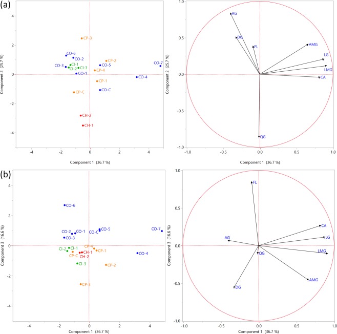
To achieve a better understanding of differences and similarities between the samples of Cecropia species, correlation and principal component analysis (PCA) was performed. With regard to the chemical composition, eight categories were considered: chlorogenic acid (CA), luteolin C-glycosydes/luteolin C,O-glycosydes (LG), apigenin C-glycosydes/apigenin C,O-glycosydes (AG), luteolin malonyl-C-glycosydes (LMG), apigenin malonyl-C,O-glycosydes (AMG), diosmetin C,O-glycosydes (DG), quercetin O-glycosydes (QG) and flavonolignans (FL) (see Supplementary Table S6).
Association between variables (chemical composition) was determined by the analysis of the sample correlation coefficient (See Supplementary Fig. S5). The CA level showed positive correlations with the content of LG (0.76), LMG (0.54) and AMG (0.36). Similarly, LG revealed a high association with LMG (0.72) and AMG (0.47); and FL with AG (0.40). In contrast, a negative correlation (−0.57) between AG and QG was observed. These results indicated that high concentrations of O-glycosides correlated with a low content of apigenin C-glycosides and vice-versa.
Two-dimensional PCA score and loading plots from Fig. 7 shows that the first three components (PC1, PC2 and PC3) accounted for 79.0 % of the cumulative variability of the original variables. The PCA results showed that C. hispidissima individuals (CH-1 and CH-2) were characterized by a strongly negative score on PC2. The low score of PC2 corresponds to higher values of QG and lower levels of AG. This result allowed to distinguish C. hispidissima from other Cecropia species. PC3 was able to separate C. obtusifolia samples (except for CO-4) from the other species. High scores of PC3 corresponded to a relatively high flavonolignans content.
Figure 7.
PCA score plots and loading plots of contents in Cecropia species, (a) PC1 vs. PC2 and (b) PC1 vs. PC3.
Conclusions
The present study was designed to describe the optimal experimental conditions for maximizing the extraction efficiency of leaves from plants of the genus Cecropia based on an experimental design and to validate an appropriate method to simultaneously quantify the polyphenolic compounds, including CA, TF and FL, in authentic and commercial samples by using high-performance liquid chromatography with diode-array detection (HPLC-DAD). Although there are some reports about analytical work from Cecropia species, previous investigations have not comprehensively considered the process optimization for improved phenolic compound recovery and the quantitative determination of flavonolignans in leaves from different species of this genus. Therefore, the methodology described in this work includes for the first time several parameters, such as extraction solvent, temperature and time, among others, combined with statistical tools in order to determine optimal extraction conditions and quantification of main phenolic constituents from Cecropia. The use of response surface methodology (RSM) was selected in this research because it can broadly be applied, due to its advantages in comparison with classical approaches (one-factor-at-a-time method), such as the capability of collecting information on many quantitative variables at once, fitting an adequate mathematical function to the experimental data and assessing the interaction effect between the parameters on the response. The results of this investigation show that solvent concentration and temperature had a significant influence on extraction, then optimization of these parameters was essential to obtain accurate and efficient recovery of the Cecropia leaves constituents. The optimized parameters were determined to be a temperature of 64 °C, methanol fractions (70%, v/v), extraction time of 30 min, 3 extractions a mass/solvent ratio of 1:30 (w/v) and particle size of ≤125 µm. Variations between predicted and measured recoveries below 2.0% were observed.
The HPLC–DAD method showed adequate validation parameters such as specificity, linearity, precision, accuracy, and limits of detection and quantification on ng/mL scale. It could be concluded that the HPLC–DAD method is reliable and adequate for the determination of the chemical composition of Cecropia samples, as an important tool for the quality control of derived commercial products. Our investigation may be useful for the discrimination of C. obtusifolia and C. hispidissima from the other species, based on their content of flavonolignans, apigenin C-glycosides, and quercetin O-glycosides.
Methods
Reagents
Methanol, acetonitrile, acetone, hexane (all HPLC grade), absolute ethanol and glacial acetic acid (both analytical reagent grade) were acquired from Fisher Chemical (Leicestershire, UK). Formic acid (98+%, pure, analytical reagent grade) was obtained from Acros OrganicsTM (Geel, Belgium). Ultrapure water with a resistivity of 18.2 MΩ • cm at 25 °C (Milli-Q, Waters) was used as extraction solvent and for mobile phase preparation.
Chlorogenic acid (CA) (99.0%) and rutin (RU) (96.9%) were obtained from Sigma-Aldrich (St. Louis, MO), while isovitexin (IV), orientin (OT), isoorientin (IO) (all with purity ≥99%) were from Extrasynthese (Genay, France). Vitexin (99.7%) was purchased from Adipogen (Liestal, Switzerland). All these compounds/substances were used as external standards. Flavonolignan 1, flavonolignan 2 and flavonolignan 3 (isolated from C. obtusifolia CO-1) were used as reference material.
Plant material
The leaves of four Cecropia species were collected in West Panama, Panama and Chiriqui Provinces, Republic of Panama (See supplementary Fig. S1). The species were identified by the botanist Orlando O. Ortiz. The plant materials were collected in October 2015, July 2016 and July 2017. Vouchers of each collection were deposited at the Herbarium of the University of Panama
The plant material was air-dried in a general protocol oven (HerathermTM, Thermo Scientific, MA, USA) at 40 °C and subsequently grounded using a mill (MF 10 Basic, IKA, Staufen, Germany). For extraction optimization and the validation of analytical methodology steps, all samples (C. obtusifolia, C. peltata, C. insignis and C. hispidissima) were pooled in equal amounts.
In addition, three commercial products were purchased online: Guarumbo Tea 200 g (Nopalife, batch No. 5000 603, México, Cecropia obtusifolia leaves) (CO-C), Embauba tea powder (NaturVitae, batch No. GEMPWD11123, Perú, Cecropia peltata leaves) (CP-C), and Umbaúba (Embaúba) leaves and stems (Chá & Cía, batch No. 052016, Brazil, C. hololeuca leaves and stems) (CHO-C).
Sample preparation
The powdered plant material was sieved through appropriate sieves (mesh ≤710, ≤355 or ≤125 µm) and weighed in a 50 mL conical tube (VWR, Radnor, USA). Each extraction was carried out by placing the samples in an ultrasonic bath (42 kHz, 100 W) (Branson 3510, Danbury, USA). Parameter combinations were decided by a design of experiments and the extractions conducted under these designed conditions. After extraction was completed, the extract was centrifuged for 5 min at 3000 g using a Heraeus Labofuge 400 Centrifuge (Langenselbold, Germany). The supernatants were cleaned-up with an Extract CleanTM 15 mL reservoir (Grace, Deerfield, IL, USA) containing 2 g Diaion HP20 resin (Supelco, Bellefonte, PA, USA). Samples were collected in a 50.0 mL volumetric flask and subsequently diluted in a ratio 1:2 with 10% methanol. Samples were stored at −20 °C prior to analysis, if necessary.
HPLC-DAD and HPLC-DAD-MS analysis
HPLC-DAD analysis was carried out on an Agilent 1200 series system with degasser, quaternary pump, automatic injection, thermostatic column compartment and a DAD (Agilent Technologies, Santa Clara, CA, USA). For analysis, 20 µL sample extract was injected on an RP-18 Kinetex column (2.10 × 100 mm, 2.6 μm, Phenomenex, Torrence, CA, USA). Aqueous formic acid (0.1%, v/v) and acetonitrile with 0.1% formic acid were used as mobile phases A and B, respectively. The gradient program was set as follows: 10% B (0–5 min), 10–15% B (5–20 min), 15% B (20–30 min), 15–25% B (30–40 min), 25% B (40–45 min), 25–40% B (45–55 min), 40% B (55–60 min), 40–100% B (60–65 min), 100% B (65–70 min), 100–10% B (70–75 min), 10% B (75–85 min). A flow rate of 0.7 mL/min was used. The column temperature was maintained at 26 °C. The DAD signal was recorded between 190 and 500 nm. TF and CA were monitored at 340 nm, while FL was detected at 390 nm. The analyte peak areas were used as responses for the optimization of the extraction process, while mass fraction (m/m) of CA, TF and FL from the plant material were used for the validation of the analytical method. TF was reported as the sum of all responses corresponding to flavonoids (Fig. 1).
An LC-DAD-MS system was used for the evaluation of the specificity of the HPLC-UV method and to confirm the chemical composition of the Cecropia species. Mass spectra were recorded using a Finnigan LXQ Mass spectrometer (Thermo Fisher Scientific, CA, USA) coupled with a Finnigan Surveyor LC system (LC Pump Plus, Autosampler Plus and PDA Plus detector). The same chromatographic conditions as described above were applied. During analysis, full scan data were recorded in ESI (−) mode from m/z 100 to 1800. The source, capillary and tube lens voltages; sheath and auxiliary gas flows; and the capillary temperature were set as follows: −4.0 kV, −8.0 V and −75.49 V; 65.0 L/h and 14.0 L/h; and 350 °C. DAD spectra were recorded between 200 and 400 nm. Data was analyzed using Thermo Xcalibur software (version 3.0).
Extraction optimization
Ultrasound-Assisted Extraction (UAE)
An ultrasound instrument (Branson 3510, Danbury, USA) with a volume of 5.7 L, a frequency of 42 kHz and an input power of 100 W was used in the experiments. The temperature and time of extraction were monitored with a thermometer (Testo 925, Almere, Netherlands) and a timer (Fisher Scientific, CA, USA).
Experimental Designs
Factors affecting UAE were screened using a Fractional Factorial Design (FFD) (27-3). Afterwards, based on the results of the screening step a Central Composite Design (CCD) was applied to determine the best combination of the important extraction variables32,33.
Fractional Factorial Design (FFD): Seven factors, two extreme levels per factor and a center point (all in duplicate) were selected. The factors were methanol fraction (MeOH %) (50 or 90%, v/v), extraction time (30 or 90 min), number of extractions with methanol (1 or 3), extraction temperature (20 or 60 °C), plant solvent ratio (1:20 or 1:100, m/v), number of extractions with acetone (0 or 2), and particle size (≤710 or ≤125 µm). FFD is shown in Supplementary Table S1.
The statistical interpretation of the estimated effect of each factor (Ex) was performed as described by Klein-Junier et al.34. Briefly, a t-test was used to evaluate which factors have significant effects. The standard error of the effect (SE)e was estimated based on Dong’s algorithm using the 75% lowest absolute factor effects. Unimportant factors are selected by an initial error estimation (S0). Finally, the critical effect (Ecritical) for the response was estimated based on (SE)e.
Central Composite Design (CCD): After the most important factors for extraction were determined in the screening step, variables were further optimized. Two factors at five levels were varied according to a CCD design in order to optimize the extraction process. The two factors were methanol concentration (%, v/v) (X1) and extraction temperature (°C) (X2). The factor levels with α = 1.414 are shown in Supplementary Table S2, while the design and the responses are given in supplementary Table S3.
A quadratic polynomial model used in the response surface analysis was established for each response using the following equation:
| 1 |
where y is the response variable, and b0, bi, bii and bij are the regression coefficients of the model representing intercept, linear, quadratic, and interaction terms, respectively; Xi and Xj are the independent variable functions; and k the number of variables (k = 2).
Validation of the analytical method
Validation studies were carried out in accordance with the guidelines of the International Conference on the Harmonization (ICH) of Technical Requirements for the Registration of Pharmaceuticals for Human Use, about the validation of analytical procedures35. Validation was performed in terms of specificity, linearity, precision, accuracy, limit of detection and limit of quantification.
Specificity
Specificity was evaluated in order to identify potential interferences by other analytes or other compounds at the chromatographic region of interest. Analytical standards (CA, VX, IV, OT, IO and RU) and sample extracts from different plant collections were analyzed to assess the selectivity of the method at the retention times of CA, flavonoids and flavonolignans within the analytical conditions established. Mass spectrometry was used to identify co-eluting substances and to determine their relevance under routine conditions. Additionally, as an indirect approach, specificity was evaluated by demonstrating acceptable accuracy (see Accuracy).
Linearity
The linearity of the method was evaluated by the calibration curves obtained by the HPLC analysis of CA, VX and RU. For the preparation of the standard solutions, a stock solution was prepared by transferring 5 mg of CA, VX and RU in a 5.0 mL volumetric flask, adding MeOH to dissolve and adjust to volume. The working standard solutions were obtained by diluting the stock solution in 10% MeOH solution to produce 0.02, 0.2, 1.6, 8.0, 20, 50, 125, 200, 350, 450, 550 and 650 µg/mL aliquots. The stock solution was stored at −20°C and the working standard solutions were prepared daily. Three determinations (n = 3) were carried out for each solution and each calibration point was fitted by linear regression. The most appropriate concentration range was determined. First-order calibration curve equations (y = a + bx), coefficients of determination (r2), residual values, 95% confidence intervals (CI) of the intercept, and t-tests (N-2, α = 0.05) for slope and intercept were calculated.
Additionally, the Mandel’s fitting test was used for mathematical verification of the first-order regression model36. See supplementary S. Methods.
Precision
The precision of the method was assessed for each analyte as repeatability (intra-day precision) and the intermediate precision (between days and concentration levels) during four different days and three concentration levels (50, 100 and 150%). Six replicates (n = 6) were performed for each working solution. The results were expressed as percentage relative standard deviation (% RSD).
The acceptability of the results was evaluated by the Horwitz equation37,38:
| 2 |
| 3 |
where RSDr is the expected coefficient of variation under repeatability conditions; RSDR is the expected coefficient of variation under intermediate precision conditions; and C is the concentration of analyte expressed as a dimensionless mass fraction (m/m, where numerator and denominator have the same units).
Accuracy
Accuracy was determined by recovery (%) at three concentration levels (75, 100 and 125%) in triplicate (n = 3). The experiment was performed by adding known quantities of analytical standards (CA, VX and RU) to plant material extracts (mixture of Cecropia species) prepared at 50% of center point of the linear range. The recovery and 95% confidence interval (CI) were estimated.
The % RSD should be in the order of coefficient of variation for intermediate precision conditions (RSDR), and the recovery % is not significantly different from 100% when the 95% CI includes 100%. Additionally, a nominal range from 98 to 102% was set as recovery %.
Limit of detection (LoD) and limit of quantification (LoQ)
The limits of detection and of quantification were estimated based on analytical calibration curves containing the analytes (CA, VX and RU) spiked to the sample extracts. The standard deviation of the intercept was used to express the experimental error.
Additionally, these mathematical estimations of LoD and LoQ were subsequently validated by analyzing samples known to be near detection and quantification limits. Six determinations (n = 6) were performed for each concentration. Then, the lower concentrations with appropriate signal/noise ratio (S/N > 2–3.5) and precision (% RSD ≤ 5.0) were adopted as LoD and LoQ for each analyte, respectively.
Application of the HPLC-DAD method
The proposed HPLC-DAD method was used to assess the contents of CA, TF and FL in fourteen authentic Cecropia leaves and three commercial products. CA, TF and FL were determined by using an external standard method, using CA (20.12 µg/mL), VX (20.32 µg/mL) and RU (18.69 µg/mL) as references. Samples were prepared in triplicate. Analytical results were reported by ± SD, where is the mean of results and SD is the standard deviation of measurements. Results were expressed in µg/g of dried-leaves weight.
Identity of chemical constituents was determined by comparison of their retention times with authentic reference standards or isolated compounds, and by UV/DAD spectral data. Additionally, individual flavonoids identity was confirmed by analysis of their mass spectral data.
Statistical analysis
Data processing, calculations and graphic plotting were performed using Microsoft Office Excel 2010 (version 14.0.7), MODDE® Pro Software (version 11.0.1, MKS Umetrics, Malmö, Sweden) and GraphPad Prism for Windows (version 6.01, La Jolla, CA, USA).
The CA, TF and FL contents of Cecropia species were analyzed applying Principal component analysis (PCA) using the software JMP Pro 13 (SAS Institute, Cary, NC, USA).
Supplementary information
Acknowledgements
The authors gratefully acknowledge the scholarship received for Mr. Rivera’s Doctoral studies from the National Secretariat for Science, Technology and Innovation (SENACYT) and the Institute for Training and Development of Human Resources (IFARHU) of the Republic of Panama. Thanks are also due to SENACYT for the financial support to Dr. Caballero-George through the incentive program of the National Investigation System (SNI) as well as through grant FID14-116.
Author Contributions
L.P., S.A. and C.C. conceived the project; A.R. performed the main experiments, data analysis, and wrote the manuscript; T.N. assisted with validation procedures; Y.V.H., G.B. and F.K. contribute with the design of experiment (DOE); E.F. assisted with principal component analysis (PCA). L.P., S.A., S.B. and K.F. supervised the work. All authors reviewed and approved the manuscript.
Competing Interests
The authors declare no competing interests.
Footnotes
Publisher’s note: Springer Nature remains neutral with regard to jurisdictional claims in published maps and institutional affiliations.
Electronic supplementary material
Supplementary information accompanies this paper at 10.1038/s41598-018-37607-2.
References
- 1.Franco-Rosselli P, Berg CC. Distributional patterns of Cecropia (Cecropiaceae): A panbiogeographic analysis. Caldasia. 1997;19:285–296. [Google Scholar]
- 2.Rivera-Mondragón A, et al. Selection of chemical markers for the quality control of medicinal plants of the genus Cecropia. Pharm. Biol. 2017;55:1500–1512. doi: 10.1080/13880209.2017.1307421. [DOI] [PMC free article] [PubMed] [Google Scholar]
- 3.Revilla-Monsalve MC, Andrade-Cetto A, Palomino-Garibay MA, Wiedenfeld H, Islas-Andrade S. Hypoglycemic effect of Cecropia obtusifolia Bertol aqueous extracts on type 2 diabetic patients. J. Ethnopharmacol. 2007;111:636–640. doi: 10.1016/j.jep.2007.01.014. [DOI] [PubMed] [Google Scholar]
- 4.Caballero-George C, et al. Biological screening of selected medicinal Panamanian plants by radioligand-binding techniques. Phytomedicine. 2001;8:59–70. doi: 10.1078/0944-7113-00011. [DOI] [PubMed] [Google Scholar]
- 5.Müller SD, et al. Anti-inflammatory and antioxidant activities of aqueous extract of Cecropia glaziovii leaves. J. Ethnopharmacol. 2016;185:255–262. doi: 10.1016/j.jep.2016.03.007. [DOI] [PubMed] [Google Scholar]
- 6.Nayak BS. Cecropia peltata L (Cecropiaceae) has wound-healing potential: a preclinical study in a Sprague Dawley rat model. Int. J. Low. Extrem. Wounds. 2006;5:20–26. doi: 10.1177/1534734606286472. [DOI] [PubMed] [Google Scholar]
- 7.Uchôa VT, de Paula RC, Krettli LG, Santana AEG, Krettli AU. Antimalarial activity of compounds and mixed fractions of Cecropia pachystachya. Drug Dev. Res. 2010;71:82–91. [Google Scholar]
- 8.Luengas-Caicedo PE, Braga FC, Brandão GC, De Oliveira AB. Seasonal and intraspecific variation of flavonoids and proanthocyanidins in Cecropia glaziovi Sneth. leaves from native and cultivated specimens. Zeitschrift fur Naturforsch. C., J. Biosci. 2007;62:701–709. doi: 10.1515/znc-2007-9-1013. [DOI] [PubMed] [Google Scholar]
- 9.Ospina Chávez J, Rincón Velanda J, Guerrero Pabón M. Perfil neurofarmacológico de la fracción butanólica de las hojas de Cecropia peltata L. Rev. Colomb. Cienc. Quím. Farm. 2013;42:244–259. [Google Scholar]
- 10.Andrade-Cetto A, Wiedenfeld H. Hypoglycemic effect of Cecropia obtusifolia on streptozotocin diabetic rats. J. Ethnopharmacol. 2001;78:145–149. doi: 10.1016/S0378-8741(01)00335-X. [DOI] [PubMed] [Google Scholar]
- 11.Pacheco NR, et al. Cecropia pachystachya: A species with expressive in vivo topical anti-inflammatory and in vitro antioxidant effects. Biomed Res. Int. 2014;2014:1–10. doi: 10.1155/2014/301294. [DOI] [PMC free article] [PubMed] [Google Scholar]
- 12.Lacaille-Dubois MA, Franck U, Wagner H. Search for potential angiotensin converting enzyme (ACE)-inhibitors from plants. Phytomedicine. 2001;8:47–52. doi: 10.1078/0944-7113-00003. [DOI] [PubMed] [Google Scholar]
- 13.Costa GM, Ortmann CF, Schenkel EP, Reginatto FH. Seasonal variations in the amount of isoorientin and isovitexin in Cecropia glaziovii Sneth. leaves over a two-year period. Rev Colomb Cienc Quím Farm. 2014;43:162–172. doi: 10.15446/rcciquifa.v43n1.45471. [DOI] [Google Scholar]
- 14.Arend DP, et al. Experimental design as a tool to evaluate chlorogenic and caffeic acids extracted from Cecropia glaziovii Sneth. J. Pharm. Biomed. Anal. 2011;54:58–68. doi: 10.1016/j.jpba.2010.08.008. [DOI] [PubMed] [Google Scholar]
- 15.Tanae MM, Lima-Landman MT, De Lima TC, Souccar C, Lapa A. Chemical standardization of the aqueous extract of Cecropia glaziovii Sneth endowed with antihypertensive, bronchodilator, antiacid secretion and antidepressant-like activities. Phytomedicine. 2007;14:309–313. doi: 10.1016/j.phymed.2007.03.002. [DOI] [PubMed] [Google Scholar]
- 16.Beringhs AO, Souza FM, de Campos AM, Ferraz HG, Sonaglio D. Technological development of Cecropia glaziovi extract pellets by extrusion-spheronization. Brazilian J. Pharmacogn. 2013;23:160–168. doi: 10.1590/S0102-695X2012005000123. [DOI] [Google Scholar]
- 17.Oliveira RR, et al. High-speed countercurrent chromatography as valuable tool to isolate C -glycosylflavones from Cecropia lyratiloba Miquel. Phytochem. Anal. 2003;14:96–99. doi: 10.1002/pca.688. [DOI] [PubMed] [Google Scholar]
- 18.Brango-Vanegas J, et al. Glycosylflavonoids from Cecropia pachystachya Trécul are quorum sensing inhibitors. Phytomedicine. 2014;21:670–675. doi: 10.1016/j.phymed.2014.01.001. [DOI] [PubMed] [Google Scholar]
- 19.Duque AP, et al. In vivo wound healing activity of gels containing Cecropia pachystachya leaves. J. Pharm. Pharmacol. 2016;68:128–138. doi: 10.1111/jphp.12496. [DOI] [PubMed] [Google Scholar]
- 20.Andrade-Cetto A, Vázquez RC. Gluconeogenesis inhibition and phytochemical composition of two Cecropia species. J. Ethnopharmacol. 2010;130:93–97. doi: 10.1016/j.jep.2010.04.016. [DOI] [PubMed] [Google Scholar]
- 21.Costa GM, Ortmann CF, Schenkel EP, Reginatto FH. An HPLC-DAD method to quantification of main phenolic compounds from leaves of Cecropia species. J. Braz. Chem. Soc. 2011;22:1096–1102. doi: 10.1590/S0103-50532011000600014. [DOI] [Google Scholar]
- 22.da Silva Mathias M, Rodrigues de Oliveira R. Differentiation of the phenolic chemical profiles of Cecropia pachystachya and Cecropia hololeuca. Phytochem. Anal. 2018;24:1–10. doi: 10.1002/pca.2791. [DOI] [PubMed] [Google Scholar]
- 23.Kurt, H. Handbook of Chemical and Biological Plant Analytical Methods. (Wiley, 2014).
- 24.Andersen, O. M. & Markham, K. R. Flavonoids. Chemistry, Biochemistry and Applications. (Taylor & Francis, 2006).
- 25.Chemat F, et al. Ultrasound assisted extraction of food and natural products. Mechanisms, techniques, combinations, protocols and applications. A review. Ultrason. Sonochem. 2017;34:540–560. doi: 10.1016/j.ultsonch.2016.06.035. [DOI] [PubMed] [Google Scholar]
- 26.Pico Y. Ultrasound-assisted extraction for food and environmental samples. Trends Anal. Chem. 2013;43:84–99. doi: 10.1016/j.trac.2012.12.005. [DOI] [Google Scholar]
- 27.Vinatoru M. An overview of the ultrasonically assisted extraction of bioactive principles from herbs. Ultrason. Sonochem. 2001;8:303–313. doi: 10.1016/S1350-4177(01)00071-2. [DOI] [PubMed] [Google Scholar]
- 28.Do Q, et al. Effect of extraction solvent on total phenol content, total flavonoid content, and antioxidant activity of Limnophila aromatica. J. Food Drug Anal. 2014;22:296–302. doi: 10.1016/j.jfda.2013.11.001. [DOI] [PMC free article] [PubMed] [Google Scholar]
- 29.Grujic N, Lepojevic Z, Srdjenovic B, Vladic J, Sudji J. Effects of different extraction methods and conditions on the phenolic composition of mate tea extracts. Molecules. 2012;17:2518–2528. doi: 10.3390/molecules17032518. [DOI] [PMC free article] [PubMed] [Google Scholar]
- 30.Ermer, J. & Miller, J. H. M. Method Validation in Pharmaceutical Analysis: A guide to best practice. (Wiley-VCH, 2005).
- 31.Ortmann CF, et al. LC/QTOF profile and preliminary stability studies of an enriched flavonoid fraction of Cecropia pachystachya Trécul leaves with potential antidepressant-like activity. Biomed. Chromatogr. 2017;31:1–13. doi: 10.1002/bmc.3982. [DOI] [PubMed] [Google Scholar]
- 32.NIST/SEMATECH. Engineering Statistics Handbook. Available at, http://www.itl.nist.gov/div898/handbook/. (Accessed: 9th February 2017) (2003).
- 33.Bezerra M, Santelli R, Oliveira E, Villar L, Escaleira L. Response surface methodology (RSM) as a tool for optimization in analytical chemistry. Talanta. 2008;76:965–977. doi: 10.1016/j.talanta.2008.05.019. [DOI] [PubMed] [Google Scholar]
- 34.Klein-Júnior LC, et al. The use of chemometrics to study multifunctional indole alkaloids from Psychotria nemorosa (Palicourea comb. nov.). Part I: Extraction and fractionation optimization based on metabolic profiling. J. Chromatogr. A. 2016;1463:60–70. doi: 10.1016/j.chroma.2016.07.030. [DOI] [PubMed] [Google Scholar]
- 35.International Conference on Harmonisation (ICH) Q (R1), Validation of analytical procedures: Text and methodology. (2005).
- 36.Funk, W., Dammann, V. & Donnevert, G. Quality Assurance in Analytical Chemistry: Applications in Environmental, Food and Materials Analysis, Biotechnology, and Medical Engineering. (Wiley-VCH, 10.1002/9783527609444 2007).
- 37.Decleer M, Rajkovic A, Sas B, Madder A, De Saeger S. Development and validation of ultra-high-performance liquid chromatography–tandem mass spectrometry methods for the simultaneous determination of beauvericin, enniatins (A, A1, B, B1) and cereulide in maize, wheat, pasta and rice. J. Chromatogr. A. 2016;1472:35–43. doi: 10.1016/j.chroma.2016.10.003. [DOI] [PubMed] [Google Scholar]
- 38.European Commission. 2002/657/EC: Commissiom decision of 12 August 2002 implementing Council Directive 96/23/EC concerning the performance of analytical methods and the interpretation of results. Available at, https://publications.europa.eu/en/publication-detail/-/publication/ed928116-a955-4a84-b10a-cf7a82bad858/language-en. (Accessed: 14th July 2018). (2002).
Associated Data
This section collects any data citations, data availability statements, or supplementary materials included in this article.


