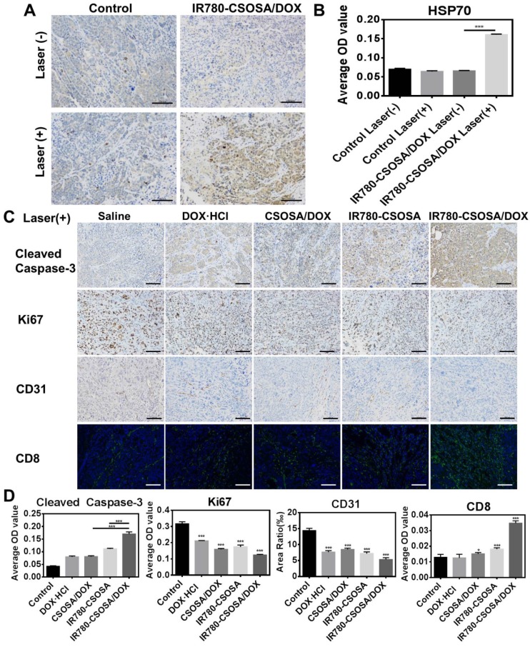
Figure 8.
In vivo immunohistochemical analysis in MCF-7 xenograft tumors treated with IR780-CSOSA/DOX micelles-mediated chemo-photothermal therapy. (A) HSP70 protein expression (brown) in saline and IR780-CSOSA/DOX with or without laser groups. Scale bar: 100 μm. (B) The average OD value of HSP70 levels. (n = 3). (C) Under laser irradiation, the induction of apoptosis by staining with cleaved caspase-3 antibody (brown). Cell proliferation evaluation by Ki67 staining (brown). Tumor blood vessel staining with CD 31 antibody (brown). CD 8+ T cells detected by immunofluorescent staining (green). Scale bar: 100 μm. (D) Under laser irradiation, the quantitative analysis of cleaved caspase-3, Ki67, CD31 and CD8 levels. (n = 3). The conditions of laser irradiation are all at 808 nm with 0.5 W/cm2 for 3 min. ***P < 0.001, *P < 0.05

