Abstract
Background
The ongoing Middle East respiratory syndrome coronavirus (MERS-CoV) infections pose threats to public health worldwide, making an understanding of MERS pathogenesis and development of effective medical countermeasures (MCMs) urgent.
Methods
We used homozygous (+/+) and heterozygous (+/−) human dipeptidyl peptidase 4 (hDPP4) transgenic mice to study the effect of hDPP4 on MERS-CoV infection. Specifically, we determined values of 50% lethal dose (LD50) of MERS-CoV for the 2 strains of mice, compared and correlated their levels of soluble (s)hDPP4 expression to susceptibility, and explored recombinant (r)shDPP4 as an effective MCM for MERS infection.
Results
hDPP4+/+ mice were unexpectedly more resistant than hDPP4+/− mice to MERS-CoV infection, as judged by increased LD50, reduced lung viral infection, attenuated morbidity and mortality, and reduced histopathology. Additionally, the resistance to MERS-CoV infection directly correlated with increased serum shDPP4 and serum virus neutralizing activity. Finally, administration of rshDPP4 led to reduced lung virus titer and histopathology.
Conclusions
Our studies suggest that the serum shDPP4 levels play a role in MERS pathogenesis and demonstrate a potential of rshDPP4 as a treatment option for MERS. Additionally, it offers a validated pair of Tg mice strains for characterizing the effect of shDPP4 on MERS pathogenesis.
Keywords: Middle East respiratory syndrome coronavirus, MERS pathogenesis, human DPP4, transgenic mice, medical countermeasures for MERS
We demonstrated that elevated levels of circulating soluble human (sh)DPP4 positively correlated with the resistance to MERS-CoV infection and identified the potential of recombinant shDPP4 as a treatment option for MERS.
Middle East respiratory syndrome (MERS) is an emerging infectious disease caused by a coronavirus (MERS-CoV), first identified in Saudi Arabia in 2012, that has since spread to 27 mostly surrounding countries, resulting in more than 2229 laboratory-confirmed cases of infection and 791 deaths (approximately 36%), as of June 2018 [1]. The pandemic potential of this infection calls for a better understanding of MERS pathogenesis and the development of effective medical countermeasures (MCMs) for humans.
Like other human CoVs, MERS-CoV uses an exoaminopeptidase, human dipeptidyl peptidase 4 (DPP4), as the entry receptor for infection of permissive cells [2]. DPP4, also known as CD26, is involved in many physiological functions via its ubiquitous expression in a variety of tissues, its propensity to interact with adenosine deaminase and other important regulatory molecules of the immune system, and its intrinsic proteolytic activity that cleaves many biologically active peptides or proteins that contain proline or alanine at the penultimate position [3, 4]. Not only is DPP4 expressed as a type II transmembrane glycoprotein, primarily on endothelial and epithelial cells and subsets of immune cells, but it is also present in a functionally intact soluble form (sDPP4) in the circulation and other body fluids [3, 4].
Because wild type mice are not susceptible to MERS-CoV, we established a heterozygous (+/−) transgenic (Tg) mouse model globally expressing hDPP4 for studies of MERS pathogenesis and development of MCMs against MERS-CoV infection [5, 6]. To ensure a steady and cost-effective supply of the animals, a Tg mouse model with homozygous expression of hDPP4, designated hDPP4+/+ Tg mice, was developed through mating of hDPP4+/− mice and used as breeders for generating offspring of both genotypes of hDPP4 Tg mice. Because hDPP4 is the functional MERS-CoV receptor, the doubling of encoded hDPP4 gene in hDPP4+/+ Tg mice could render them more susceptible than their hDPP4+/− counterparts to MERS-CoV infection and disease. To our surprise, we found that hDPP4+/+ mice are more resistant than their hDPP4+/− counterparts to MERS-CoV infection. We subsequently found that an increased expression of functionally intact soluble hDPP4 (shDPP4) in the circulation of hDPP4+/+ Tg mice, relative to that of hDPP4+/− mice, was associated with this increased resistance and might be, at least in part, accountable for the seemingly counterintuitive findings on susceptibility to MERS-CoV infection. This notion was supported by studies showing that elevated shDPP4 levels, brought about by administration of recombinant shDPP4 (rshDPP4), resulted in increased resistance of recipient hDPP4+/− mice to MERS-CoV. Together, our results indicate that manipulation of shDPP4 might serve as a strategy for counteracting MERS-CoV infection and disease in humans.
METHODS
Human DPP4 Transgenic Mice
hDPP4+/− transgenic mice were established, as previously reported [5, 6]. hDPP4+/+ breeder mice were derived by mating 2 parental hDPP4+/− mice. The homozygosity was determined by quantitative polymerase chain reaction analysis of tail DNA (data not shown) and verified by their subsequent mating with wild-type (wt) mice. Only those mice uniformly yielding heterozygous offspring were selected as hDPP4+/+ breeders. Interbreeding between hDPP4+/+ mice produced additional hDPP4+/+ mice, whereas backcrossing them to wt mice generated hDPP4+/− mice.
Viral Infection, Isolation, and Titration
All of the in vitro and animal studies involving infectious MERS-CoV were conducted at the biosafety level 3 (BSL3) laboratory and animal BSL3 facilities at the Galveston National Laboratory in accordance with approved protocols and the guidelines and regulations of the National Institutes of Health (NIH) and Association for Assessment and Accreditation of Laboratory Animal Care (AAALAC). Detailed methodologies for viral infection, isolation from infected lungs and brain, and determination of infectious viral loads have been established and routinely used in our laboratory [5, 6]. The original stock of MERS-CoV EMC-2012 strain, a gift of Heinz Feldmann (NIH, Hamilton, MT) and Ron A. Fouchier (Erasmus Medical Center, Rotterdam, Netherlands), was expanded in Vero E6 cells 3 times consecutively. Passage 3 containing a titer of approximately 5 × 10⁶ 50% cell culture infectious dose (TCID50)/mL of virus was used throughout the study.
Determination of 50% Lethal Dose
The 50% lethal dose (LD50) values for hDPP4+/+ and hDPP4+/− mice was determined by using traditional virus dilution assays and the Reed-Muench method, as we previously described [6]. Briefly, groups of 4 young (6–8 weeks) or old (7–10 months) hDPP4+/+ and hDPP4+/− mice were inoculated, via intranasal route, with dosages of EMC-2012 MERS-CoV in 10-fold decrements from 102 to 10–1 TCID50 in a volume of 60 µL. Mice were monitored daily for clinical manifestations (weight loss) and mortality for at least 21 days postinfection (dpi). LD50 values for each strain of mice were estimated based on the ratio of the surviving mice to the total inoculated mice, as previously described [6]. Those surviving for more than 21 days were also evaluated for specific antibody responses to MERS-CoV receptor binding domain (RBD) protein by enzyme-linked immunosorbent assay (ELISA) [6]. Only those showing specific antibody to RBD were considered as “MERS-CoV infected.”
Quantification of Circulating Soluble Human DPP4 in Tg Mouse Sera
To quantify the circulating shDPP4 in the sera of naive DPP4+/+, DPP4+/−, and DPP4−/− mice, a commercial ELISA-based assay was used, following the manufacturer’s instructions (eBioscience catalog No. BMS235). Absorbance at 450 nm in 96-well plates was read in an ELISA plate reader (Molecular Device).
Serological and Microneutralization Assays
ELISA-based and Vero E6 cell-based microneutralization assays, previously described [7], were used to determine the titers of MERS-CoV RBD-specific serum IgG and neutralizing antibodies in hDPP4 Tg mice in response to MERS-CoV infection.
Binding Specificity and Anti-MERS-CoV Activity of rshDPP4 in Tissue Cultures
Purified insect cell-derived human DPP4 ectodomain (residues 39–766; GenBank accession no. NP_001926.2) containing an N-terminal human CD5 signal peptide and a C-terminal His6 tag, as we previously described and characterized [7, 8], was prepared and used for treatment studies. Testing binding specificity of the rshDPP4 to the RBD proteins of MERS-CoV and severe acute respiratory syndrome coronavirus (SARS-CoV; both RBDs were generous gifts of Drs Du and Jiang at New York Blood Center, NY) was determined in ELISA-based assays [7, 9]. For determining the capacity of rshDPP4 to inhibit MERS-CoV infection in vitro we initially used our standard microneutralization procedure with cytopathic effect (CPE) inhibition as the endpoint. These assays revealed a dose-dependent reduction of CPE at 72 hours, ranging from less than 5% for 100, 50, and 25 μg/mL and gradually increased to approximately 30% for 12.5 μg/mL of rshDPP4. In addition to standard microneutralization assay with a CPE endpoint, we measured the antiviral effect of rshDPP4 using virus yield of each rshDPP4 dilution from 100 to 0.8 μg/mL, expressed as log10 TCID50/mL.
Administration of rshDPP4 to Mice Before and After Challenge With MERS-CoV
The effect of rshDPP4 for inhibiting MERS-CoV infection in Tg mice was determined using hDPP4+/− mice in 2 pilot studies with 2 different batches of rshDPP4 showing similar, but not identical, binding capacity to MERS-CoV RBD and in vitro neutralizing activity. Briefly, groups of hDPP4+/− mice (n = 3 per group) were treated twice with either 100 µg or 400 µg of rshDPP4 or phosphate-buffered saline (PBS) as control, via the intraperitoneal route 2 hours before (−2 hours) and 24 hours after (+24 hours) infection (intranasal) with 103 TCID50 of MERS-CoV. Mice were sacrificed at 3 dpi to assess infectious viral loads and histopathology in the lungs.
Histopathology
Inflated lung specimens and brain tissues were fixed in 10% neutral buffered formalin for 48 hours before paraffin embedding and processing for routine hematoxylin and eosin stain (H&E) to assess the histopathology, as we previously described [5, 6].
Statistical Analysis
Statistical analyses were performed using GraphPad Prism software. Neutralizing antibody titers and virus titers were averaged for each group of mice and compared using Students t test, 1-way ANOVA, or others as indicated.
RESULTS
hDPP4+/+ Mice are More Resistant Than DPP4+/− Mice to MERS-CoV Infection and Disease
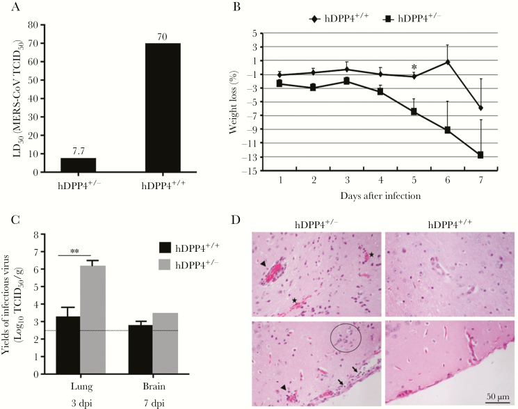
For the initial comparison of the susceptibility of hDPP4+/+ and hDPP4+/− mice to MERS-CoV, we determined the LD50 values for mice 7–10 months of age, as we previously described [6]. Because hDPP4 is the functional receptor of MERS-CoV, we anticipated that DPP4+/+ mice might be more, or at least equally, permissive as DPP4+/− mice to MERS-CoV infection. To our surprise, we found that hDPP4+/+ mice were more resistant than hDPP4+/− mice as indicated by LD50 values of 4.3 and 32.4 TCID50 of MERS-CoV for hDPP4+/− and hDPP4+/+ mice, respectively. To confirm this seemingly counterintuitive finding and rule out any potential effect of age and gender, we repeated the study using age- (6–8 weeks old) and sex-matched Tg mice of both genotypes. Shown in Figure 1A is a representative of 2 independently performed experiments that confirmed the LD50 difference; values for the hDPP4+/− and hDPP4+/+ mice were 7.7 and 70.0 TCID50 of MERS-CoV, respectively, indicating that hDPP4+/+ mice are more resistant than their age- and sex-matched hDPP4+/− counterparts to MERS-CoV infection.
Figure 1.
Homozygous human dipeptidyl peptidase (hDPP4+/+) transgenic (Tg) mice are more resistant than heterozygous hDPP4 (hDPP4+/−) Tg mice to Middle East respiratory syndrome-associated coronavirus (MERS-CoV) infection and disease. A, The LD50 values of age- and sex-matched hDPP4+/+ and hDPP4+/− mice. Data shown are representative of 2 independently performed studies. B, Kinetics of weight loss of hDPP4+/+ (n = 9) and hDPP4+/− (n = 11) mice in response to infection with 103 TCID50 of MERS-CoV. *P < 0.05. C, Titers of infectious virus recovered from MERS-CoV (103 TCID50)-infected hDPP4+/+ and hDPP4+/− mice at 3 (lungs) and 7 (brains) dpi. **P < .01 (t test, lung titers at 3 dpi), D, Histopathological changes were detectable in MERS-CoV (103 TCID50)-infected hDPP4+/−, but not hDPP4+/+, mice at 7 dpi, and consisted of infiltration of mononuclear cells within the meninges (lower panel, arrows), perivascular cuffing (upper and lower panel, arrow head), microglial nodule (lower panel, circle), microhemorrhage (upper panel, asterisks), and cell death at the junctions of gray-white matter (upper panel). Abbreviations: dpi, days postinfection; LD50, 50% lethal dose; TCID50, 50% cell culture infectious dose.
Using sera collected 21 dpi from each strain of Tg mice that survived the lower challenge dosages, we quantified MERS-CoV RBD-specific IgG antibodies by ELISA. We found that infection had occurred in Tg mice of both strains. Infection rates for those given 100 TCID50 were similar (1/1 for DPP4+/− mice and 2/2 for DPP4+/+ mice) and 10 TCID50 (2/2 for each strain) but were greater for DPP4+/− mice (3/3) than DPP4+/+ mice (1/4) for 1 TCID50. The difference in infection rates is consistent with the increased resistance of DPP4+/+ mice described earlier.
To further verify the difference in susceptibility to MERS-CoV infection, we infected (intranasal) hDPP4+/+ (n = 9) and hDPP4+/− (n = 11) Tg mice with an equal dose of MERS-CoV (103 TCID50/per mouse) and monitored them daily for morbidity (weight loss) and mortality. Three mice of each strain, unless indicated otherwise, were euthanized at 3, 5, and 7 dpi to assess infectious viral titers and the histopathology of lungs and brains. In contrast to DPP4+/− mice, which exhibited marked weight loss, starting at 3–4 dpi, and 2 deaths at 6 dpi (data not shown), infected hDPP4+/+ mice exhibited minimal weight changes and uniformly survived through 7 dpi when the experiment was terminated (Figure 1B). When the viral loads were measured at 3 dpi, we readily recovered infectious virus from the lungs, but not the brains, of all 3 hDPP4+/− mice examined, but from the lung of only 1 of 3 hDPP4+/+ mice. Although we usually recover virus from lungs of some mice, efforts to recover infectious virus from both lung and brain specimens of both strains of Tg mice at 5 dpi were unsuccessful (data not shown); however, we were able to retrieve infectious virus from the brain (but not lungs) of the sole hDPP4+/− survivor and from all 3 hDPP4+/+ mice that survived to 7 dpi. The virus titer in the brain was 106.2/g for the single hDPP4+/− mouse, a titer significantly higher than the average of 103.7/g for 3 hDPP4+/+ mice (Figure 1C). This ability to recover infectious virus from the lungs approximately 2–3 days earlier than from the brains is consistent with the pattern, kinetics, and tissue distribution of MERS-CoV infection in DPP4 Tg mice we have previously reported [6]. We also compared the histopathology of lungs and brains, 2 of the prime targets of MERS-CoV infection of hDPP4 Tg mice [5]. Although infected DPP4+/− mice elicited mild-to-moderate histopathological changes within the lungs at 3 dpi after a dose of 103 TCID50 of MERS-CoV infection, as in our earlier study [6], infected DPP4+/+ mice exhibited reduced or no lung histopathology (data not shown). Brain histopathology at 7 dpi for the sole hDPP4+/− survivor had infiltrations of mononuclear cells in the meninges (Figure 1D, below), perivascular cuffing (Figure 1D, above and below), microglial nodules (Figure 1D, below), microhemorrhage (Figure 1D, above) and cell death at the junctions of gray and white matter (Figure 1D, above) but not in DPP4+/+ mice. Collectively, the significant differences between these 2 strains of mice in their LD50 values, seroconversion rates, viral loads, weight loss, and histopathology support the notion that DPP4+/+ mice are more resistant than DPP4+/− mice to MERS-CoV infection.
hDPP4+/+ Mice Exhibit Significantly Higher Levels of Soluble hDPP4 in Sera Than Those of hDPP4+/− Mice
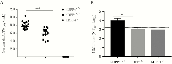
Because functionally active sDPP4 exists in the circulation and other body fluids of humans [10, 11], we explored whether the levels shDPP4 expression in the sera of these 2 strains of hDPP4 Tg mice could be different, thereby contributing to their difference in susceptibility to MERS-CoV infection. Using a commercial ELISA-based analysis, we were unable to detect shDPP4 in age- and sex-matched hDPP4-negative (hDPP4−/−) littermates. However, as shown in Figure 2A, a representative of 2 independently performed studies, an average of 8.1 ± 0.2 μg/mL (mean ± SD) and 6.2 ± 0.4 μg/mL of shDPP4 was detected in hDPP4+/+ (n = 16) and DPP4+/− mice (n = 10), respectively. As shDPP4 retains its binding specificity to MERS-CoV RBD protein [7], the significantly different expression of shDPP4 in the sera of these 2 strains of Tg mice (P < .001) prompted us to examine if elevated shDPP4 expression might relate to the higher resistance of DPP4+/+ to MERS-CoV infection by possibly acting like a decoy that binds MERS-CoV RBD and prevents virus infection. Using microneutralization tests, we noted that the 50% neutralization titers (NT50), expressed as the geometric mean titers (GMT), were 3.9 ± 0.3 and 3.1 ± 0.1 for hDPP4+/+ and hDPP4+/−mice, respectively (P = .026), compared to those of hDPP4−/− mice which were uniformly below the limit of detection (i.e., ~3.0) (Figure 2B).
Figure 2.
Sera of naive human dipeptidyl peptidase 4 (hDPP4+/+ ) mice contain significantly higher levels of soluble hDPP4 (shDPP4) that exhibit higher levels of neutralizing antibody-like activity than those of naive hDPP4+/− mice. A, Groups of at least 10 age- and sex-matched hDPP4+/+, hDPP4+/−, and their transgene-negative (hDPP4−/−) littermates were subjected to retroorbital bleeding to assess the contents of shDPP4, using commercially available ELISA kits that quantify specific hDPP4 (hDPP4+/+ vs hDPP4 +/−, P < .001 t test). B, Sera obtained from naive hDPP4+/+ and hDPP4+/− mice (n = 10 of each) and hDPP4−/− mice (n = 6) were subjected to the standard Vero E6-based microneutralization tests to determine their potential to neutralize MERS-CoV. Data are presented as the geometric mean neutralization titers (GMT log2) of 50% neutralization titer (NT50). A 1–10 dilution of sera is approximately 3.4 log2. (* P = .026, t test and Mann-Whitney Rank Sum Test, when compared to those of hDPP4+/− mice). The GMT titers of hDPP4−/− mice were uniformly below the limit of detection. Abbreviations: ELISA, enzyme-linked immunosorbent assay; MERS-CoV, Middle East respiratory syndrome-associated coronavirus. Dotted line, limit of detection.
Administration of Recombinant Soluble hDPP4 Significantly Inhibits MERS-CoV Infection in hDPP4+/− Mice
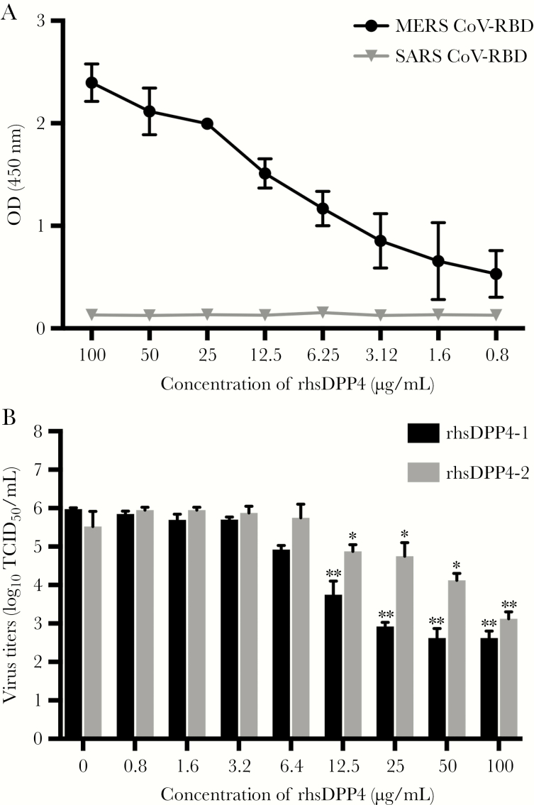
Because the significantly higher expression of shDPP4 with better neutralizing activity might contribute to the increased resistance of hDPP4+/+ mice to MERS-CoV infection, we explored whether an increased shDPP4 expression might increase resistance of naive hDPP4+/− mice to MERS-CoV infection. Using our limited amount of insect cell-derived rshDPP4, known to specifically bind to RBD of MERS-CoV but not SARS-CoV, and to neutralize MERS-CoV in vitro in a dose-dependent manner (Figure 3A and 3B), we administered 100 μg of rshDPP4, via the intraperitoneal route, into each of 3 DPP4+/− mice 2 hours before (−2 hours) and 24 hours after (+ 24 hours) infection with 103 TCID50 of MERS-CoV. The effect of the rshDPP4 against MERS-CoV infection was assessed at 3 dpi by using the titers of infectious virus within the lungs as the end point for this pilot study. While all of 3 PBS-treated mice exhibited moderate titers of live virus, we were unable to recover infectious virus from any of 3 rshDPP4-treated mice (Table 1, Experiment 1). Encouraged by the preliminary data, we generated another batch of rshDPP4 shown to inhibit MERS-CoV infection in a dose-dependent manner (Figure 3B) to repeat the experiment. We gave groups of 3 DPP4+/− mice either 100 or 400 μg of rshDPP4, or PBS (as control) 2 hours before and 24 hours after virus infection as before. To determine if administration of rshDPP4 would increase the circulating levels of shDPP4, serum levels of shDPP4 in mice prior to and 2 hours after the first administration and before challenge with MERS-CoV were measured. We found that titers at 0 and 2 hours of Tg mice given 100 μg (6.6 ± 0.4 versus 6.6 ± 0.9 μg/mL), were similar to those of PBS-treated mice (7.5 ± 0.8 versus 7.3 ± 1.0 μg/mL). However, mice treated with 400 μg of rshDSPP4 showed significantly increased titers 2 hours after treatment (6.6 ± 0.6 increased to 13.7 ± 0.8 μg/mL; P = .002, t test). These results suggest that an increased serum level of shDPP4 can be achieved by administration of rshDPP4 in a dose-dependent manner.
Figure 3.
Recombinant soluble human dipeptidyl peptidase 4 (rshDPP4) proteins derived from insect cells specifically bind to receptor binding domain (RBD) protein of Middle East respiratory syndrome-associated coronavirus (MERS-CoV), but not severe acute respiratory syndrome coronavirus (SARS-CoV), and are capable of neutralizing MERS-CoV in a dose-dependent manner. A, Binding specificity of rshDPP4 to MERS-CoV RBD. ELISA 96-well plates, precoated with RBD protein of MERS-CoV or SARS-CoV, were used to determine the binding specificity of rshDPP4 by using the standard ELISA-based assays. Absorbance was measured at a wavelength of 450 nm. B, The dose-dependent neutralizing activities of 2 different batches of rshDPP4 (rshDPP4-1 and -2) against MERS-CoV. The modified Vero E6-based microneutralization test (virus yield) was used to quantify the neutralizing antibody-like capacity of rshDPP4, as described earlier [12] and in the Methods. *P < .05; **P < .01. Abbreviations: ELISA, enzyme-linked immunosorbent assay; OD, optical density; TCID50, 50% cell culture infectious dose.
Table 1.
Effect of Administration of Recombinant Soluble Human DPP4 on MERS-CoV Infection Within the Lungs of Infected DPP4+/− Micea
| Treatments | Experiment 1 | Experiment 2 |
|---|---|---|
| Lung Viral Titers (TCID50/mL) | ||
| PBS | 2.7 | 3.7 |
| 3.6 | 4.5 | |
| 3.4 | 4.8 | |
| 3.2 ± 0.3b | 4.3 ± 0.3b | |
| rshDPP4 (100 μg) | ≤2.4c | 2.8 |
| ≤2.4 | 3.7 | |
| ≤2.4 | 3.6 | |
| ≤2.4b,d (P = .038) | 3.4 ± 0.3b (P = .077) | |
| rshDPP4 (400 μg) | NT | ≤2.4c |
| 2.7 | ||
| 2.8 | ||
| 2.6 ± 0.1e (P = .011) | ||
Abbreviations: dpi, days postinfection; LD50, 50% lethal dose; MERS-CoV, Middle East respiratory syndrome coronavirus; NT, not tested; PBS, phosphate-buffered saline; rshDPP4, recombinant soluble human dipeptidyl peptidase 4; TCID50, 50% cell culture infectious dose.
aMice (n = 3 each group) were given either 100 μL of PBS or PBS containing 100 or 400 μg of rshDPP4/per mouse by the intraperitoneal route 2 hours before and 24 hours after intranasal challenge with 100 LD50 (approximately 103 TCID50) of MERS-CoV. Lung infectious viral titers were quantified at 3 dpi using a standard Vero E6 cell-based infectivity assay.
bMean ± SE.
cNone detected (limit of detection was 2.5 log10 TCID50/g).
d P = .038 (t test) using 2.4 as the value for nondetectable samples.
e P = .011, 1-way ANOVA.
In addition to the viral loads within the lungs at 3 dpi, the pulmonary histopathology was examined to investigate the effect of rshDPP4 on MERS-CoV infection. Unlike the first study in which treatment with 2 doses of 100 μg (at −2 hours and +24 hours) of rshDPP4 fully protected against MERS-CoV infection, we were able to recover reduced titers of infectious virus from each of 3 mice given 100 μg of the second batch of rshDPP4 when compared to those of PBS-treated controls (P = .077, t test). However, as shown in Table 1, Experiment 2, the titers of infectious virus in mice treated with 400 μg of rshDPP4 were significantly reduced from an average of 4.3 ± 0.3 (mean ± SE) in control mice to 2.6 ± 0.1 TCID50/g (P = .011, t test). This finding is consistent with the reduced potency of batch 2 of rshDPP4, as shown in Figure 3B. However, the histopathology in mice treated with the high dose of rshDPP4 (ie, 400 μg) was reduced as well when compared to that of the PBS controls (data not shown).
DISCUSSION
In this study we found that hDPP4+/+ mice were more resistant than hDPP4+/− mice to MERS-CoV infection, as evidenced by approximately 10-fold increases of LD50, reduced infectious viral yields, and seroconversion rates, as well as less weight loss and lower mortality than their age- and sex-matched hDPP4+/− counterparts (Figure 1). We also found that hDPP4+/+ mice had significantly higher levels of shDPP4 in their circulation than did hDPP4+/− mice (Figure 2). Moreover, these higher serum levels of shDPP4 exhibited higher titers of neutralizing activity against MERS-CoV in Vero E6 cell-based assays. Finally, we showed that administration with functionally active rhsDPP4 proteins (Figure 3) enabled hDPP4+/− mice to better resist MERS-CoV infection in a dose-dependent manner (Table 1), a finding in accordance with increased levels of shDPP4 in their circulation. Taken together, these results support the notion that increasing the levels of shDPP4 is a potential option for counteracting MERS-CoV infection and disease in humans.
DPP4, ubiquitously expressed on many types of cells and tissues, has been well characterized as critically involved in regulating many important physiological functions, in part through its intrinsic enzymatic activity and propensity to interact with other key regulatory molecules of the immune system [3, 13]. As the functional receptor that mediates entry of MERS-CoV to permissive host cells, the membrane-associated hDPP4 plays a pivotal role in MERS-CoV infection and disease. However, specific role(s) that shDPP4 might have in MERS pathogenesis remain much less understood. While the levels of sDPP4 vary significantly, even among healthy individuals, it has been shown that the intensities of sDPP4 expression in the circulation, along with its intrinsic enzymatic activity, could be a factor in dictating the severity of many human diseases, including malignancies, autoimmune and inflammatory diseases, diabetes mellitus and other metabolic syndromes, and chronic infectious disease such as AIDS and hepatitis C [4, 10, 11, 14, 15]. It has been recently reported that serum levels of sDPP4 expression in confirmed MERS patients were significantly reduced when compared to those of healthy individuals [16]; however, the suggestion that these reduced levels could serve as biomarkers for susceptibility requires knowledge regarding the levels in MERS cases before onset of infection and disease. In addition, further studies of the therapeutic value of shDPP4 as either a significant resistance factor or a potential countermeasure for MERS-CoV in humans is warranted. Of note, the soluble forms of the viral receptors for several other viruses, including those caused by SARS-CoV, rhinovirus, and HIV, have been proposed as potentially effective antiviral therapeutics [17–19].
We showed in this study that sera derived from naive hDPP4 Tg mice of either strain, especially hDPP4+/+, possess detectable neutralizing antibody-like activity against MERS-CoV (Figure 2B). Whether the significantly higher shDPP4 expression of hDPP4+/+ mice could be solely accountable for its greater resistance to MERS-CoV infection through functioning as receptor decoys seems unlikely, because it took at least 12.5 μg of rshDPP4, a level higher that the approximately 8 μg/mL in sera of hDPP4+/+ mice, to significantly inhibit MERS-CoV infection in Vero E6 cells (Figure 3B). Additional studies are needed to better understand the shDPP4-related protective mechanisms against MERS-CoV, especially those of the immune system. However, the validated direct correlation between the level of shDPP4 and the susceptibility to MERS-CoV infection, as shown in this study, may provide a possible genetic basis for the observed wide spectrum of diseases, ranging from asymptomatic, mild-to-moderate, to severe infection and death, in MERS patients [20, 21].
With the limited supplies of rshDPP4, we have shown in 2 independently performed proof-of-principle studies that administration of exogenous rshDPP4 might be a treatment option for MERS-CoV infection (Table 1). Additional studies are required to determine if increasing shDPP4 levels by rshDPP4 treatment could be a useful treatment option for human MERS. The study presented in this report demonstrates the usefulness of this homozygous and heterozygous pair of hDPP4 Tg mice to fully explore the interactions between hDPP4 and MERS-CoV infection and disease, studies that could lead to identification of novel molecular and cellular targets for MCMs against MERS-CoV infection and disease in humans.
Presented in part: International Meeting for NIDO Virus, Kansas City, 2017; and Annual Meeting of American Society of Virology, College Park, MD, 2018.
Notes
Financial support. This work was supported by the National Institute of Allergy and Infectious Diseases, National Institutes of Health (grant numbers R21AI113206 to C.-T. K. T. and R01AI110700 to F.L).
Potential conflicts of interest. All authors: No reported conflicts of interest. All authors have submitted the ICMJE Form for Disclosure of Potential Conflicts of Interest. Conflicts that the editors consider relevant to the content of the manuscript have been disclosed.
References
- 1. World Health Organization Regional Office for the Eastern Mediterranean. Epidemic and pandemic-prone diseases. MERS situation update, June 2018 http://www.emro.who.int/pandemic-epidemic-diseases/mers-cov/mers-situation-update-june-2018.html. Accessed 12 July 2018. [Google Scholar]
- 2. Raj VS, Mou H, Smits SL, et al. . Dipeptidyl peptidase 4 is a functional receptor for the emerging human coronavirus-EMC. Nature 2013; 495:251–4. [DOI] [PMC free article] [PubMed] [Google Scholar]
- 3. Klemann C, Wagner L, Stephan M, von Hörsten S. Cut to the chase: a review of CD26/dipeptidyl peptidase-4’s (DPP4) entanglement in the immune system. Clin Exp Immunol 2016; 185:1–21. [DOI] [PMC free article] [PubMed] [Google Scholar]
- 4. Wagner L, Klemann C, Stephan M, von Hörsten S. Unravelling the immunological roles of dipeptidyl peptidase 4 (DPP4) activity and/or structure homologue (DASH) proteins. Clin Exp Immunol 2016; 184:265–83. [DOI] [PMC free article] [PubMed] [Google Scholar]
- 5. Agrawal AS, Garron T, Tao X, et al. . Generation of a transgenic mouse model of Middle East respiratory syndrome coronavirus infection and disease. J Virol 2015; 89:3659–70. [DOI] [PMC free article] [PubMed] [Google Scholar]
- 6. Tao X, Garron T, Agrawal AS, et al. . Characterization and demonstration of the value of a lethal mouse model of Middle East respiratory syndrome coronavirus infection and disease. J Virol 2016; 90:57–67. [DOI] [PMC free article] [PubMed] [Google Scholar]
- 7. Du L, Kou Z, Ma C, et al. . A truncated receptor-binding domain of MERS-CoV spike protein potently inhibits MERS-CoV infection and induces strong neutralizing antibody responses: implication for developing therapeutics and vaccines. PLoS One 2013; 8:e81587. [DOI] [PMC free article] [PubMed] [Google Scholar]
- 8. Yang Y, Du L, Liu C, et al. . Receptor usage and cell entry of bat coronavirus HKU4 provide insight into bat-to-human transmission of MERS coronavirus. Proc Natl Acad Sci U S A 2014; 111:12516–21. [DOI] [PMC free article] [PubMed] [Google Scholar]
- 9. Lu G, Hu Y, Wang Q, et al. . Molecular basis of binding between novel human coronavirus MERS-CoV and its receptor CD26. Nature 2013; 500:227–31. [DOI] [PMC free article] [PubMed] [Google Scholar]
- 10. Mamgain S, Mathur S, Kothiyal P. Imunomodulatory activity of DPP4. J Pharmacol Clin Toxicol 2013; 1:1006. [Google Scholar]
- 11. Lambeir AM, Durinx C, Scharpé S, De Meester I. Dipeptidyl-peptidase IV from bench to bedside: an update on structural properties, functions, and clinical aspects of the enzyme DPP IV. Crit Rev Clin Lab Sci 2003; 40:209–94. [DOI] [PubMed] [Google Scholar]
- 12. Agrawal AS, Tao X, Algaissi A, et al. . Immunization with inactivated Middle East respiratory syndrome coronavirus vaccine leads to lung immunopathology on challenge with live virus. Hum Vaccin Immunother 2016; 12:2351–6. [DOI] [PMC free article] [PubMed] [Google Scholar]
- 13. Morimoto C, Schlossman SF. The structure and function of CD26 in the T-cell immune response. Immunol Rev 1998; 161:55–70. [DOI] [PubMed] [Google Scholar]
- 14. Nohtomi K, Terasaki M, Kohashi K, Hiromura M, Nagashima M, Hirano T. A dipeptidyl peptidase-4 inhibitor directly suppresses inflammation and foam cell formation in monocytes/macrophages beyond incretins. Diabetologia 2014; 57(Suppl 1):S373. [Google Scholar]
- 15. Itou M, Kawaguchi T, Taniguchi E, Sata M. Dipeptidyl peptidase-4: a key player in chronic liver disease. World J Gastroenterol 2013; 19:2298–306. [DOI] [PMC free article] [PubMed] [Google Scholar]
- 16. Inn KS, Kim Y, Aigerim A, et al. . Reduction of soluble dipeptidyl peptidase 4 levels in plasma of patients infected with Middle East respiratory syndrome coronavirus. Virology 2018; 518:324–7. [DOI] [PMC free article] [PubMed] [Google Scholar]
- 17. Hofmann H, Geier M, Marzi A, et al. . Susceptibility to SARS coronavirus S protein-driven infection correlates with expression of angiotensin converting enzyme 2 and infection can be blocked by soluble receptor. Biochem Biophys Res Commun 2004; 319:1216–21. [DOI] [PMC free article] [PubMed] [Google Scholar]
- 18. Huguenel ED, Cohn D, Dockum DP, et al. . Prevention of rhinovirus infection in chimpanzees by soluble intercellular adhesion molecule-1. Am J Respir Crit Care Med 1997; 155:1206–10. [DOI] [PubMed] [Google Scholar]
- 19. Gardner MR, Kattenhorn LM, Kondur HR, et al. . AAV-expressed eCD4-Ig provides durable protection from multiple SHIV challenges. Nature 2015; 519:87–91. [DOI] [PMC free article] [PubMed] [Google Scholar]
- 20. Saad M, Omrani AS, Baig K, et al. . Clinical aspects and outcomes of 70 patients with Middle East respiratory syndrome coronavirus infection: a single-center experience in Saudi Arabia. Int J Infect Dis 2014; 29:301–6. [DOI] [PMC free article] [PubMed] [Google Scholar]
- 21. Arabi YM, Balkhy HH, Hayden FG, et al. . Middle East respiratory syndrome. N Engl J Med 2017; 376:584–94. [DOI] [PMC free article] [PubMed] [Google Scholar]


