As our knowledge of biologic therapies used in cardiovascular regenerative medicine grows, our focus is turning to developing a bioartificial heart that can alleviate the shortage of donor hearts. Using knowledge gained during 5 decades' worth of work on mechanical total artificial hearts (TAHs), we are developing tissue-engineered hearts for transplantation.
The field of cardiovascular regenerative medicine began in the 1990s, when the first attempts were made to treat advanced heart disease with the use of genes or cells. Since then, biologic therapies have shown promise in slowing the progression of cardiovascular disease, so their use is gradually being accepted; however, they are unlikely to reverse end-stage organ failure. Heart transplantation remains the definitive treatment for the failing heart.
As early as the 1960s, when the heart was perceived only as a pump, mechanical devices were being designed to replace a failing organ. The need to build an efficient, functional replacement for transplantable organs has become urgent because clinicians are able to prolong the lives of patients who have acute myocardial infarctions or chronic heart failure, but they cannot cure them. Therefore, an engineered bioartificial heart—a living organ manufactured for transplantation—is viewed as a viable way to overcome shortages of donor organs.
Many insights from mechanical TAH development can be applied to engineering a bioartificial heart. Appropriate sizing, interdependency of the organ's left and right sides, antithrombogenicity, and durability are important design criteria for any transplantable heart. However, the use of cells as an energy source, the need for vascularization, and the capability for endogenous repair are unique to a bioartificial organ. Another essential component is a sterile bioreactor system that can feed and support the organ as it matures in a sterile environment at controlled temperatures and atmospheric pressures.
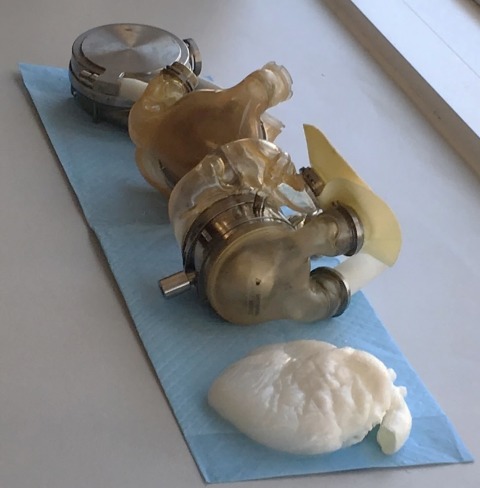
A normal, healthy heart comprises specialized cells (cardiomyocytes, endothelium, fibroblasts, neurons, and so on) that are integrated into a scaffold of extracellular matrix (ECM) (Fig. 1), richly infiltrated by a dense network of cardiac vessels.1 Much research has focused on finding cell sources and a suitable vascularized scaffold. Stem cells, initially envisioned for cardiac repair, are a natural source for this milieu. As stem cell biology has progressed, so have methods of growing stem cells, which are being used in vitro to populate cardiac patches and even whole organs. Now that inducible pluripotent stem cell technology has been introduced, autologous tissue engineering is imaginable. However, hearts need hundreds of billions of cardiomyocytes and other cells, and generating that number of cells is a hurdle.
Fig. 1.

Photograph shows mechanical heart-assist devices tested at the Texas Heart Institute (THI) next to a bioartificial decellularized heart (front) with extracellular matrix developed at THI.
Of course, cells alone do not constitute an organ. During development, cells coalesce with ECM in a given orientation to form functional units that are unique to each organ. From our perspective, bioengineering an organ involves recombining cells with ECM of an adult organ from which cells have been removed. This decellularized ECM provides appropriate architectural and biological cues to support form and function.2 It retains organ complexity and structure, including fiber orientation and macro- and microarchitecture, as well as vascular channels to deliver nutrients,3,4 thus enabling full-thickness tissue generation. Furthermore, this ECM contains biologically active molecules that support cell migration, proliferation, and differentiation. Finally, the dynamic reciprocity between cells and the ECM enables both to evolve as functional needs change.
To overcome the challenges of generating enough cells to build a whole heart that must then remain sterile in vitro, we are exploring in vivo organ engineering involving decellularized heart scaffolds (with or without re-endothelialized vessels) and the body's capacity to repopulate the scaffolds with cells. Specifically, we have implanted decellularized porcine hearts into living pigs in a heterotopic position. We have found that the surgical circuit created, generated a pressure gradient for, and promoted flow crucial to prevent coagulation and increase graft patency. In addition, short-term implantation promoted endothelial cell adhesion to the vessel lumina, and long-term implantation promoted tissue formation with evidence of cardiomyocytes and recellularized vessels within the graft.5 By adding load and electrical input, possibly with use of temporary mechanical assistance or a pacemaker, we think that vascular and parenchymal cell maturation can occur.
We hope that these and other efforts will enable tissue engineering to contribute to heart transplantation in the future.
References
- 1.Taylor DA, Parikh RB, Sampaio LC. Bioengineering hearts: simple yet complex. Curr Stem Cell Rep. 2017;3(1):35–44. doi: 10.1007/s40778-017-0075-7. [DOI] [PMC free article] [PubMed] [Google Scholar]
- 2.Taylor DA, Sampaio LC, Ferdous Z, Gobin AS, Taite LJ. De-cellularized matrices in regenerative medicine. Acta Biomater. 2018;74:74–89. doi: 10.1016/j.actbio.2018.04.044. [DOI] [PubMed] [Google Scholar]
- 3.Rana D, Zreiqat H, Benkirane-Jessel N, Ramakrishna S, Ramalingam M. Development of decellularized scaffolds for stem cell-driven tissue engineering. J Tissue Eng Regen Med. 2017;11(4):942–65. doi: 10.1002/term.2061. [DOI] [PubMed] [Google Scholar]
- 4.Ott HC, Matthiesen TS, Goh SK, Black LD, Kren SM, Netoff TI, Taylor DA. Perfusion-decellularized matrix: using nature's platform to engineer a bioartificial heart. Nat Med. 2008;14(2):213–21. doi: 10.1038/nm1684. [DOI] [PubMed] [Google Scholar]
- 5.Taylor DA, Frazier OH, Elgalad A, Hochman-Mendez C, Sampaio LC. Building a total bioartificial heart: harnessing nature to overcome the current hurdles. Artif Organs. 2018;42(10):970–82. doi: 10.1111/aor.13336. [DOI] [PubMed] [Google Scholar]


