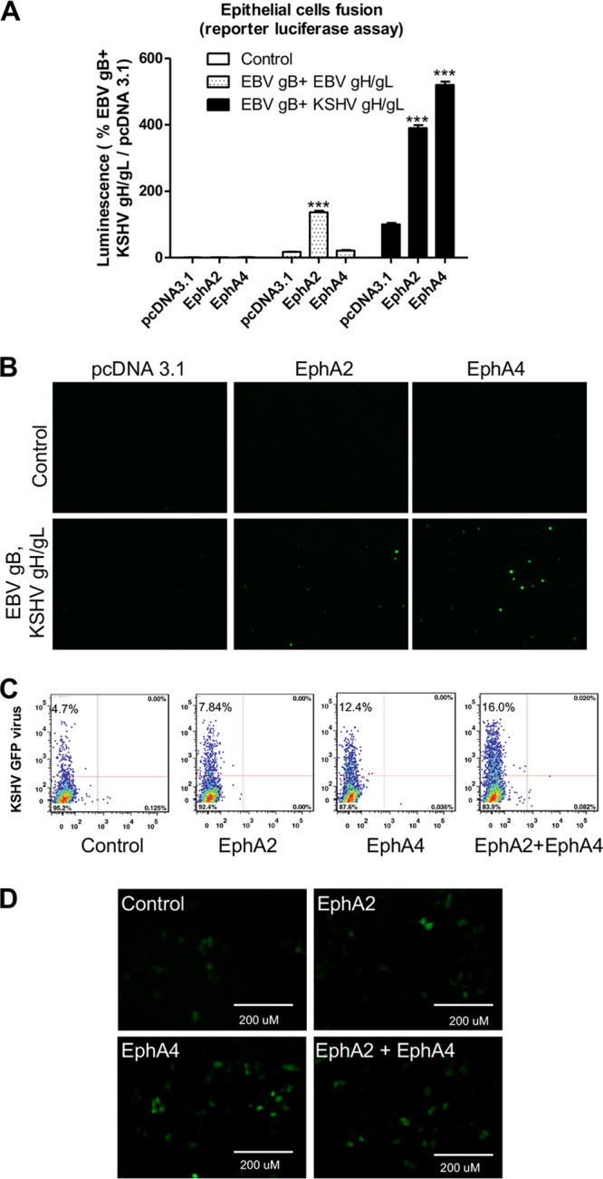
FIG 1.

EphA4 promotes both KSHV infection and virus-free cell-cell fusion. (A) CHO-K1 cells were transfected with T7 luciferase plasmid and either control plasmid or EBV gH/gL, EBV gB, or KSHV gH/gL. Transfected CHO-K1 cells were overlaid with HEK293T cells transfected with pcDNA3.1, EphA2, or EphA4 together with T7 polymerase. Fusion activity was standardized to EBV gB and KSHV gH/gL fusion with HEK293T cells transfected with control pcDNA3.1, which was set to 100%. ***, P < 0.001 (ANOVA followed by post hoc Tukey’s multiple-comparison test), compared to pcDNA 3.1. (B) A total of 2.5 × 10⁵ CHO-K1 cells transfected with Rluc81-7 plasmid together with either control plasmid, EBV gH/gL with EBV gB, or KSHV gH/gL with EBV gB, were overlaid with 2.5 × 10⁵ CHO-K1 cells transfected with pcDNA3.1, EphA2, or EphA4 together with Rluc88-11. Green cells, indicative of fusion, were visualized and captured with an EVOS fluorescence microscope. (C) HEK293T cells were transfected with pcDNA3.1, EphA2, or EphA4. At 24 h posttransfection, 5 × 10⁴ cells were seeded into a 48-well plate. Twenty-four hours later, the cells were infected with concentrated KSHV. After an additional 24 h, the infected cells were analyzed by flow cytometry (C) or visualized by microscopy and images captured with an EVOS fluorescence microscope (D).
