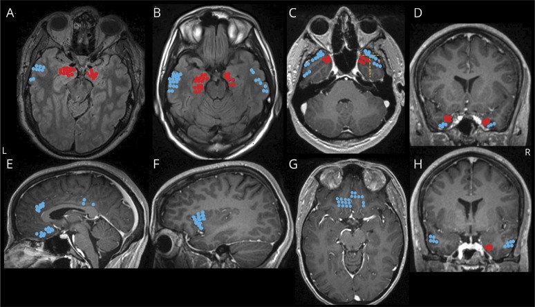
Figure 1. Locations of electrode contacts investigated for structures that produce stimulation-induced apnea.
(A–H) Electrode locations of stimulated electrodes, represented in different views of a single MRI brain for convenience. Locations that produced apnea are shown in red (amygdala [A], hippocampus head and body [B], mesial temporo-polar cortex [C, D, and H], and its subregions marked with a yellow dashed line: temporal tip (anterior), lateral and mesial temporo-polar cortices [C], and parahippocampal gyrus [H]). Locations that did not produce apnea are shown in blue (lateral temporal [A, B, C, and H], orbitofrontal cortex [G], anterior insular [F], anterior and posterior cingulate and subcallosal gyri [H]).

