Abstract
[Purpose] There have been many study ipsilateral erector spinae in regard of prone hip extension (PHE). However, mediating methods have been focusing on the reinforcement of gluteus. Hereupon, this study is intended to identify how an increase of gluteus maximus influences on posterior oblique sling (POS) and suggest a mediating method to effectively reinforce them. This study shows the seclective POS strength exercise. [Participants and Methods] This study has been conducted on normal male (13) and female (13), and participants were asked to proceed PHE exercise and prone hip extension with hip abduction with knee flexion (PHEAKF). Surface electromyography (EMG) was recorded from the contralateral latissimus dorsi, contralateral erector spinae, ipsilateral erector spinae, ipsilateral gluteus maximus, and ipsilateral biceps femoris. A paried t-test was used to compare muscle activity POS. [Results] EMG activity of the contralateral latissimus dorsi, ipsilateral erector spinae, and ipsilateral gluteus maximus was significantly greater performed PHEAKF than PHE. As for ipsilateral biceps femoris, muscle activation was lower in PHEAKF than PHE. [Conclusions] According to the results of this study, increase in muscular activation from the direction of muscular fiber and posterior oblique sling seems to be an important factor that influencontralateral crector spinae on muscular activation of POS.
Key words: Posterior oblique sling, Electromyography, Prone hip extension
INTRODUCTION
Muscular chain is referred to as when muscles are applied together or influence on the kinetic patterns. In the muscular chain, there are synergists, muscle slings, and myofascial chain, and each of them has interdependent relationship with neuron organs and joints. Among them, POS for maintaining the stability of bodipsilateral erector spinae during the cross-walk and delivering the power from lower to upper limb of the body is comprised of contralateral latissimus dorsi, contralateral erector spinae, thoracolumbar fascia, ipsilateral gluteus maximus, and ipsilateral biceps femoris1). POS contributes to the stability of lumbar pelvis, and training for such muscles influencontralateral crector spinae on power of sacroiliac joint. Therefore, it is said to contribute to the stability of lumbar pelvis. If POS is activated while moving the body, it contributes to dynamic stability of lumbar pelvis with small muscles2, 3).
Ipsilateral gluteus maximus and contralateral latissimus dorsi are functionally connected by thoracolumbar fascia that considers mechanism delivered with weight to the diagonal direction towards opposite legs from arms. Such models provide dynamic momentum for exercise program for patients with backpain3). During the functional movement, weakened gluteus maximus and reduced activation might cause excessively activated erector spine muscles4). In order to reinforce gluteus maximus, PHE (prone hip extension) is generally much used5,6,7). In the PHE exercise, exercising for the extension in abduction by 30° over the abduction by 0° on hip joint minimizes the dominance of hamstring reinforcing the muscle of mesogluteus power8). In other studies, it was reported that exercising on extension with abdcution of hip joint with bending knees activated gluteus maximus while reducing the activation of hamstring9). Posture of abduction by 30° on hip joint requires more power on gluteus maximus, and 30° PHEAKF (prone hip extension with abduction with knee flexion) shortens the occurrence of EMG of gluteus maximus8). In the meantime, there were many studies about how to reinforce gluteus maximus. However, there has not been any study dealing with posterior oblique sling from the activation of gluteus maximus. Therefore, it was intended to make changes in activation of muscles from posterior oblique sling from gluteus maximus with PHE that was frequently used in the exercise for gluteus maximus. In this study, selective POS reinforcement can be applied to patients with back pain to porvide adequate exercise.
PARTICIPANTS AND METHODS
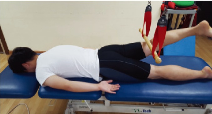
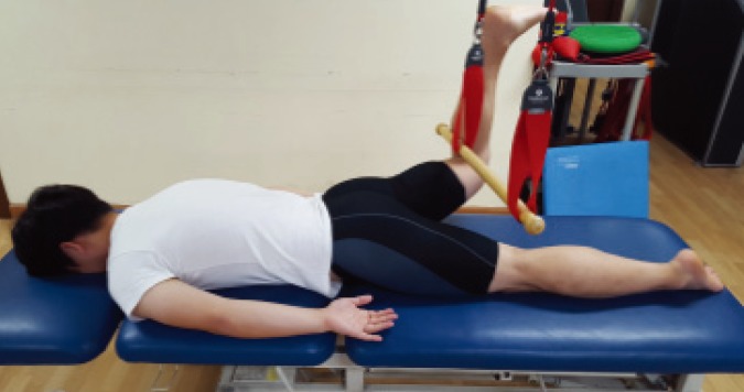
Participants for this study are 26 normal persons who are in D College located at Dae-gu Province, and the researcher classified the participants for study in random allocation method. Participants have general characteristics of average age of 23.15 ± 2.68, average height of 169.03 ± 8.27 cm, and average weight of 62.26 ± 10.20 kg (Table 1). Research purpose and method were explained to all the participants prior to participating in the study, and all participants provided written informed consent according to the ethical standards of the Declaration of Helsinki and agreed to participate in the study. Prior to the experiment, participantss were educated about how to proceed PHE and PHEAKF for about a half an hour10). Dominant leg was determined for the one that was used to mostly kick a ball11). Experiment was proceeded only on participantss with right dominant leg. Muscular activation was randomly proceeded with 10° of extension in hip joint from PHE (Fig. 1). In addition, muscular activation was randomly measured in 10° of extension in hip joint with 90° of knee joints with 30° of abduction on hip joint in PHEAKF (Fig. 2). After proceeding it for 5 seconds, data in the middle 3 seconds except for the first and last 1 second were used. As fatigue accumulated on the muscle when measuring electromyogram could influence on the measured data, there was a rest period for 60 seconds when each experiment ended to prevent them. Each of the methods was to use sealed envelope for randomization, and each of the experiments was repeated for three times. For standardization of each muscular action potential, we used maximal voluntary isometric contraction (MVIC), and employed the method of Kendall et al11). In order to analyze the activation of latissimus dorsi muscle on the opposite side, erector spine muscle on the opposite side and on the same side, gluteus maximus on the same side, and biceps muscle of thigh, radio electromyogram (Telemyo 2400T-G2, Noraxon, USA) was used. The bipolar snap electrode composed with ground electrode, active electrode, and reference electrode is connected as a measuring electrode with the system of electromyogram. Surface electromyogram signals were received from 5 channels, transformed into digital signals by remote control system, and transmitted to the computer in the Bluetooth method. Sampling rate of electromyogram signals was 1,024 Hz, and we used a notch filter for the band pass filter between 20 Hz and 500 Hz and for 60 Hz, and treated and analyzed the collected signals by root mean square (RMS) after full wave rectification. To decrease skin resistance, we sandpapered 3 to 4 times and removed the dead skin cells on the areas where electrodes are to be attached, cleaned the skin by soaked absorbent cotton with rubbing alcohol, placed ground electrodes on the anterior superior iliac spine (ASIS), and attached bipolar surface electrodes on the dominant muscles of the experiment participants depending on the driving direction of muscular fiber. Surface electrodes (Trigno sensors; Delsys) were placed on the latissimus dorsi (4 cm below the inferior tip of the scapula and half the distance between the spine and lateral edge of the torso), erector spinae (approximately 2 cm lateral to the L1 spinous procontralateral crector spinaes and aligned parallel to the spine), gluteus maximus (half the distance between the greater trochanter and second sacral vertebra and on an oblique angle to or slightly above the level of the trochanter), and ipsilateral biceps femoris (2 cm from the lateral border of the thigh and two-thirds the distance between the trochanter and the back of the knee12)).
Table 1. General characteristics of the participants.
| Age (yrs) | Height (cm) | Weight (kg) | |
| participants (n=26) | 23.1 ± 2.6 | 169.0 ± 8.2 | 62.2 ± 10.2 |
All values are mean ± standard deviation (SD).
Fig. 1.
Prone hip extension.
Fig. 2.
Prone hip extension with hip abduction with knee flexion.
Data collected in this study were procontralateral crector spinaesed by using statistical program, Window SPSS ver 20.0 calculating and comparing average and standard deviation of each variable. As for muscular activation, paired t-test was used to identify the difference of posterior oblique sling between PHE and PHEAKF. Set up p<0.05 for the significant level (α).
RESULTS
Latissimus dorsi muscle on the opposite side, erector on the same side, and gluteus maximus turned out to represent higher muscular activation in PHEAKF (p<0.05), and hamstring represented higher muscular activation in PHE (p<0.05) (Table 2).
Table 2. Activity in the muscles of the posterior oblique sling (%MIVC) during PHE and PHEAKF (N=26).
| Muscle | Mean %MVIC(SD) | |
| PHE | PHEAKF | |
| CLD | 14.93 ± 7.25* | 24.00 ± 8.53* |
| CES | 6.82 ± 3.34 | 7.95 ± 3.18 |
| IES | 7.36 ± 4.99* | 9.55 ± 5.84* |
| IGM | 15.00 ± 8.74* | 38.84 ± 13.11* |
| IBF | 55.85 ± 12.58* | 14.36 ± 8.36* |
CLD: contralateral latissimus; CES: contralateral erector spinae; IES: ipsilateral erector spinae; IGM: ipsilateral gluteus maximus; IBF: ipsilateral biceps femoris; PHE: prone hip exension; PHEAKF: prone hip extension with hip abduction with knee flexion.
*p<0.05.
DISCUSSION
In order to prevent and cure back pain, it is required to keep waist from excessively moving while maintaining the spine and pelvis in the optimal position. In addition, muscular contraction on the body is required for stabilization of limbs while bodies move and also for the right patterns of muscular activies. Among muscles that provide stability on the pelvis, imbalance of gluteus maximus, medius, and hamstring might lead to the issue during expansion of hip joint4). Many of the studies have reported that causes of chronic back pain are the imbalance on abdominal areas. In many studies, the role of gluteus was emphasized for the cause of imbalance of abdominal conditions13, 14). Weakened gluteus maximus is frequently seen among patients with dominance of hamstring or changes in the anaplastia of hamstring15). The direction of arrangement of muscular fiber contributes to muscular contraction, and it is efficient when the direction of muscular arrangement and contraction is identical to maximize the muscular activation16,17,18). In order to optimize muscular activation, lever arm length is important. If lever arm becomes longer, muscular strength improves in regard of mechanical advantages. This has been proved in the previous studies19). The length of hamstring in PHEAKF that could change the muscular activation decreased in gluteus maximus, and muscular activation significantly increased with the consistent direction of arrangement of muscular fiber of gluteus maximus (p<0.05), and the muscular activation of hamstring was reduced (p<0.05). As shown in the results by Sakamoto et al.9), extension exercise in the abduction of hip joint while bending knees and lying on belly activated gluteus maximus activation, and the activation, and the activities of hamstring were reduced. Vlemming et al.2) indicated that contralateral latissimus dorsi and ipsilateral gluteus maximus among POS are connected to thoracolumbar fascia delivering the weight through the center of the body and providing stability on the lumbar pelvis. Fascia located between muscle and bone can use the tensile power connecting the body with all limbs as a connecting system for producing power towards outside from the muscular contraction and helping many muscles interact with each other for the movement. In addition, muscular contraction creates power for activated muscle to spread towards outside the origin while delivering such power to other muscles, fascia ligament, articular capsule, and skeletal frame that are located vertically or in parallel. With such a method, power occurs where is away from the origin of muscular contraction3). With such influence, it seems that muscular activation of erector spine muscles on the same side and latissimus dorsi muscle on the opposite side increased. In the study by Kim et al.20), ipsilateral gluteus maximus, ipsilateral erector spinae, and contralateral latissimus dorsi as POS muscles had their muscular activation increased during the PHE on the unstable side, and this result was consistent with this experiment.
Due to the importance of how to reinforce selective muscular power of gluteus maximus, there were many studies conducted frequently to deal with PHE that was good for patients with pain on joints on lower limb because of great stability. However, there was no study dealing with muscular activation of POS from gluteus maximus among PHE exercises in the past. According to the aforementioned results, PHEAKF arranged the direction of muscular fiber of gluteus maximus increasing the muscular activation of latissimus dorsi muscle on the opposite side from the connection of thoracolumbar fascia with increased muscular activation in gluteus maxima. In addition, it seems that fleixon knees lower the activation of hamstring improving the activation of selective latissimus dorsi muscle and gluteus maximus. Therefore, it is believed to be a good exercise program for reinforcing gluteus maximus with degraded function in articulatio sacroiliaca. There are several limitations on this study. First of all, healthy adults were the participantss. Secondly, angle of pelvis and waist was not measured that it was not possible to accurately identify whether muscular activation of gluteus maximus influenced on the movement of lumbar and anteversion of pelvis. Third, speed for lifting hip joint during the expansion was not controlled. Fourth, since exercise was performed in the open chain, there might be different results when exercising in the close chain such as standing or walking. In the follow-up study, these limitations are recommended to be considered to perform the exercise.
Conflict of interest
None.
REFERENCES
- 1.Page P, Frank C, Lardner R: Assessment and treatment of muscle imbalance. The Janda Approach, Human Kinetics, 2010. [Google Scholar]
- 2.Vleeming A, Pool-Goudzwaard AL, Stoeckart R, et al. : The posterior layer of the thoracolumbar fascia. Its function in load transfer from spine to legs. Spine, 1995, 20: 753–758. [PubMed] [Google Scholar]
- 3.Mooney V, Pozos R, Vleeming A, et al. : Exercise treatment for sacroiliac pain. Orthopedics, 2001, 24: 29–32. [DOI] [PubMed] [Google Scholar]
- 4.Sahrmann S: Diagnosis and treatment of movement impairment syndromes, Elsevier Health Sci, 2002. [DOI] [PMC free article] [PubMed] [Google Scholar]
- 5.Cappozzo A, Felici F, Figura F, et al. : Lumbar spine loading during half-squat exercises. Med Sci Sports Exerc, 1985, 17: 613–620. [PubMed] [Google Scholar]
- 6.Distefano LJ, Blackburn JT, Marshall SW, et al. : Gluteal muscle activation during common therapeutic exercises. J Orthop Sports Phys Ther, 2009, 39: 532–540. [DOI] [PubMed] [Google Scholar]
- 7.Wilson J, Ferris E, Heckler A, et al. : A structure review of the role of gluteus maximus in rehabilitation. NZ J Physiother, 2005, 33: 95–100. [Google Scholar]
- 8.Kang SY, Jeon HS, Kwon O, et al. : Activation of the gluteus maximus and hamstring muscles during prone hip extension with knee flexion in three hip abduction positions. Man Ther, 2013, 18: 303–307. [DOI] [PubMed] [Google Scholar]
- 9.Sakamoto AC, Teixeira-Salmela LF, de Paula-Goulart FR, et al. : Muscular activation patterns during active prone hip extension exercises. J Electromyogr Kinesiol, 2009, 19: 105–112. [DOI] [PubMed] [Google Scholar]
- 10.Oh JS, Cynn HS, Won JH, et al. : Effects of performing an abdominal drawing-in maneuver during prone hip extension exercises on hip and back extensor muscle activity and amount of anterior pelvic tilt. J Orthop Sports Phys Ther, 2007, 37: 320–324. [DOI] [PubMed] [Google Scholar]
- 11.Kendall FP, McCreary EK, Provance PG, et al. : Testing and function with posture and pain, muscles, 5th ed. Blatimore, 2005. [Google Scholar]
- 12.Criswell E: Cram’s introduction to surface electromyographyn, 2nd ed. Sudbury, 2010. [Google Scholar]
- 13.O’Sullivan PB: Lumbar segmental ‘instability’: clinical presentation and specific stabilizing exercise management. Man Ther, 2000, 5: 2–12. [DOI] [PubMed] [Google Scholar]
- 14.Panjabi MM: The stabilizing system of the spine. Part I. Function, dysfunction, adaptation, and enhancement. J Spinal Disord, 1992, 5: 383–389, discussion 397. [DOI] [PubMed] [Google Scholar]
- 15.Grimaldi A, Richardson C, Stanton W, et al. : The association between degenerative hip joint pathology and size of the gluteus medius, gluteus minimus and piriformis muscles. Man Ther, 2009, 14: 605–610. [DOI] [PubMed] [Google Scholar]
- 16.Landers KA, Hunter GR, Wetzstein CJ, et al. : The interrelationship among muscle mass, strength, and the ability to perform physical tasks of daily living in younger and older women. J Gerontol A Biol Sci Med Sci, 2001, 56: B443–B448. [DOI] [PubMed] [Google Scholar]
- 17.Smidt GL, Rogers MW: Factors contributing to the regulation and clinical assessment of muscular strength. Phys Ther, 1982, 62: 1283–1290. [DOI] [PubMed] [Google Scholar]
- 18.Soderberg GL: Muscle mechanics and pathomechanics. Their clinical relevance. Phys Ther, 1983, 63: 216–220. [DOI] [PubMed] [Google Scholar]
- 19.Otis JC, Jiang CC, Wickiewicz TL, et al. : Changes in the moment arms of the rotator cuff and deltoid muscles with abduction and rotation. J Bone Joint Surg Am, 1994, 76: 667–676. [DOI] [PubMed] [Google Scholar]
- 20.Kim SY, Chae JB, Kwon JH: Physical therapy session duration in patients with low back pain: descriptive research. J Korea Society phys ther, 2001, 7: 51–66. [Google Scholar]


