Abstract
Background
Cortical bone trajectory (CBT) screws are an alternative to traditional pedicle screws (PS) for lumbar fixation. The proposed benefits of CBT screws include decreased approach-related morbidity and greater cortical bone contact to prevent screw pullout. Relatively little data is published on this technique. Here, we compare the midline lumbar fusion (MIDLF) approach for CBT screw placement to transforaminal lumbar interbody fusion (TLIF) for traditional PS placement.
Methods
A prospectively maintained institutional database was retrospectively reviewed for all patients undergoing lumbar spinal fusion using CBT screws over the past 5 years. Controls were identified from the same database as patients undergoing lumbar spinal fusion with traditional PS placement and matched based on age, sex, and number of levels fused. Exclusion criteria included prior lumbar instrumentation. The electronic health record was retrospectively reviewed for demographic, perioperative, and postoperative data.
Results
A total of 23 patients who underwent CBT screw placement and 35 controls who received traditional PS were included in the study. The median follow-up time was 52.5 months. The CBT screw group had significantly less mean estimated blood loss than the PS group (186 mL versus 414 mL respectively; P = .008). Both groups experienced significant improvements in preoperative Oswestry Disability Index (ODI) and visual analog scale (VAS) scores for back and leg pain. However, there was no significant difference between the groups in regard to operative time and amount of improvement in VAS pain score or ODI. The CBT group was associated with a significantly shorter mean length of stay (LOS). There were 2 instances of screw pullout in each group.
Conclusions
The MIDLF approach with CBT screw placement is associated with less intraoperative blood loss and shorter LOS than traditional PS placement. There is no difference between the 2 techniques in regard to improvement in pain or disability.
Keywords: cortical bone trajectory, lumbar fusion, midline lumbar fusion, pedicle screw, transforaminal lumbar fusion
INTRODUCTION
Lumbar fusion is a common procedure used to treat a variety of spine pathologies that has increased in frequency by nearly 3-fold over the past 20 years.1 In the posterior approach, arthrodesis is most commonly performed with fixation using pedicle screws (PSs). Cortical bone trajectory (CBT) screws are emerging as an alternative for instrumenting the lumbar spine. These screws differ from PSs in that their placement follows a lateral path in the transverse plane and caudocephalad path in the sagittal plane using a simple laminectomy. This approach differs from the wider posterior exposure required to place a PS. The CBT screws are thinner and shorter than PSs, which enables them to access higher-density cortical bone.2
The proposed benefits of CBT screws include decreased approach-related morbidity and greater cortical bone contact, which theoretically reduces screw pullout.3 The latter is especially important in patients with low bone mineral density who are at higher risk of pseudarthrosis. The wide posterior approach required for PS placement likely yields greater blood loss compared to CBT screw placement.4
Despite the proposed benefits of CBT screws, relatively little data is published on its technique or outcomes. The goal of this study was to compare the midline lumbar fusion (MIDLF) approach for CBT screw placement to transforaminal lumbar interbody fusion (TLIF) for traditional PS placement. We hypothesized that the MIDLF approach would be associated with less blood loss, shorter length of stay, and decreased rates of screw pullout compared to TLIF for PS placement.
METHODS
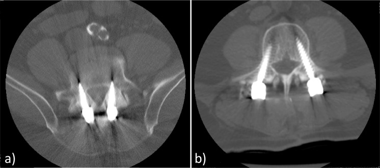
After obtaining institutional review board approval a prospectively maintained institutional database was retrospectively reviewed for all patients undergoing posterior lumbar spinal fusion using CBT screws over the past 5 years. Controls were identified from the same database as patients undergoing lumbar spinal fusion with PS placement and matched based on age, sex, and number of levels fused. The decision regarding which technique to use for screw placement was based on surgeon discretion. The MIDLF approach was chosen when there was concern about the patient's bone quality or ability to tolerate blood loss intraoperatively, but no formal selection criteria were enforced. The CBT screws were placed according to previously described techniques starting in the caudal and medial part of the pedicle and advanced in the caudo-cranial direction.5 Figure 1 contrasts the more medial starting point and medio-lateral trajectory of CBT screws with traditional PS. Exclusion criteria included a history of prior lumbar surgery, bleeding diathesis, and age < 18 years. The electronic health record was retrospectively reviewed for demographic, perioperative, and postoperative data. The visual analog scale (VAS) pain score and Oswestry Disability Index (ODI) were used to determine the degree of pain and disability, respectively, before and after surgery. The ODI was used due to its recognition as the gold standard for examining functional outcomes following lumbar surgery.6
Figure 1.
Computed tomography sequences of cortical bone trajectory (CBT) screws (a) and traditional pedicle screws (b). Note the more medial starting point of CBT screws and their medio-lateral trajectory.
Preoperative data were collected from the electronic health record and included type of lumbar pathology, VAS score for back and leg pain, ODI, and presence of neurologic deficit. Perioperative data included operative time, estimated blood loss (EBL), number of segments fused, and presence of intraoperative complication. Postoperative data included number of postoperative days until ambulation, length of hospital stay, presence of hardware-related complication, follow-up time after the procedure, VAS score for back and leg pain, and ODI.
Statistical Methods
Multiple stepwise backward elimination linear regression models were used to evaluate the difference in improvement of VAS and ODI scores, EBL, operative time, and length of stay (LOS) between the 2 operative techniques. Age, sex, number of segments operated on, and whether or not an interbody cage was placed were adjusted for in each model. IBM SPSS Statistics 24.0 (IBM Corp., Armonk, New York) was used for all statistical analysis. A P value of < .05 was considered significant.
RESULTS
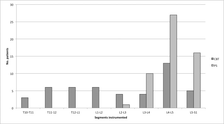
A total of 24 consecutive patients who underwent CBT screw placement and 39 controls who received traditional PS were identified. One patient who underwent CBT screw placement died prior to outpatient follow-up and was excluded from the analysis. Four patients who underwent traditional PS placement were unable to be reached for follow-up. A total of 23 patients in the CBT group and 35 patients in the PS group were included in the study. The mean age of the cohort was 51.5 ± 12.1 years. There were 26 females and 32 males. The majority of the patients presented with back and/or leg pain, and claudication. Additionally, 3 patients presented with lower extremity paresis and 1 presented with lower extremity hypesthesia. One patient in the CBT group had osteoporosis, and none in the PS group carried this diagnosis. Six patients who underwent CBT screw placement had a history of trauma. The demographic data and presenting characteristics dichotomized by treatment group are summarized in Table 1. The majority of patients had 1 segment operated on (range: 1 to 4). The distribution of levels operated on is shown in Figure 2. The number of patients in the CBT and PS groups that underwent interbody cage placement was 10 (42%) and 31 (88%), respectively.
Table 1.
Univariate analysis of preoperative data for each treatment group.
| CBT group (%) |
PS group (%) |
P |
|
| Total no. of patients | 23 | 35 | — |
| Mean age | 48.5 ± 13.4 | 53.4 ± 10.85 | .70 |
| Sex | .07 | ||
| M | 16 (69.6) | 16 (46) | |
| F | 7 (30.4) | 19 (54) | |
| No. of segments operated on | .34 | ||
| 1 | 12 (52.2) | 22 (62.9) | |
| 2 | 3 (13) | 6 (17.1) | |
| 3 | 3 (13) | 5 (14.3) | |
| 4 | 5 (21.7) | 2 (5.7) | |
| Median no. segments operated on | 1 | 1 | — |
| Symptoms present | — | ||
| Back pain | 23 (100) | 35 (100) | |
| Leg pain | 15 (65.2) | 29 (82.8) | |
| Claudication | 0 | 6 (17.1) | |
| Neurologic deficit | 0 | 6 (17.1) | |
| Preoperative VAS | |||
| Back | 7.9 ± 1.7 | 8.2 ± 2.4 | .83 |
| Leg | 4.7 ± 3.0 | 6.3 ± 3.1 | .46 |
| Preoperative ODI | 52.0 ± 16.9 | 55.8 ± 16.4 | .87 |
Abbreviations: CBT, cortical bone trajectory; PS, pedicle screws; VAS, visual analog scale; ODI, Oswestry Disability Index.
Figure 2.
Comparison of segments instrumented in each group. Abbreviations: CBT, cortical bone trajectory; PS, pedicle screw.
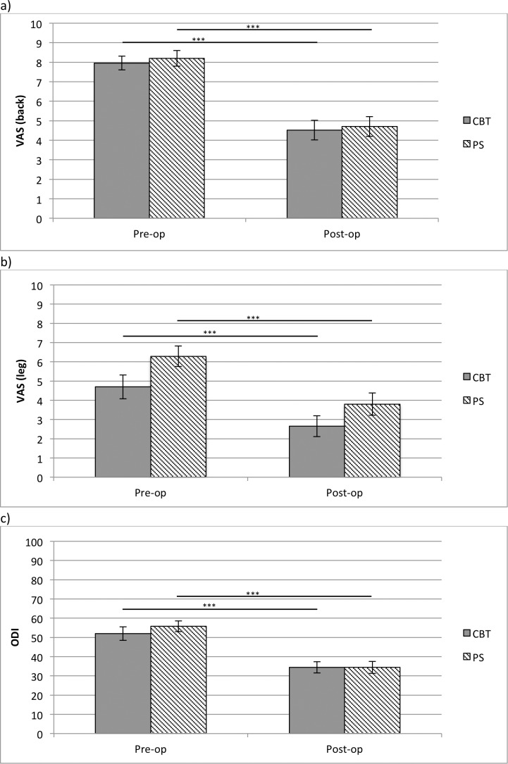
The mean EBL per level operated on was 327 ± 260 mL and the mean operative time per level was 215 ± 105 minutes. The median follow-up time was 52.5 months (range: 8 to 74). As shown in Figure 3, there were statistically significant improvements in preoperative ODI and VAS scores for back and leg pain in each treatment group. Outcomes compared between treatment groups are summarized in Table 2. When adjusting for age, sex, number of segments operated on, and interbody cage utilization, the mean EBL was significantly lower in the CBT group (186 ± 114.3 mL) compared to the PS group (413.7 ± 290.0 mL; P = .008). Two patients in the PS group required a blood transfusion in the immediate postoperative setting. No patients in the CBT group received a blood transfusion. When adjusting for age, sex, and number of segments operated on, there was no difference in the amount of improvement in preoperative ODI score or preoperative VAS scores for back or leg pain. However, the mean LOS in the CBT group was significantly shorter than in the PS group (3.6 versus 4.6 days, respectively; P = .02). There was no difference in the operative time between groups.
Figure 3.
Comparison of preoperative and postoperative visual analog scale (VAS) back scores (a), VAS leg scores (b), and Oswestry Disability Index (ODI), (c) in each group. *** indicates P < .001. Abbreviations: CBT, cortical bone trajectory; PS, pedicle screw.
Table 2.
Multivariate linear regression analysis comparing postoperative outcomes. P values are adjusted for age, sex, number of segments operated on, and whether or not an interbody cage was placed.
| CBT group |
PS group |
P |
|
| Mean improvement in VAS score (%) | |||
| Back | 43.8 ± 31 | 34.8 ± 55.7 | .85 |
| Leg | 44.2 ± 36.4 | 36.3 ± 50.6 | .99 |
| Improvement in ODI (%) | 33.2 ± 18.7 | 30.1 ± 53.3 | .56 |
| Mean LOS (days) | 3.6 ± 1.7 | 4.6 ± 2.3 | .02 |
| Mean EBL (mL) | 186 ± 114.3 | 413.7 ± 290.0 | .008 |
| Median POD of ambulation | 1 | 2 | .91 |
| Mean operative time (minutes) | 192 ± 131.7 | 226 ± 84.7 | .56 |
| No. of patients with screw pullout | 2 | 2 | — |
Abbreviations: CBT, cortical bone trajectory; PS, pedicle screw; VAS, visual analog scale; ODI, Oswestry Disability Index; LOS, length of stay; EBL, estimated blood loss; POD, postoperative day.
There were 2 hardware complications in the group that underwent CBT screw placement and 3 in the group that received traditional PS placement. Two patients in each group had screw loosening or pullout on follow-up. None of them had osteoporosis. In the PS group, 1 patient had screw malposition necessitating return to the operating room for revision. Furthermore, 2 patients in the PS group developed pseudarthrosis. Two patients in the CBT group had postoperative cerebrospinal fluid leaks compared to 1 patient in the PS group. No patients in the CBT group underwent CBT screw placement as a salvage technique for a failed PS.
DISCUSSION
There are many approaches for instrumentation of the lumbar spine. The limitations of PSs include relatively poor access to higher-density cortical bone and the need for a wide exposure to place them. The emergence of CBT screws, however, may help alleviate some of these drawbacks. The threads of these thinner and shorter screws have greater contact with cortical bone, which is especially important for preventing screw pullout in the osteoporotic patient.5 Given their caudomedial starting point, the approach for CBT screws stays more medial than that required for PS placement. This reduces dissection of the facet joints and retraction of the paraspinal muscles. Furthermore, the medial-to-lateral and caudal-to-cephalad path of CBT screws theoretically reduces the risk of neural injury. Thus, it is believed that the use of these screws can decrease blood loss and potentially reduce pain in the immediate postoperative setting.7 CBT screws can also be used as a salvage technique in the instance of failed PS placement or in patients with a pedicle too small to accept a screw. The biomechanical evidence supporting their use is strong. Santoni et al8 demonstrated a 30% increase in uniaxial yield pullout load compared to PSs in cadaveric models. In a cadaveric study by Baluch et al,9 CBT screws had greater resistance to toggle testing and required a greater force for displacement than traditional PSs. However, there is a dearth of clinical evidence confirming their efficacy. Most of the data are in the form of small case series, and only a few studies have compared CBT screws to a group receiving traditional PSs.
Our results suggest that MIDLF and TLIF yield similar improvements in back pain, leg pain, and disability. This is similar to the results seen in a comparison by Sakaura et al10 of PLIF using CBT screws to traditional PSs. They observed similar, but statistically significant, improvements in Japanese Orthopaedic Association scale scores among each group. Although they have reported the largest cohort of patients undergoing CBT screw fixation, their primary clinical outcome, the Japanese Orthopaedic Association score, does not distinguish between back and leg symptoms. Furthermore, it is weighted towards myelopathic symptoms. Our results are also similar to those reviewed by Phan et al,3 in which they concluded that CBT screws have comparable safety and efficacy as traditional PSs. Okudaira et al11 also reported similar pain relief and functional outcomes in patients undergoing MIDLF and conventional open PLIF, though like Sakura et al,10 they used the Japanese Orthopaedic Association scale to measure functional outcomes. Overall, our study results are congruent with those of other published reports, suggesting that CBT screws and traditional PSs are associated with similar improvements in pain and function.
There was no statistically significant difference in operative time between the 2 groups, but the MIDLF technique was associated with decreased LOS. The existing data regarding these outcomes are mixed. Dabbous et al reported a shorter operative time and LOS with MIDLF; however, they did not have a control group for direct comparison.12 Okudaira et al11 reported a shorter operative time in patients undergoing CBT compared to open PLIF. Conversely, Gonchar et al13 did not find a difference in operative time between patients undergoing CBT screw or PS placement. Given the CBT technique is relatively new compared to the traditional PS procedure, it is possible that the surgeon's lack of familiarity with the technique may obscure potential time saved by the more limited exposure required for CBT screw placement. The mean LOS in a study by Rodriguez et al14 study was 2.8 days, which is very close to our mean LOS of 3.6 days. It has been shown that increased muscle damage during spine surgery is correlated with longer LOS.15 Therefore, CBT screw placement may have yielded a shorter LOS in our cohort by way of decreased pain and muscle damage associated with its less extensive dissection. A prospective randomized trial is needed to confirm this finding.
In our study the amount of blood loss in the CBT group was significantly less than in the PS group. Although more interbody cages were placed in the PS group, the difference in EBL remained statistically significant after adjusting for this factor. This was expected, because traditional PS placement requires a more extensive exposure. Although this difference has been seen in some studies,11,13,16 others have reported similar EBL.17 As with operative time, differences in surgeon familiarity with the technique and body mass index among patient cohorts can potentially obscure the perceived benefits of the less invasive exposure associated with CBT screws. Increased blood loss during lumbar fixation has been correlated with increased muscle damage.18 Another theoretical advantage of decreased blood loss is a subsequent decreased risk of blood transfusion and other complications in patients with comorbid conditions who are more sensitive to lower postoperative hemoglobin levels. Indeed, no patients in the CBT group required a postoperative blood transfusion.
The incidence of screw pullout was the same in each group. Since this is not a common complication, it is likely that our cohort was too small to detect a difference. Additionally, the mean age of our cohort was relatively young, and only 1 patient had osteoporosis. It is possible that an older cohort with a greater incidence of osteoporosis would be needed to identify a difference in screw pullout. Gonchar et al13 found only a 1% incidence in screw loosening in the CBT group, compared to 25% in the PS group. Unfortunately, the largest CBT screw cohort to date did not report on the incidence of screw pullout or loosening.17 Larger comparison studies are needed to determine whether or not the biomechanical advantages of CBT screws can be replicated in the clinical setting.
Due to the small sample size in our study, there was a possibility of Type II statistical error. Given the number of patients in each group, the difference in percentage of improvement of VAS (back pain), VAS (leg pain), and ODI to identify a significant difference (α = 0.05, β = 0.2) would have needed to be 23.7%, 27.5%, and 14.3%, respectively. The difference in operative time would have needed to be 100.8 minutes. Based on the effect sizes in our study, in order to achieve a statistical power of 0.8 with an enrollment ratio of 1 and α = 0.05, future studies would require N = 130, N = 162, and N = 110 to detect differences in percentage of improvement of VAS (back pain), percentage of improvement of VAS (leg pain), and percentage of improvement of ODI, respectively. To detect a difference in mean operative time, 472 patients would need to be enrolled.
Our study has a few limitations. The sample size was small; however, most of the published case series of patients undergoing CBT screw placement have fewer than 20 patients. The largest series to date is published by Sakaura et al,10 which did not include VAS or ODI as outcomes. Given screw pullout is a relatively uncommon event, our study was likely not powered to detect a difference in its incidence between the 2 treatments. Furthermore, our cohort was relatively young, and the incidence of osteoporosis was low. It is possible that an older population with poorer bone mineral density is required to realize any benefit derived from CBT screws' greater contact with cortical bone. Future studies may select for these patients to better determine if CBT screws have lower pullout rates. Finally, our study is limited by its retrospective nature. A randomized prospective study is required to establish any superiority of one technique over the other. However, our results identify important associations between each technique that merit investigation in a prospective manner.
CONCLUSION
In conclusion, the MIDLF approach with CBT screw placement has similar efficacy as traditional PS placement but is associated with less EBL and shorter LOS. We did not identify a difference in hardware-related complications between the 2 groups.
REFERENCES
- 1.Rajaee SS, Bae HW, Kanim LE, Delamarter RB. Spinal fusion in the United States: analysis of trends from 1998 to 2008. Spine (Phila Pa 1976) 2012;37(1):67–76. doi: 10.1097/BRS.0b013e31820cccfb. [DOI] [PubMed] [Google Scholar]
- 2.Bielecki M, Kunert P, Prokopienko M, Nowak A, Czernicki T, Marchel A. Midline lumbar fusion using cortical bone trajectory screws. Preliminary report. Wideochir Inne Tech Maloinwazyjne. 2016;11(3):156–163. doi: 10.5114/wiitm.2016.62289. [DOI] [PMC free article] [PubMed] [Google Scholar]
- 3.Phan K, Hogan J, Maharaj M, Mobbs RJ. Cortical bone trajectory for lumbar pedicle screw placement: a review of published reports. Orthop Surg. 2015;7(3):213–221. doi: 10.1111/os.12185. [DOI] [PMC free article] [PubMed] [Google Scholar]
- 4.Glennie RA, Dea N, Kwon BK, Street JT. Early clinical results with cortically based pedicle screw trajectory for fusion of the degenerative lumbar spine. J Clin Neurosci. 2015;22(6):972–975. doi: 10.1016/j.jocn.2015.01.010. [DOI] [PubMed] [Google Scholar]
- 5.Mizuno M, Kuraishi K, Umeda Y, Sano T, Tsuji M, Suzuki H. Midline lumbar fusion with cortical bone trajectory screw. Neurol Med Chir (Tokyo) 2014;54(9):716–721. doi: 10.2176/nmc.st.2013-0395. [DOI] [PMC free article] [PubMed] [Google Scholar]
- 6.Fairbank JC, Pynsent PB. The Oswestry Disability Index. Spine (Phila Pa 1976) 2000;25(22):2940–2953. doi: 10.1097/00007632-200011150-00017. [DOI] [PubMed] [Google Scholar]
- 7.Lee GW, Son JH, Ahn MW, Kim HJ. JS Y. The comparison of pedicle screw and cortical screw in posterior lumbar interbody fusion: a prospective randomized noninferiority trial. Spine J. 2015;15(7):1519–1526. doi: 10.1016/j.spinee.2015.02.038. [DOI] [PubMed] [Google Scholar]
- 8.Santoni BG, Hynes RA, McGilvray KC, et al. Cortical bone trajectory for lumbar pedicle screws. Spine J. 2009;9(5):366–373. doi: 10.1016/j.spinee.2008.07.008. [DOI] [PubMed] [Google Scholar]
- 9.Baluch DA, Patel AA, Lullo B, et al. Effect of physiological loads on cortical and traditional pedicle screw fixation. Spine (Phila Pa 1976) 2014;39(22):E1297–E1302. doi: 10.1097/BRS.0000000000000553. [DOI] [PubMed] [Google Scholar]
- 10.Sakaura H, Miwa T, Yamashita T, Kuroda Y, Ohwada T. Posterior lumbar interbody fusion with cortical bone trajectory screw fixation versus posterior lumbar interbody fusion using traditional pedicle screw fixation for degenerative lumbar spondylolisthesis: a comparative study. J Neurosurg Spine. 2016;25(5):591–595. doi: 10.3171/2016.3.SPINE151525. [DOI] [PubMed] [Google Scholar]
- 11.Okudaira T, Konishi H, Baba H, Hiura K, Yamashita K, Yamada S. Comparison study of lumbar interbody fusion with cortical bone trajectory screws versus conventional open posterior lumbar interbody fusion. Proceedings of the 2014 Society for Minimally Invasive Spine Surgery Global Forum; September 19–21 2014; Miami, FL. In.
- 12.Dabbous B, Brown D, Tsitlakidis A, Arzoglou V. Clinical outcomes during the learning curve of MIDline Lumbar Fusion (MIDLF) using the cortical bone trajectory. Acta Neurochir. 2016;158(7):1413–1420. doi: 10.1007/s00701-016-2810-8. [DOI] [PubMed] [Google Scholar]
- 13.Gonchar I, Kotani Y, Matsui Y, Miyazaki T, Kasemura T. T M. Experience of 100 consecutive spine reconstructions using cortical bone trajectory (CBT) screws vs traditional pedicle screws. Proceedings of the 2014 Society for Minimally Invasive Spine Surgery Global Forum; September 19–21 2014; Miami, FL. In.
- 14.Rodriguez A, Neal MT, Liu A, Somasundaram A, Hsu W, Branch CL., Jr Novel placement of cortical bone trajectory screws in previously instrumented pedicles for adjacent-segment lumbar disease using CT image-guided navigation. Neurosurg Focus. 2014;36(3):E9. doi: 10.3171/2014.1.FOCUS13521. [DOI] [PubMed] [Google Scholar]
- 15.Fan S, Hu Z, Zhao F, Zhao X, Huang Y, Fang X. Multifidus muscle changes and clinical effects of one-level posterior lumbar interbody fusion: minimally invasive procedure versus conventional open approach. Eur Spine J. 2010;19(2):316–324. doi: 10.1007/s00586-009-1191-6. [DOI] [PMC free article] [PubMed] [Google Scholar]
- 16.Kasukawa Y, Miyakoshi N, Hongo M, Ishikawa Y, Kudo D, Shimada Y. Short-term results of transforaminal lumbar interbody fusion using pedicle screw with cortical bone trajectory compared with conventional trajectory. Asian Spine J. 2015;9(3):440–448. doi: 10.4184/asj.2015.9.3.440. [DOI] [PMC free article] [PubMed] [Google Scholar]
- 17.Sakaura H, Miwa T, Yamashita T, Kuroda Y, Ohwada T. Cortical bone trajectory screw fixation versus traditional pedicle screw fixation for 2-level posterior lumbar interbody fusion: comparison of surgical outcomes for 2-level degenerative lumbar spondylolisthesis. J Neurosurg Spine. 2018;28(1):57–62. doi: 10.3171/2017.5.SPINE161154. [DOI] [PubMed] [Google Scholar]
- 18.Marengo N, Ajello M, Pecoraro MF, et al. Cortical bone trajectory screws in posterior lumbar interbody fusion: minimally invasive surgery for maximal muscle sparing—a prospective comparative study with the traditional open technique. Biomed Res Int. 2018. pp. 1–7. [DOI] [PMC free article] [PubMed]


