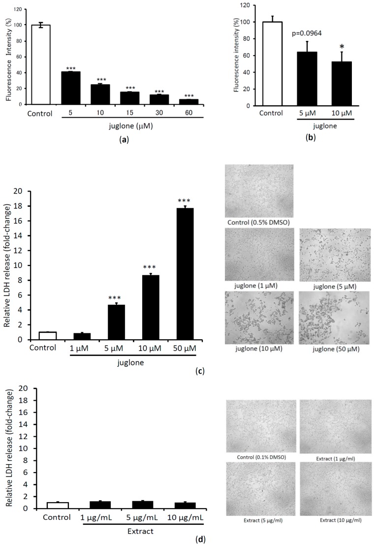
Figure 5.
The amyloid fibril disrupting activity of different concentration of juglone when incubated with V30M-TTR tetramer for 24 h and then quantified with Thioflavin T assay (a) and PROTEOSTAT® Protein aggregation assay (b). LDH release (left panel) and images of HEK293 cells treated with the different concentration of juglone (c) and 50% acetone extract of immature fruits of J. mandshurica var. cordiformis (d). Data sets were analyzed by Tukey-Kramer multiple comparison test. * p < 0.05; ** p < 0.01; *** p < 0.001 as compared with control (TTR treated with DMSO in each experiment for (a,b), and DMSO for (c,d)); All experiments were performed in triplicate (means ± SEM, n = 3).

