



Abstract
Genome duplication is carried out by pairs of replication forks that assemble at origins of replication and then move in opposite directions. DNA replication finishes when converging replication forks meet. During this process, called replication termination, DNA synthesis is completed, the replication machinery is disassembled and daughter molecules are resolved. In this Review, we outline the steps that are likely to be common to replication termination in most organisms, namely fork convergence, synthesis completion, replisome disassembly and decatenation. We then briefly review the mechanism of termination in the bacterium Escherichia coli and in simian virus 40 (SV40) before focusing on recent advances in eukaryotic replication termination. In particular, we discuss the recently discovered E3 ubiquitin ligases that control replisome disassembly in yeast and higher eukaryotes, and how their activity is regulated to avoid genome instability.
Introduction
Genomic DNA replication can be divided into three general phases: (1) Initiation, in which the origin of DNA replication is unwound by the replicative DNA helicase (Figure 1A-B). (2) Elongation, in which forks copy the chromosome using semi-conservative DNA synthesis (Figure 1C-D). (3) Termination, when converging replication forks meet (Figure 1D-G). From bacteria to eukaryotic cells, replication initiation is regulated such that genome duplication is limited to a single round per cell cycle1,2. Unlike initiation and elongation, which have been studied extensively3,4, replication termination has received relatively little attention, especially in eukaryotic cells. This is a major gap in our knowledge of genome duplication, especially because termination events are just as abundant as initiations, occurring approximately 50,000 times during a typical S phase of mammalian cells5.
Figure 1: Steps in DNA replication.

Generic illustration of replication initiation (A-B), elongation (C-D), and five events that are unique to replication termination (D-G). The replicative DNA helicase is depicted without reference to a specific translocation mechanism; RNA primers are in red. The order of the termination events is hypothetical.
At least five processes are unique to the final phase of replication and thus can be considered part of replication termination. The first concerns the resolution of topological stress. Unwinding of the parental duplex leads to overwinding of the unreplicated DNA, leading to the formation of positive supercoils ahead of the fork (Figure 1C). If too many supercoils accumulate, further unwinding becomes energetically unfavorable and replication ceases. There are two ways to dissipate positive supercoils. The first involves relaxation of supercoils by type I or type II DNA topoisomerases 6. Alternatively, the entire fork can rotate clockwise relative to the direction of fork movement. This rotation counteracts the overwinding of unreplicated DNA and causes the two replicated sisters to cross over each other, leading to the formation of “pre-catenanes” 7,8 (Figure 1E), which can be resolved by Type II, but not Type I, topoisomerases. As replication proceeds, the region of parental DNA that can be supercoiled decreases in size, whereas the region of replicated DNA that can undergo pre-catenation increases. If supercoils and pre-catenanes are energetically equivalent, their relative abundance during replication should reflect the ratio of unreplicated versus replicated DNA in a topologically constrained domain9. In this view, as replication progresses, the resolution of topological stress would become increasingly reliant on the formation and subsequent removal of pre-catenanes. Importantly, at some point, the parental DNA between converging forks becomes too short to supercoil (Figure 1D) owing to the inherent stiffness of DNA. At this stage, which occurs when 150 bp or less of parental DNA remains,10 relief of topological stress becomes absolutely dependent on the formation of pre-catenanes (Figure 1E). This phase of replication is unique to termination and is defined as replication fork “convergence.” An important question is whether replication forks slow down or require accessory factors as replication becomes dependent on the formation of pre-catenanes to manage topological stress. If so, one might expect a gradual slowing of DNA replication forks as they approach one another. Moreover, if the formation or removal of pre-catenanes were disrupted, forks would stall at a very late stage of replication due to accumulation of topological stress.
The second process unique to replication termination is the meeting of converging replication forks, which we call “encounter” (Figure 1E). It is presently unclear whether this involves a steric clash between replisomes and whether such a clash impedes further steps in termination. Third, replisomes dissociate from the DNA in a process called “disassembly” (Figure 1E-F). It is generally assumed that the replisome dissociates during termination to prevent re-replication and to avoid interference with other chromatin-based processes such as transcription or the next round of replication. Active disassembly pathways are likely required because key replisome components, such as replicative helicases and processivity factors, are clamped tightly around DNA. Crucially, to prevent fork stalling, any disassembly mechanism must not act on replisomes still engaged in replication. Key questions are thus whether the replisome is actively disassembled, when this happens and what is the effect of defective disassembly. Fourth, DNA synthesis is completed through gap filling (Figure 1E-F). At the moment of replisome encounter, a single-stranded gap exists between the 3’ end of the leading strand and the downstream Okazaki fragment of the opposing fork. This gap is filled in, and the last Okazaki fragment is processed. Presently, it is unclear whether gap filling requires replisome disassembly and whether maturation of the last Okazaki fragment occurs via the same mechanism as during replication elongation. Finally, copying the last turn of parental duplex creates a new catenane and also converts any pre-catenanes to catenanes (Figure 1F). All of these catenanes have to be decatenated (resolved) prior to chromosome segregation (Figure 1F-G). Another important issue is whether most termination events are sequence-specific or stochastic, and whether these two modes are mechanistically distinct. Furthermore, the exact order of these processes during termination remains unclear.
In this review, we begin by summarizing current models of termination in the bacterium Escherichia coli and in simian virus 40 (SV40). We then discuss recent advances in our understanding of replication termination in eukaryotes, which include the first evidence of an active replisome unloading mechanism.
Termination in Escherichia coli
The circular E. coli chromosome, comprising 4.6 million basepairs of DNA, is replicated from a single origin of replication, oriC (Figure 2A)2. Two forks are established, each containing a hexameric replicative helicase, DNA synthesis protein B (DnaB), which unwinds parental DNA by encircling it and translocating on the lagging strand template. Each DnaB helicase binds to at least two molecules of DNA polymerase III (Pol III), which synthesize the leading and lagging strands in association with the processivity clamp β. The two replication forks emanating from oriC travel around the chromosome in opposite directions at a rate of ~60 kb/min and terminate in a specialized region across from the origin. This termination zone contains 10 ter sites (A-J), which can bind the DNA replication terminus site-binding protein (Tus) and comprise potent and polar replication fork barriers (Figure 2A, reviewed in11). The ter sites are oriented such that the leftward fork can pass the first five ter sites it encounters, which are marked as red arrowheads in Figure 2A, but stalls at the five blue sites. Conversely, the rightward fork passes through the ter sites marked as blue arrowheads but stalls at the red sites. In this way, forks can enter but not leave the termination zone.
Figure 2: Replication termination in Escherichia coli.

(A) Depiction of the Escherichia coli chromosome, including the origin of replication oriC, and the ten ter sites shown as red and blue arrowheads. The termination zone is underlined in red. In the box, the green arrow shows a replication fork passing through a ter site in the permissive orientation, and the red arrow shows a fork stalling at a ter site in the non-permissive orientation. (B) Two scenarios of fork stalling in the termination zone. (a) The rightward fork (Fork 1) arrives first and stalls at terC, followed by the arrival of the leftward (Fork 2). (b) The two forks arrive to the termination zone contemporaneously and meet between terC and terA. (C) Possible mechanism of E. coli replication termination. (a) Forks converge between ter sites with the formation of pre-catenanes. (b) Two DNA synthesis protein B (DnaB) replicative helicase complexes pass each other and collide with the downstream leading strand, generating a 3’ flap. DnaB dissociates, the 3’ flap is removed, gaps are filled in and the final Okazaki fragment is processed by DNA polymerase I (Pol I). (c) Nicks are ligated and the final catenane, which is generated during the completion of DNA synthesis is removed (not shown). (D) Possible mechanism of replication re-initiation. (a) If the 3’ flaps are not removed or remodeled, a new replication fork is established, which prevents the completion of termination. (b) The free end re-invades the sister chromatid using recombination protein A (RecA) and RecBCD, which establish a new replication fork. (c) The Holliday junction is resolved and DnaB is re-loaded onto the fork by primosomal proteins A (PriA) and PriB.
The function of Tus–ter complexes in replication termination remains unclear. It has been debated11 whether fork encounter occurs after one of the two forks has already stalled at a ter site (Figure 2Ba), or whether fork encounter occurs between two ter sites (Figure 2Bb). Some forks clearly collide with the non-permissive face of a Tus–ter complex, as judged by 2-D gel electrophoresis12. However, when the tus gene is deleted (Δtus), the location of most fork fusions remains ~10,000 bp to the right of terC13,14. Therefore, most forks appear to converge between ter sites C and A (Figure 2Bb). Surprisingly, Δtus strains have no abnormal growth phenotypes15, suggesting that the Tus-ter system is not an integral part of the termination machinery, but instead has other roles (see below).
How do the five steps of replication termination outlined in Figure 1 unfold between ter sites? The two type II topoisomerases in E. coli are DNA gyrase and topoisomerase IV (TopoIV). Consistent with their substrate specificities in vitro, gyrase relaxes positive supercoils ahead of the fork during the elongation stage of DNA replication, whereas TopoIV is required for decatenation of the fully replicated daughter molecules16 (Figure 2Ca,Cb). Evidence also indicates that TopoIV is required to resolve pre-catenanes and thereby allow fork convergence 17,18. Whether converging forks clash during encounter is unknown. A recently suggested model for gap filling is based on the observation that in cells lacking 3’-flap removal activity, replication re-initiates, as judged by deep sequencing of genomic DNA14. This DNA amplification in the termination zone is suppressed by the absence of primosomal protein A (priA), which promotes oriC-independent loading of DnaB. Together with other reports19,20, these data imply that when replication forks meet in wild type cells, a 3’ flap is generated (Figure 2Cb). The flap is normally degraded or remodeled and the gap is subsequently filled in (Figure 2Cc). Pol I may use its 5’ to 3’ exonuclease activity to remove the RNA primer of the last Okazaki fragment21, as seen during replication elongation, to facilitate ligation (Figure 2Cc). If the flap is not removed, two replication forks are established following strand invasion and priA-dependent loading of DnaB (Figure 2D). Notably, the extent of re-replication is greatly increased in the absence of Tus14,19,21. Therefore, it appears that the primary function of Tus is not to promote site-specific termination, but rather to limit the extent of any re-replication after aberrant initiation in the termination zone.
To validate this model of replication termination, it will be essential to determine whether the 3’ flap is generated in unperturbed cells. If so, how does this occur? It has been proposed that when DnaB reaches the 3’ end of the opposing fork’s leading strand, it unwinds this strand (Figure 2Cb). However, biochemical studies indicate that in this situation, DnaB would pass over the 3’ end and keep translocating along dsDNA without further DNA unwinding22. Therefore, either DnaB behaves differently as part of the replisome, or the flap is generated by another DNA helicase. A related issue concerns replisome disassembly. Does the encounter of two Pol III holoenzymes, in which leading and lagging strand polymerases are physically coupled to DnaB, cause a steric clash that requires replisome disassembly before gaps can be filled? The model that DnaB creates a 3’ flap implies that DnaB is unloaded late in replication termination, after the DnaB molecules of converging forks have passed each other. In this view, the polymerases would need to either be unloaded or disengage from the leading strands to allow converging DnaB complexes to pass each other (Figure 2Cb). Further work is needed to address these issues, including why E.coli termination appears to be so susceptible to re-initiation.
Termination in simian virus 40
Replication termination has been studied extensively in the context of the mammalian DNA tumor virus SV40, whose small circular (plasmid) chromosome comprises 5,200 bp (Figure 3A). SV40 encodes its own replicative helicase, large T antigen (T-ag), which cooperates with mammalian host replication factors to replicate the SV40 chromosome23. Two replication forks are established at the origin and terminate on the opposite side of the plasmid (Figure 3A). Relocation of the origin leads to a corresponding shift in the termination zone24, indicating that SV40 lacks genetically encoded termination sites.
Figure 3: Model for simian virus 40 DNA replication termination.
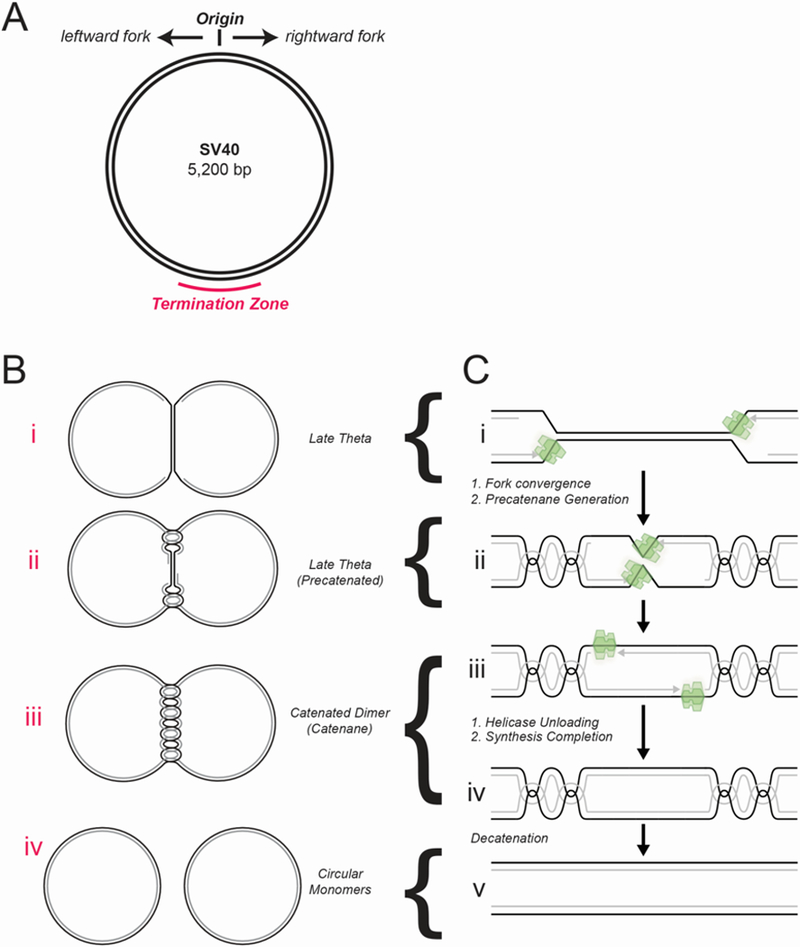
(A) The simian virus 40 (SV40) chromosome is a plasmid that includes the origin of replication and termination zone (underlined in red). (B) Late stages of SV40 DNA replication. (a) Late theta intermediate, when fork convergence begins. (b) Fork convergence, when superhelical stress is dissipated by the formation of pre-catenanes. (c) Catenated dimers are generated when pre-catenanes are converted to catenanes at the end of replication. (d) Decatenation produces two circular monomers. (C) Hypothetical mechanism of SV40 replication termination. (a) When forks come within 450 bp of each other, they stall, possibly owing to reduced formation of pre-catenanes. (b) Forks converge, leading to the formation of pre-catenanes and the encounter of the large T antigen (T-ag) helicases. Whether the helicases stall upon encounter is not known. (c) The helicases pass each other and stall at the downstream Okazaki fragment, accounting for the single-stranded DNA gap that persists on replicated molecules. (d) T-ag is unloaded, the remaining gaps are filled in. (e) The catenanes are removed, yielding fully replicated and decatenated daughter chromosomes.
Two long-lived intermediates have been detected during SV40 replication termination. The first is a ‘late theta’ structure25–28, in which all but the final ~450 bp of the SV40 chromosome is replicated (Figure 3Ba)29. The accumulation of this intermediate might be explained if, during fork convergence, the removal of supercoils ahead of the fork becomes inefficient, and kinetically slower formation of pre-catenanes takes over (Figure 3Bb). In cell-free extracts, replicated SV40 plasmid dimers contain 5–20 catenanes (Figure 3Bc) 30,31, consistent with the formation of precatenanes during fork convergence (Figure 3Bb). Catenated plasmid dimers are ultimately resolved into circular monomers (Figure 3Bd). The final stage of SV40 DNA synthesis requires topoisomerase II (Topo II)32,33, indicating that the removal of pre-catenanes allows convergence, as seen in bacteria. The second long-lasting intermediate is a single-stranded DNA (ssDNA) gap of ~60 nucleotides, which is observed in the termination region after forks converge (Figure 3Cc)34, but what causes this gap is a matter of speculation (see below). Importantly, decatenation (Figure 3Cc) and gap filling (Figure 3Cd) appear to be mechanistically independent events30.
Many concepts in SV40 termination are linked to models of how T-ag functions. Early results suggested that T-ag dissociates from DNA when replication is only 80% complete, possibly at the onset of convergence35. However, such a model does not explain how the last 20% of parental DNA is unwound, and it is incompatible with evidence that complete SV40 DNA replication can be reconstituted in a defined system where T-ag is the only DNA helicase36. An early model of DNA unwinding by T-ag proposed that it encircles the DNA duplex and translocates along it37. In this view, T-ag molecules would stall at encounter and would have to be disassembled prior to gap filling. However, more recent work indicates that T-ag, like other replicative DNA helicases, translocates along one strand of the DNA38 (Figure 3Ca), suggesting that converging T-ag molecules should be able to pass each other (Figure 3Cb,c). T-ag stalling at the downstream Okazaki fragment might inhibit ligation and explain the persistence of the 60 nt ssDNA after decatenation34. To understand SV40 termination more fully, it will be crucial to determine when exactly T-ag dissociates from DNA, whether this is an active process, and what are the consequences of disrupting its unloading. In summary, SV40 replication termination involves at least two long-lived intermediates (late theta structures and gapped molecules), and future studies will be required to address how they are linked to replisome disassembly.
Replication termination in eukaryotes
The mechanism of replication in eukaryotic cells is complex3,4,37,39 (Box 1). The process begins in the G1 phase of the cell cycle when six minichromosome maintenance ATPases (MCM2-MCM7), which together form the MCM2–7 replicative DNA helicase motor, are recruited to each origin of replication. In S phase, MCM2–7 is converted to the active CMG helicase, which is composed of the cell division cycle 45 (CDC45), MCM2–7 and the four-subunit complex Go-Ichi-Ni-San (GINS). CMG unwinds the origin, followed by the assembly of two replisomes that copy the DNA using distinct leading and lagging strand DNA polymerases. Budding yeast replication was recently reconstituted in vitro using purified components40–42. This system supports rapid initiation and elongation, but termination is inefficient43. Given that termination is supported by frog egg extracts44,45, it is likely that the yeast reconstituted system is missing one or more termination proteins. Below, we discuss recent insights into the events that underlie eukaryotic replication termination, including the active disassembly of the replisome.
Box 1: Eukaryotic replication initiation and elongation: the basics.

Here we provide a brief summary of eukaryotic DNA replication initiation and elongation (reviewed in 3,4,37,39). ‘Licensing’ of DNA replication occurs in the G1 phase of the cell cycle, when the origin recognition complex (ORC), the ATPase cell division cycle 6 (CDC6) and CDC10 dependent transcript 1 (CDT1) cooperate to recruit two minichromosome maintenance 2–7 (MCM2–7) complexes to each origin of replication, thereby forming the pre-replicative complex (pre-RC) (see the figure). MCM2–7 is a heterohexamer composed of the related ATPases MCM2–MCM7 and serves as the motor of the replicative helicase. Within pre-RCs, two inactive MCM2–7 complexes encircle double-stranded DNA, with their N-terminal tiers oriented towards each other to form a tight dimer interface.
In S phase, a subset of pre-RCs undergoes activation by cyclin-dependent kinase (CDK), Dbf4-dependent kinase (DDK) and many accessory factors, leading to the binding of two helicase co-factors, CDC45 and the four-subunit Go-Ichi-Ni-San (GINS) complex, to each MCM2–7 complex, thereby forming the active CDC45–MCM–GINS (CMG) helicase (see the figure). CMG encircles the leading strand and translocates along it in the 3’ to 5’ direction; the trailing edge of CMG is formed by the C-terminal lobe of MCM2–792. CMG unwinds the origin, allowing the assembly of two DNA replication forks that travel away from the origin. Cells prevent re-replication by blocking licensing in S phase1.
In yeasts, although origin sequences are well-defined, initiation is partly stochastic, so that the program of origin firing is probably unique in every cell. In higher eukaryotes, the DNA sequences of origins are poorly defined and initiation is inefficient and frequently occurs in large zones, resulting in replication programs that are even more stochastic than in yeasts.
The replisome is a macromolecular assembly composed of multiple protein complexes. The leading strand is synthesized continuously by DNA polymerase ε (Pol ε), whereas the lagging strand is composed of Okazaki fragments synthesized by Pol δ93 (see the figure). Pol δ acquires processivity by its association with the ring-shaped protein proliferating cell nuclear antigen, which is deposited around DNA by replication factor C. The leading strand and every Okazaki fragment are primed by Pol α−primase, which synthesizes a ~10 nucleotide RNA primer and then extends it by 20–30 nucleotides of DNA before the switch to the more processive Pol ε or Pol δ occurs. When the 3’ end of one Okazaki fragment reaches the 5’ of another, Pol δ performs strand displacement synthesis (Figure 4D). The resulting flap is removed by flap endonuclease 1. Long flaps are degraded by the helicase-nuclease DNA synthesis defective protein 2. The replisome also contains topoisomerase I, chromatin remodeling factors, checkpoint signaling proteins and cohesion establishment factors. CMG binds directly to Pol ε and indirectly to Pol α through chromosome transmission fidelity protein 4. Therefore, unlike in bacteria, leading and lagging strand polymerases appear not to form a stable complex in eukaryotes.
Genomic distribution of termination sites
We first consider where on eukaryotic chromosomes termination events occur. Using a fork synchronization protocol, ~70 of the ~300 termination events mapped in budding yeast reproducibly occurred in the same chromosomal location, possibly owing to genetically encoded elements46. More recently, Okazaki fragments were mapped genome-wide in unsynchronized budding yeast cells to identify fork merger zones47. This analysis showed that termination events generally occur midway between origins and that the more active two origins are, the better defined the termination zone between them. Alteration of origin firing (activation) caused predictable changes in termination zones, consistent with the large majority of termination events being sequence non-specific and largely dictated by initiation patterns47,48. Similar conclusions were reached from Okazaki fragment mapping in mammalian cells49. The absence of specific termination sites is well suited to deal with the significant level of stochasticity observed in eukaryotic origin firing. Only in rare instances are termination events site-specific (see below).
Replication fork convergence
The SV40 termination model suggests that DNA synthesis slows down during fork convergence, and studies in both SV40 and E. coli suggested that fork convergence requires the activity of Topo II. To study eukaryotic replication termination in frog egg extracts, forks were stalled at the outer edges of a ~500 bp-long array of LacI molecules, followed by IPTG addition to induce locus-specific and synchronous termination events44. No slowing in the rate of DNA synthesis was observed during duplication of the final 500 bp of DNA. Although some topological stress may have dissipated prior to disruption of the LacI barrier, completion of replication still required the removal of ~50 supercoils. These results suggest that in this cell-free system, the resolution of topological stress is not rate-limiting for fork convergence, presumably owing to the efficient formation of pre-catenanes (Figure 4A). Accordingly, DNA catenation was detected immediately following fork convergence44, as previously reported50,51. Replicon size does not affect the number of catenanes formed52, suggesting precatenanes are primarily formed during replication termination rather than elongation9,53. Strikingly, unlike what is observed in E. coli and SV40, fork convergence and synthesis completion do not require Topo II in egg extracts or yeast44,54,55. In the absence of Topo II, fully replicated daughter plasmids are generated, and they are highly catenated, indicating that resolution of precatenanes is not essential for convergence in eukaryotic systems. In the future, it will be important to understand the differential requirements for type II topoisomerases in eukaryotic and prokaryotic systems.
Figure 4: Model of eukaryotic replication termination.

(A) After copying most of the replicon, forks come too close to each other to allow formation of supercoils in the unreplicated DNA, leading to the onset of convergence. During convergence, which lasts until forks encounter each other, topological stress is relieved by the formation of precatenanes. An end-on view of CMG illustrates the presence of single-stranded DNA in its central channel. (B) The encounter causes no detectable fork stalling, implying that converging CMGs bypass each other. After bypass, CMG helicases keep translocating until they reach a downstream Okazaki fragment. (C) The CMG helicases pass over the ssDNA–dsDNA junction and keep moving on dsDNA (see end-on view). (D) The leading strand is extended to the downstream Okazaki fragment. The last Okazaki fragment is processed, possibly by de novo recruitment of DNA polymerase δ (Pol δ) and by 3’ flap processing by flap endonuclease 1 (FEN1). (E) Once CMG encircles dsDNA, it undergoes polyubiquitylation on its MCM7 subunit by SCFDia2 or CRL2Lrr1. The ubiquitylated MCM7 is extracted from chromatin by the ATPase p97 ATPase. (F) Catenanes are removed. CMG, CDC45–MCM–GINS
Replisome encounter
A critical question is what happens when converging CMG complexes meet. In frog egg extracts, nascent leading strands pass each other without detectable pausing, followed by rapid ligation of all nascent strands (Figure 4), indicating that during fork encounter, there is no steric clash or if there is, it is very short-lived. This is consistent with our current understanding of replisome architecture. First, replisome-associated CMG appears to interact primarily with the leading strand template 56. Therefore, the converging CMG complexes will encounter each other on different strands (Figure 4A), thereby facilitating bypass. Second, unlike in E. coli, in eukaryotic cells there is little evidence for the existence of a stable complex between the leading strand replication machinery (CMG, Pol ε, etc.) and the lagging strand replication machinery (Pol δ, proliferating cell nuclear antigen (PCNA), flap endonuclease 1 [FEN1], DNA ligase, etc.), although Pol α–primase (Pol α) does bind weakly to a CMG complex in yeast 57. Such a separation between leading and lagging strand machineries allows the CMG of one fork to pass unobstructed onto the lagging strand template of the converging fork. The absence of pausing during encounter is attractive as any instances of fork stalling would likely be deleterious for genome stability. Curiously, when CMG encounters a covalent DNA–protein (~40 kDa) complex on the lagging strand template, it stalls for a few minutes58. This observation raises the interesting possibility of specific evolutionary adaptations that prevent a clash during encounter of two CMGs during termination.
Replisome disassembly
The removal of the CMG helicase from chromatin is emerging as a key event in eukaryotic replication termination. Late in the S phase of budding yeast and frogs, K48-linked ubiquitin chains are assembled on MCM745,59 (Figure 4E). In budding yeast, MCM7 polyubiquitylation and CMG dissociation both require the E3 ubiquitin ligase SCFDia2 (Skp, Cullin, F-box containing complex associated with Digs Into Agar 2 [Dia2]) 59, strongly suggesting that MCM7 ubiquitylation is causally linked to CMG unloading. Recently, CRL2Lrr1 (Cullin RING Ligase 2 associated with Leucine Rich Repeats 1 [Lrr1]) was identified as the ubiquitin ligase that promotes MCM7 ubiquitylation and CMG unloading at the end of S phase in worms and frogs60,61. SCFDia2 may be constitutively associated with replisomes in yeast 62, whereas frog CRL2Lrr1 only binds to terminating replisomes60. Once CMG is ubiquitylated, it is removed from chromatin by the ATPase p97 (also known as VCP) 45,59,63 (Figure 4E,F), which cooperates with diverse co-factors to extract ubiquitylated proteins from their local environments64. Data from frogs indicate that ubiquitylated MCM7 is not degraded by the proteasome after chromatin extraction, suggesting that ubiquitylated CMG is recycled after disassembly65. Yeast cells lacking Dia2 exhibit constitutive activation of the replication checkpoint, sensitivity to DNA damaging agents and gross chromosomal rearrangements66–68. In the absence of CRL2Lrr1, worm CMG persists on chromatin until prophase, when it is unloaded by a second pathway involving p97 and UBX domain containing 3 (UBXN-3, the worm ortholog of human FAS-associated factor 1, FAF1)61. Importantly, combined knock-down of LRR-1 and UBXN-3 stabilizes CMG on chromatin until metaphase and is synthetically lethal, suggesting replisome unloading is essential for viability61. Whether vertebrates also have a mitotic CMG unloading mechanism is unknown. It will also be of great interest to elucidate the consequences of defective CMG unloading and whether these underlie any human diseases.
Interestingly, CMG is unloaded only after the gap between the leading strand of one fork and the lagging strand of the converging fork has been filled in and ligated (Figure 4D-F)44. As such, CMG unloading is one of the latest known events in eukaryotic replication termination, occurring once CMG is associated with dsDNA. Most likely, converging CMGs pass each other and keep translocating along the leading strand template until they reach the downstream Okazaki fragment, whereupon they pass onto dsDNA (Figure 4C). This model is consistent with the observation that when purified CMG reaches a ssDNA–dsDNA junction, it passes over the junction and keeps moving along dsDNA without further DNA unwinding, which requires a 5’ flap, as reported also for DnaB69,70. It will be important to examine whether CMG can also pass over a junction containing an RNA–DNA hybrid, as would be the case for an Okazaki fragment. Together, the data suggest that MCM7 ubiquitylation occurs when CMG encircles double-stranded DNA. Importantly, synthesis completion is unaffected when CMG unloading is inhibited, consistent with unloading being a later event in termination44,60.
A critical unresolved issue concerns the trigger for CMG unloading. Given that MCM2–7 loading (and therefore CMG assembly) in S phase is prohibited to prevent re-replication1, premature CMG removal from active forks must be prevented to avoid fork stalling and breakage. An attractive model is that the presence of dsDNA in the central channel of CMG leads to recruitment of CRL2Lrr1 (or in yeast activation of already bound SCFDia2) owing to a conformational change of CMG. Remarkably, induction of SCFDia2 expression in G1 can unload CMG complexes that remained on chromatin from the previous S phase59, suggesting that SCFDia2 can target CMG that is associated with dsDNA. The advantage of this mechanism is that it cannot operate on active replication forks, where CMG encircles ssDNA56 (Box 1). To discriminate between CMG complexes during termination and licensed MCM2–7 complexes, which also encircle dsDNA, we speculate that the ligase might detect the presence of CDC45 and/or GINS, or be inhibited by the dimerization of licensed MCM2–7 complexes. Alternatively, the MCM7 ubiquitin ligase might detect the juxtaposition of converged CMG complexes or the collision of CMG with the rear face of PCNA molecules from the converging fork. Interestingly, CMG complexes are also actively unloaded when forks converge on a DNA inter-strand crosslink (ICL) 56,71. Although this pathway involves MCM7 ubiquitylation and p97 (REF.64)65, it operates on CMG complexes that encircle single-stranded DNA on either side of an ICL, and unlike termination-dependent CMG unloading, it requires BRCA1 bound to BRCA1-associated RING domain protein 1 (BARD1) (REFS 64, 70)65,71. Therefore, the mechanisms of CMG unloading during termination and ICL repair are clearly distinct.
How does the rest of the replisome dissociate from chromatin during termination? CMG makes direct or indirect contact with numerous proteins at the replication fork, including the components of the replisome progression complex and Pol ε39,62,72. It is therefore likely that numerous replication proteins are unloaded passively, as an indirect consequence of CMG unloading. Consistent with this idea, CMG and Pol ε dissociate with similar kinetics 44, and blocking CMG unloading leads to the retention of most RPC components at the fork, including Pol ε 60. To determine whether SCFDia2 or CRL2LRR1 target other replisome components, it will be important to test whether loss of these E3 ligases mimics the elimination of ubiquitylation sites on MCM7. Proteins that do not interact with CMG are probably removed from chromatin independently of replication termination. For example, PCNA is continually unloaded by ELG1 (also known as ATAD5)73,74, which should also lead to the removal of PCNA-interacting proteins such as FEN1, DNA ligase and Pol δ at the lagging-strand maturation machinery.
Gap filling
In frog egg extracts, leading strands are extended past each other without visible pausing until they come within a few nucleotides of the downstream Okazaki fragment, whereupon the two are rapidly ligated44 (in contrast to the situation in SV40 DNA, where gaps persist). This observation implies that neither the resolution of topological stress nor CMG unloading are rate-limiting for synthesis completion. Indeed, by passing over the downstream Okazaki fragment, CMG would vacate the ssDNA–dsDNA junction and make room for the enzymes that carry out Okazaki fragment processing. CMG may also drag Pol ε, with which it forms a stable complex 72, away from the junction60. This is potentially advantageous, because unlike Pol δ, Pol ε does not functionally cooperate with FEN1 for Okazaki fragment processing 75. Thus, removal of Pol ε should make room for Pol δ and facilitate processing. This mechanism has a potential disadvantage. If CMG is able to translocate a significant distance along dsDNA before being unloaded, it would promote re-replication if it encounters a downstream Okazaki fragment with a 5’ flap, analogous to the situation in E. coli (Figure 2Da). In possible agreement with this possibility, loss of CRL2Lrr1 in worms appears to induce re-replication76. In the future, it will be crucial to assess whether short stretches of re-replication normally occur in healthy cells or in cells where a component of the Okazaki maturation machinery, such as FEN1, has been compromised.
Decatenation of replicated chromosomes
When the last turn of parental DNA duplex is unwound, the daughter molecules end up catenated via one interwinding (Figure 1F). In addition, any pre-catenanes are automatically converted into catenanes9. The resolution of catenanes is unlikely to be mechanistically distinct from the unlinking of precatenanes behind replication forks18. We note that both E. coli and yeast are capable of carrying out a mechanistically distinct decatenation process, which is driven by supercoiling of the chromosome77,78. The mechanism described in yeast requires Condensin loading and spindle formation78, which only occur during mitosis. Therefore, it is unlikely to be extensively employed during replication termination, which occurs during S phase.
Site-specific replication termination
Although the vast majority of eukaryotic termination events appear to be sequence non-specific (see above), at least two classes of site-specific termination exist 79,80. The first class is caused by sequence-specific replication fork barriers (RFBs) that stall one fork long enough for a converging fork to arrive. The best-characterized example is the polar RFB in the ribosomal DNA (rDNA) locus, which contains tandem repeats of highly transcribed ribosomal gene clusters. Each rDNA cluster contains an RFB comprising a termination element (Ter in budding yeast) that is bound by a ‘terminator’ protein (fork blocking protein 1 (Fob1) in budding yeast). Like the Tus–ter complex in E. coli, the Fob1–Ter complex creates a polar RFB and thus prevents head-on collisions between the replication and transcription machineries, which have the potential to cause genome instability81. Although the mechanism of fork arrest at the rDNA RFB is quite well defined82,83, how termination unfolds at this locus, or any other RFB, has not been examined. In frog extracts, stalled replication forks can readily restart and terminate replication44. Therefore it is likely that the replisome from the fork stalled at the rDNA RFB can restart once the converging. fork displaces the terminator protein. Most likely, replisomes then pass each other and terminate replication using the same mechanism employed at other loci (Figure 4b-f). In budding yeast, the helicase rDNA recombination mutation protein 3 (Rrm3) is required for progression of replication forks past protein–DNA complexes. Interestingly, although rrm3Δ strains exhibit a ~2-fold increase in replication fork stalling at Fob1–Ter, they show a ~10-fold accumulation of converged forks at this RFB, suggesting a specific role for Rrm3 in fork convergence84. Importantly, rrm3Δ strains exhibit no genome-wide defects in replisome disassembly59, suggesting a specialized role for Rrm3 at the rDNA RFB, perhaps in displacing Fob182. In the future, it will be interesting to determine whether the mechanism of termination at site-specific RFBs differs in any fundamental way from the general mechanism that occurs at most genomic termination sites. The mechanism of fork stalling at RFBs, as well as their roles in other processes such as gene silencing, imprinting and aging are not discussed as they have been reviewed elsewhere 79,80.
The second class of site-specific termination events occurs at telomeres85. Here, replication ceases when the fork reaches the end of the chromosome. Importantly, when the fork copies the telomere, the leading strand appears to be extended to within a few nucleotides of the chromosome end86–88, similar to the run-off DNA synthesis exhibited by by purified Pol ε89. Together, these observations suggest that CMG slides off the end of the leading strand template, allowing Pol ε to reach the end of the chromosome90. Given the evidence that Pol α is tethered to the replisome through CMG57,60, Pol α should dissociate with CMG, thereby preventing new priming. This might explain why the amount of DNA lost on the lagging strand in humans is roughly equivalent to the size of an Okazaki fragment87. The above model predicts that replisome dissociation at telomeres does not require MCM7 ubiquitylation, and it will be important to test this in the future.
Outlook
In light of the recently-acquired insights into the mechanism of eukaryotic replication termination discussed above, it will be fascinating to revisit termination mechanisms in bacteria and viruses and determine the similarities and differences between them. Based on studies in viruses and bacteria, termination is a challenging process that unfolds in fits and starts. By contrast, in eukaryotic cells, neither precatenane resolution nor helicase unloading are essential for the completion of DNA synthesis, indicating that termination is a highly robust process.
The identification of an active CMG removal pathway in eukaryotes provides the first hint that the end game of DNA replication might be as highly regulated as initiation, albeit by ubiquitylation rather than by the phospho-regulatory mechanisms that are employed during replication initiation. Similarly, do bacterial and viral systems involve an active helicase removal mechanism? If not, is this because the helicase is less tightly clamped around DNA, allowing passive dissociation? Passive dissociation would likely also occur during replication elongation, which might explain the existence of helicase reloading pathways in bacteria but not eukaryotes91. Furthermore, in eukaryotes, the lack of interference between converging CMG complexes prevents the persistence of DNA gaps at the end of replication. By comparison, do converging DnaB molecules pass each other during termination? If not, and they stall upon contact, is that because of the physical coupling between the leading and lagging strand polymerases?
Another important challenge is to determine whether replication termination is as susceptible to re-initiation in eukaryotes as it is in bacteria. If so, does deregulation of termination contribute to genomic instability and human disease? How do the triggers for CMG unloading during termination and ICL repair differ? Finally, what effect does chromatin structure have on the mechanism of termination? Can a CMG that has terminated replication and is translocating on dsDNA displace nucleosomes? Answering these questions will be important to deepen our understanding of a neglected but crucial part of the DNA replication process.
Online summary
Termination of DNA replication occurs when two replication forks meet on the same stretch of DNA, during which the following events occur, though not necessarily in this order: forks converge until all intervening DNA is unwound; any remaining gaps are filled in and ligated; catenanes are removed; replication proteins are unloaded.
In eukaryotes, most termination sites are determined stochastically by the location of replication initiation sites. In prokaryotes, termination generally occurs at a specific locus.
Replication termination can be a problematic process. Termination of simian virus 40 (SV40) replication involves stalling of converging forks and in bacterial termination is prone to inducing re-replication. By contrast, fork stalling or re-replication have not been observed during unperturbed termination in eukaryotes.
Topological stress accumulates between converging forks and is relieved by generation ‘pre-catenanes’, which are removed by Type II topoisomerases. During bacterial and SV40 termination, Type II topoisomerases are required for fork convergence, but in eukaryotes they are dispensable for this purpose.
After forks converge, any remaining catenanes are removed by a Type II topoisomerase. In eukaryotes, gaps are readily filled in by extension of the leading strands, but in bacteria and SV40 this process is less defined.
In eukaryotes, a dedicated replisome removal pathway was recently identified, which operates late during termination, after the DNA is fully replicated. It is unclear whether any comparable pathway exists in prokaryotes.
Ackowledgements
We thank Emily Low for critical feedback on the manuscript and Karim Labib and Aga Gambus for sharing results before publication. J.C.W.’s work on termination is supported by NIH grant GM80676. J.C.W. is an investigator of the Howard Hughes Medical Institute.
Glossary
- Catenanes
double-stranded intertwines between two DNA molecules
- Fork stalling
a pathological situation where replication fork progression is impaired
- Okazaki fragment
a short DNA fragment synthesized on the lagging strand template
- Origin of DNA replication
the location at which replicative helicases are loaded onto DNA, which are generally site-specific in bacteria and yeast, but not in metazoa
- Pre-catenanes
catenanes that are located behind the replication fork, on recently-replicated DNA
- Replication forks
splayed DNA structures where the replisome is engaged in DNA synthesis
- Replicon
a DNA region that is replicated by two replication forks emanating from a single origin
- Replisomes
the collections of proteins involved in DNA replication at the replication fork
- Replisome progression complex
a large assembly of proteins bound directly or indirectly to the replicative CMG helicase
- Supercoil
a superhelical twist of the DNA duplex that arises in response to topological stress
- Topoisomerases
enzymes that relieve topological stress on DNA by cutting and resealing one (Type I) or both (Type II) DNA strands
- Topological stress
a structural distortion of the DNA that is caused when the two strands of the double helix are wrapped around each other too many or too few times
Biographies:
Johannes Walter obtained his Ph.D. from Yale University and was a post-doctoral fellow at UCSD, where he developed the first soluble system that supports vertebrate DNA replication. He is now a Professor of Biological Chemistry and Molecular Pharmacology at Harvard Medical School and an investigator of the Howard Hughes Medical School. His laboratory uses frog egg extracts to elucidate mechanisms of DNA replication and replication-coupled DNA repair.
James Dewar performed his graduate studies on telomere metabolism at Newcastle University, England. He was a postdoctoral fellow in Johannes Walter’s lab at Harvard Medical school, USA, where he developed a reversible replication barrier approach to study DNA replication and repair. He is now an Assistant Professor in the Department of Biochemistry at Vanderbilt University. His lab uses frog egg extracts to investigate DNA replication termination and telomere replication.
Footnotes
Publisher’s note
Springer Nature remains neutral with regard to jurisdictional claims in published maps and institutional affiliations.
Competing interests statement
The authors declare no competing interests.
References
- 1.Siddiqui K, On KF & Diffley JF Regulating DNA replication in eukarya. Cold Spring Harb Perspect Biol 5, (2013). [DOI] [PMC free article] [PubMed] [Google Scholar]
- 2.Skarstad K & Katayama T Regulating DNA replication in bacteria. Cold Spring Harb Perspect Biol 5, a012922, (2013). [DOI] [PMC free article] [PubMed] [Google Scholar]
- 3.Balakrishnan L & Bambara RA Okazaki fragment metabolism. Cold Spring Harb Perspect Biol 5, (2013). [DOI] [PMC free article] [PubMed] [Google Scholar]
- 4.Costa A, Hood IV & Berger JM Mechanisms for initiating cellular DNA replication. Annual review of biochemistry 82, 25–54, (2013). [DOI] [PMC free article] [PubMed] [Google Scholar]
- 5.Berezney R, Dubey DD & Huberman JA Heterogeneity of eukaryotic replicons, replicon clusters, and replication foci. Chromosoma 108, 471–484, (2000). [DOI] [PubMed] [Google Scholar]
- 6.Zechiedrich EL & Osheroff N Eukaryotic topoisomerases recognize nucleic acid topology by preferentially interacting with DNA crossovers. EMBO J 9, 4555–4562, (1990). [DOI] [PMC free article] [PubMed] [Google Scholar]
- 7.Champoux JJ & Been MD in Mechanistic Studies of DNA Replication and Genetic Recombination: ICN-UCLA Symposia on Molecular and Cellular Biology. (ed Alberts B) 809–815 (New York: Academic Press, Inc.). [Google Scholar]
- 8.Postow L, Crisona NJ, Peter BJ, Hardy CD & Cozzarelli NR Topological challenges to DNA replication: Conformations at the fork. P Natl Acad Sci USA 98, 8219–8226, (2001). [DOI] [PMC free article] [PubMed] [Google Scholar]
- 9.Ullsperger C, Vologodskii A, Cozzarelli NR Unlinking of DNA by Topoisomerases During DNA Replication. Nucleic Acids and Molecular Biology 9, 115–142, (1995). [Google Scholar]
- 10.Vafabakhsh R & Ha T Extreme bendability of DNA less than 100 base pairs long revealed by single-molecule cyclization. Science 337, 1097–1101, (2012). [DOI] [PMC free article] [PubMed] [Google Scholar]
- 11.Dimude JU, Midgley-Smith SL, Stein M & Rudolph CJ Replication Termination: Containing Fork Fusion-Mediated Pathologies in Escherichia coli. Genes (Basel) 7, (2016). [DOI] [PMC free article] [PubMed] [Google Scholar]
- 12.Duggin IG & Bell SD Termination structures in the Escherichia coli chromosome replication fork trap. J Mol Biol 387, 532–539, (2009). [DOI] [PubMed] [Google Scholar]
- 13.Ivanova D et al. Shaping the landscape of the Escherichia coli chromosome: replication-transcription encounters in cells with an ectopic replication origin. Nucleic Acids Res 43, 7865–7877, (2015). [DOI] [PMC free article] [PubMed] [Google Scholar]
- 14.Rudolph CJ, Upton AL, Stockum A, Nieduszynski CA & Lloyd RG Avoiding chromosome pathology when replication forks collide. Nature 500, 608-+, (2013). [DOI] [PMC free article] [PubMed] [Google Scholar]
- 15.Duggin IG, Wake RG, Bell SD & Hill TM The replication fork trap and termination of chromosome replication. Mol Microbiol 70, 1323–1333, (2008). [DOI] [PubMed] [Google Scholar]
- 16.Levine C, Hiasa H & Marians KJ DNA gyrase and topoisomerase IV: biochemical activities, physiological roles during chromosome replication, and drug sensitivities. Biochim Biophys Acta 1400, 29–43, (1998). [DOI] [PubMed] [Google Scholar]
- 17.Espeli O, Levine C, Hassing H & Marians KJ Temporal regulation of topoisomerase IV activity in E-coli. Molecular Cell 11, 189–201, (2003). [DOI] [PubMed] [Google Scholar]
- 18.Hiasa H & Marians KJ Two distinct modes of strand unlinking during theta-type DNA replication. J Biol Chem 271, 21529–21535, (1996). [DOI] [PubMed] [Google Scholar]
- 19.Hiasa H & Marians KJ Tus prevents overreplication of oriC plasmid DNA. J Biol Chem 269, 26959–26968, (1994). [PubMed] [Google Scholar]
- 20.Wendel BM, Courcelle CT & Courcelle J Completion of DNA replication in Escherichia coli. Proc Natl Acad Sci U S A 111, 16454–16459, (2014). [DOI] [PMC free article] [PubMed] [Google Scholar]
- 21.Markovitz A A new in vivo termination function for DNA polymerase I of Escherichia coli K12. Mol Microbiol 55, 1867–1882, (2005). [DOI] [PubMed] [Google Scholar]
- 22.Kaplan DL & O’Donnell M DnaB drives DNA branch migration and dislodges proteins while encircling two DNA strands. Molecular Cell 10, 647–657, (2002). [DOI] [PubMed] [Google Scholar]
- 23.Sowd GA & Fanning E A wolf in sheep’s clothing: SV40 co-opts host genome maintenance proteins to replicate viral DNA. PLoS Pathog 8, e1002994, (2012). [DOI] [PMC free article] [PubMed] [Google Scholar]
- 24.Weaver DT, Fields-Berry SC & DePamphilis ML The termination region for SV40 DNA replication directs the mode of separation for the two sibling molecules. Cell 41, 565–575, (1985). [DOI] [PubMed] [Google Scholar]
- 25.Seidman MM & Salzman NP Late replicative intermediates are accumulated during simian virus 40 DNA replication in vivo and in vitro. J Virol 30, 600–609, (1979). [DOI] [PMC free article] [PubMed] [Google Scholar]
- 26.Tapper DP & Depamphilis ML Discontinuous DNA-Replication - Accumulation of Simian-Virus 40 DNA at Specific Stages in Its Replication. Journal of Molecular Biology 120, 401–422, (1978). [DOI] [PubMed] [Google Scholar]
- 27.Sebring ED, Kelly TJ Jr., Thoren MM & Salzman NP Structure of replicating simian virus 40 deoxyribonucleic acid molecules. J Virol 8, 478–490, (1971). [DOI] [PMC free article] [PubMed] [Google Scholar]
- 28.Levine AJ, Kang HS & Billheimer FE DNA replication in SV40 infected cells. I. Analysis of replicating SV40 DNA. J Mol Biol 50, 549–568, (1970). [DOI] [PubMed] [Google Scholar]
- 29.Tapper DP & DePamphilis ML Preferred DNA sites are involved in the arrest and initiation of DNA synthesis during replication of SV40 DNA. Cell 22, 97–108, (1980). [DOI] [PubMed] [Google Scholar]
- 30.Sundin O & Varshavsky A Arrest of segregation leads to accumulation of highly intertwined catenated dimers: dissection of the final stages of SV40 DNA replication. Cell 25, 659–669, (1981). [DOI] [PubMed] [Google Scholar]
- 31.Sundin O & Varshavsky A Terminal stages of SV40 DNA replication proceed via multiply intertwined catenated dimers. Cell 21, 103–114, (1980). [DOI] [PubMed] [Google Scholar]
- 32.Ishimi Y, Sugasawa K, Hanaoka F, Eki T & Hurwitz J Topoisomerase-Ii Plays an Essential Role as a Swivelase in the Late Stage of Sv40 Chromosome-Replication Invitro. Journal of Biological Chemistry 267, 462–466, (1992). [PubMed] [Google Scholar]
- 33.Snapka RM, Powelson MA & Strayer JM Swiveling and decatenation of replicating simian virus 40 genomes in vivo. Mol Cell Biol 8, 515–521, (1988). [DOI] [PMC free article] [PubMed] [Google Scholar]
- 34.Chen MC, Birkenmeier E & Salzman NP Simian virus 40 DNA replication: characterization of gaps in the termination region. J Virol 17, 614–621, (1976). [DOI] [PMC free article] [PubMed] [Google Scholar]
- 35.Tack LC & DePamphilis ML Analysis of simian virus 40 chromosome-T-antigen complexes: T-antigen is preferentially associated with early replicating DNA intermediates. J Virol 48, 281–295, (1983). [DOI] [PMC free article] [PubMed] [Google Scholar]
- 36.Waga S & Stillman B Anatomy of a DNA replication fork revealed by reconstitution of SV40 DNA replication in vitro. Nature 369, 207–212, (1994). [DOI] [PubMed] [Google Scholar]
- 37.Takahashi TS, Wigley DB & Walter JC Pumps, paradoxes and ploughshares: mechanism of the MCM2–7 DNA helicase. Trends Biochem Sci 30, 437–444, (2005). [DOI] [PubMed] [Google Scholar]
- 38.Yardimci H et al. Bypass of a protein barrier by a replicative DNA helicase. Nature 492, 205–209, (2012). [DOI] [PMC free article] [PubMed] [Google Scholar]
- 39.Bell SP & Labib K Chromosome Duplication in Saccharomyces cerevisiae. Genetics 203, 1027–1067, (2016). [DOI] [PMC free article] [PubMed] [Google Scholar]
- 40.Kurat CF, Yeeles JT, Patel H, Early A & Diffley JF Chromatin Controls DNA Replication Origin Selection, Lagging-Strand Synthesis, and Replication Fork Rates. Mol Cell 65, 117–130, (2017). [DOI] [PMC free article] [PubMed] [Google Scholar]
- 41.Yeeles JT, Deegan TD, Janska A, Early A & Diffley JF Regulated eukaryotic DNA replication origin firing with purified proteins. Nature 519, 431–435, (2015). [DOI] [PMC free article] [PubMed] [Google Scholar]
- 42.Yeeles JT, Janska A, Early A & Diffley JF How the Eukaryotic Replisome Achieves Rapid and Efficient DNA Replication. Mol Cell 65, 105–116, (2017). [DOI] [PMC free article] [PubMed] [Google Scholar]
- 43.Devbhandari S, Jiang J, Kumar C, Whitehouse I & Remus D Chromatin Constrains the Initiation and Elongation of DNA Replication. Mol Cell 65, 131–141, (2017). [DOI] [PMC free article] [PubMed] [Google Scholar]
- 44.Dewar JM, Budzowska M & Walter JC The mechanism of DNA replication termination in vertebrates. Nature 525, 345–350, (2015). [DOI] [PMC free article] [PubMed] [Google Scholar]
- 45.Moreno SP, Bailey R, Campion N, Herron S & Gambus A Polyubiquitylation drives replisome disassembly at the termination of DNA replication. Science 346, 477–481, (2014). [DOI] [PubMed] [Google Scholar]
- 46.Fachinetti D et al. Replication termination at eukaryotic chromosomes is mediated by Top2 and occurs at genomic loci containing pausing elements. Mol Cell 39, 595–605, (2010). [DOI] [PMC free article] [PubMed] [Google Scholar]
- 47.McGuffee SR, Smith DJ & Whitehouse I Quantitative, genome-wide analysis of eukaryotic replication initiation and termination. Mol Cell 50, 123–135, (2013). [DOI] [PMC free article] [PubMed] [Google Scholar]
- 48.Greenfeder SA & Newlon CS A replication map of a 61-kb circular derivative of Saccharomyces cerevisiae chromosome III. Mol Biol Cell 3, 999–1013, (1992). [DOI] [PMC free article] [PubMed] [Google Scholar]
- 49.Petryk N et al. Replication landscape of the human genome. Nat Commun 7, 10208, (2016). [DOI] [PMC free article] [PubMed] [Google Scholar]
- 50.Lucas I, Germe T, Chevrier-Miller M & Hyrien O Topoisomerase II can unlink replicating DNA by precatenane removal. EMBO J 20, 6509–6519, (2001). [DOI] [PMC free article] [PubMed] [Google Scholar]
- 51.Charbin A, Bouchoux C & Uhlmann F Condensin aids sister chromatid decatenation by topoisomerase II. Nucleic Acids Res 42, 340–348, (2014). [DOI] [PMC free article] [PubMed] [Google Scholar]
- 52.Schalbetter SA, Mansoubi S, Chambers AL, Downs JA & Baxter J Fork rotation and DNA precatenation are restricted during DNA replication to prevent chromosomal instability. Proc Natl Acad Sci U S A 112, E4565–4570, (2015). [DOI] [PMC free article] [PubMed] [Google Scholar]
- 53.Lucas I, Germe T, Chevrier-Miller M & Hyrien O Topoisomerase II can unlink replicating DNA by precatenane removal. Embo Journal 20, 6509–6519, (2001). [DOI] [PMC free article] [PubMed] [Google Scholar]
- 54.Baxter J & Diffley JFX Topoisomerase II inactivation prevents the completion of DNA replication in budding yeast. Molecular Cell 30, 790–802, (2008). [DOI] [PubMed] [Google Scholar]
- 55.Dinardo S, Voelkel K & Sternglanz R DNA Topoisomerase-Ii Mutant of Saccharomyces-Cerevisiae - Topoisomerase-Ii Is Required for Segregation of Daughter Molecules at the Termination of DNA-Replication. P Natl Acad Sci-Biol 81, 2616–2620, (1984). [DOI] [PMC free article] [PubMed] [Google Scholar]
- 56.Fu YV et al. Selective bypass of a lagging strand roadblock by the eukaryotic replicative DNA helicase. Cell 146, 931–941, (2011). [DOI] [PMC free article] [PubMed] [Google Scholar]
- 57.Gambus A et al. A key role for Ctf4 in coupling the MCM2–7 helicase to DNA polymerase alpha within the eukaryotic replisome. EMBO J 28, 2992–3004, (2009). [DOI] [PMC free article] [PubMed] [Google Scholar]
- 58.Duxin JP, Dewar JM, Yardimci H & Walter JC Repair of a DNA-protein crosslink by replication-coupled proteolysis. Cell 159, 346–357, (2014). [DOI] [PMC free article] [PubMed] [Google Scholar]
- 59.Maric M, Maculins T, De Piccoli G & Labib K Cdc48 and a ubiquitin ligase drive disassembly of the CMG helicase at the end of DNA replication. Science 346, 1253596, (2014). [DOI] [PMC free article] [PubMed] [Google Scholar]
- 60.Dewar JM, Low E, Mann M, Raschle M & Walter JC CRL2-Lrr1 promotes unloading of the vertebrate replisome from chromatin during replication termination. Gene Dev in press, (2017). [DOI] [PMC free article] [PubMed] [Google Scholar]
- 61.Sonneville R et al. CUL-2LRR-1 and UBXN-3/FAF1 drive replisome disassembly during DNA replication termination and mitosis. Nature Cell Biology in press, (2017). [DOI] [PMC free article] [PubMed] [Google Scholar]
- 62.Gambus A et al. GINS maintains association of Cdc45 with MCM in replisome progression complexes at eukaryotic DNA replication forks. Nat Cell Biol 8, 358–366, (2006). [DOI] [PubMed] [Google Scholar]
- 63.Franz A et al. CDC-48/p97 coordinates CDT-1 degradation with GINS chromatin dissociation to ensure faithful DNA replication. Mol Cell 44, 85–96, (2011). [DOI] [PMC free article] [PubMed] [Google Scholar]
- 64.Meyer H, Bug M & Bremer S Emerging functions of the VCP/p97 AAA-ATPase in the ubiquitin system. Nat Cell Biol 14, 117–123, (2012). [DOI] [PubMed] [Google Scholar]
- 65.Fullbright G, Rycenga HB, Gruber JD & Long DT p97 Promotes a Conserved Mechanism of Helicase Unloading during DNA Cross-Link Repair. Mol Cell Biol 36, 2983–2994, (2016). [DOI] [PMC free article] [PubMed] [Google Scholar]
- 66.Pan X et al. A DNA integrity network in the yeast Saccharomyces cerevisiae. Cell 124, 1069–1081, (2006). [DOI] [PubMed] [Google Scholar]
- 67.Koepp DM, Kile AC, Swaminathan S & Rodriguez-Rivera V The F-box protein Dia2 regulates DNA replication. Mol Biol Cell 17, 1540–1548, (2006). [DOI] [PMC free article] [PubMed] [Google Scholar]
- 68.Blake D et al. The F-box protein Dia2 overcomes replication impedance to promote genome stability in Saccharomyces cerevisiae. Genetics 174, 1709–1727, (2006). [DOI] [PMC free article] [PubMed] [Google Scholar]
- 69.Kang YH et al. Interaction between human Ctf4 and the Cdc45/Mcm2–7/GINS (CMG) replicative helicase. Proc Natl Acad Sci U S A 110, 19760–19765, (2013). [DOI] [PMC free article] [PubMed] [Google Scholar]
- 70.Kaplan DL, Davey MJ & O’Donnell M Mcm4,6,7 uses a “pump in ring” mechanism to unwind DNA by steric exclusion and actively translocate along a duplex. J Biol Chem 278, 49171–49182, (2003). [DOI] [PubMed] [Google Scholar]
- 71.Long DT, Joukov V, Budzowska M & Walter JC BRCA1 promotes unloading of the CMG helicase from a stalled DNA replication fork. Mol Cell 56, 174–185, (2014). [DOI] [PMC free article] [PubMed] [Google Scholar]
- 72.Langston LD et al. CMG helicase and DNA polymerase epsilon form a functional 15-subunit holoenzyme for eukaryotic leading-strand DNA replication. Proc Natl Acad Sci U S A 111, 15390–15395, (2014). [DOI] [PMC free article] [PubMed] [Google Scholar]
- 73.Kubota T, Nishimura K, Kanemaki MT & Donaldson AD The Elg1 replication factor C-like complex functions in PCNA unloading during DNA replication. Mol Cell 50, 273–280, (2013). [DOI] [PubMed] [Google Scholar]
- 74.Dungrawala H et al. The Replication Checkpoint Prevents Two Types of Fork Collapse without Regulating Replisome Stability. Mol Cell 59, 998–1010, (2015). [DOI] [PMC free article] [PubMed] [Google Scholar]
- 75.Garg P, Stith CM, Sabouri N, Johansson E & Burgers PM Idling by DNA polymerase delta maintains a ligatable nick during lagging-strand DNA replication. Genes Dev 18, 2764–2773, (2004). [DOI] [PMC free article] [PubMed] [Google Scholar]
- 76.Merlet J et al. The CRL2LRR-1 ubiquitin ligase regulates cell cycle progression during C. elegans development. Development 137, 3857–3866, (2010). [DOI] [PMC free article] [PubMed] [Google Scholar]
- 77.Zechiedrich EL, Khodursky AB & Cozzarelli NR Topoisomerase IV, not gyrase, decatenates products of site-specific recombination in Escherichia coli. Genes Dev 11, 2580–2592, (1997). [DOI] [PMC free article] [PubMed] [Google Scholar]
- 78.Baxter J et al. Positive supercoiling of mitotic DNA drives decatenation by topoisomerase II in eukaryotes. Science 331, 1328–1332, (2011). [DOI] [PubMed] [Google Scholar]
- 79.Bastia D & Zaman S Mechanism and physiological significance of programmed replication termination. Semin Cell Dev Biol 30, 165–173, (2014). [DOI] [PMC free article] [PubMed] [Google Scholar]
- 80.Dalgaard JZ et al. Random and site-specific replication termination. Methods Mol Biol 521, 35–53, (2009). [DOI] [PubMed] [Google Scholar]
- 81.Hamperl S & Cimprich KA Conflict Resolution in the Genome: How Transcription and Replication Make It Work. Cell 167, 1455–1467, (2016). [DOI] [PMC free article] [PubMed] [Google Scholar]
- 82.Mohanty BK, Bairwa NK & Bastia D The Tof1p-Csm3p protein complex counteracts the Rrm3p helicase to control replication termination of Saccharomyces cerevisiae. Proc Natl Acad Sci U S A 103, 897–902, (2006). [DOI] [PMC free article] [PubMed] [Google Scholar]
- 83.Jaiswal R et al. Functional architecture of the Reb1-Ter complex of Schizosaccharomyces pombe. Proc Natl Acad Sci U S A 113, E2267–2276, (2016). [DOI] [PMC free article] [PubMed] [Google Scholar]
- 84.Ivessa AS, Zhou JQ & Zakian VA The Saccharomyces Pif1p DNA helicase and the highly related Rrm3p have opposite effects on replication fork progression in ribosomal DNA. Cell 100, 479–489, (2000). [DOI] [PubMed] [Google Scholar]
- 85.Martinez P & Blasco MA Replicating through telomeres: a means to an end. Trends Biochem Sci 40, 504–515, (2015). [DOI] [PubMed] [Google Scholar]
- 86.Wu P, Takai H & de Lange T Telomeric 3’ overhangs derive from resection by Exo1 and Apollo and fill-in by POT1b-associated CST. Cell 150, 39–52, (2012). [DOI] [PMC free article] [PubMed] [Google Scholar]
- 87.Chow TT, Zhao Y, Mak SS, Shay JW & Wright WE Early and late steps in telomere overhang processing in normal human cells: the position of the final RNA primer drives telomere shortening. Genes Dev 26, 1167–1178, (2012). [DOI] [PMC free article] [PubMed] [Google Scholar]
- 88.Soudet J, Jolivet P & Teixeira MT Elucidation of the DNA end-replication problem in Saccharomyces cerevisiae. Mol Cell 53, 954–964, (2014). [DOI] [PubMed] [Google Scholar]
- 89.Hogg M et al. Structural basis for processive DNA synthesis by yeast DNA polymerase varepsilon. Nat Struct Mol Biol 21, 49–55, (2014). [DOI] [PubMed] [Google Scholar]
- 90.Sfeir AJ, Chai W, Shay JW & Wright WE Telomere-end processing the terminal nucleotides of human chromosomes. Mol Cell 18, 131–138, (2005). [DOI] [PubMed] [Google Scholar]
- 91.Yeeles JT, Poli J, Marians KJ & Pasero P Rescuing stalled or damaged replication forks. Cold Spring Harb Perspect Biol 5, a012815, (2013). [DOI] [PMC free article] [PubMed] [Google Scholar]
- 92.Georgescu R et al. Structure of eukaryotic CMG helicase at a replication fork and implications to replisome architecture and origin initiation. Proc Natl Acad Sci U S A 114, E697–E706, (2017). [DOI] [PMC free article] [PubMed] [Google Scholar]
- 93.Lujan SA, Williams JS & Kunkel TA DNA Polymerases Divide the Labor of Genome Replication. Trends Cell Biol 26, 640–654, (2016). [DOI] [PMC free article] [PubMed] [Google Scholar]