The TP53 gene, encoding the critical p53 tumor suppressor, is the most commonly mutated gene in cancer. Intratumoral T cell responses to mutations occurring frequently at certain TP53 positions, termed hot spots, have not been systematically studied. The 8 most commonly mutated positions in TP53 were found in 33 (24%) of 140 common epithelial tumors analyzed. A TP53-specific screening assay was developed to evaluate T cell responses to these p53 neoepitopes presented though intracellular (tandem minigene) and extracellular (pulsed peptide) pathways on autologous antigen-presenting cells expressing all human leukocyte antigen (HLA) class I and II molecules. Tumor-infiltrating lymphocytes (TILs) from 11 patients recognized autologous p53 neoantigens, which accounted for 8% and 39% of all patients sequenced (n = 140) and screened (n = 28), respectively. These responses were restricted by a variety of HLA restriction elements, including common class I (A*02:01) and class II (DPB1*02:01 and DRB1*13:01) alleles. T cell receptors (TCRs) were identified from TP53 mutation–reactive helper (CD4) and cytotoxic (CD8) T cells, and TIL and TCR gene–engineered T cells recognized tumor cell lines endogenously expressing HLA and mutant TP53. Thus, the most commonly mutated gene in cancer, TP53, appears to be immunogenic and represents an attractive candidate for evaluating targeted immune cancer therapies.
Introduction
The adoptive cell transfer of select tumor-infiltrating lymphocytes (TILs) for personalized immunotherapy can mediate long-term objective tumor regressions in patients with metastatic melanoma, bile duct, cervix, colon, and breast cancers (1–5). These clinical responses are likely mediated by recognition of mutated gene products termed neoantigens because they are absent in normal tissues. Intratumoral T cell responses to mutated neoantigens identified thus far have been unique to the patient, i.e., they are private mutations not identified in other patients’ tumors, except for a neoepitope arising from the KRASG12D driver mutation (1, 3). However, it is largely unknown whether naturally occurring T cells within tumors recognize other shared mutated neoantigens expressed by cancers of unrelated patients.
Across all cancer types, TP53 is the most commonly mutated gene, but mutations occur throughout the gene with preference in the DNA binding domain (6, 7). Therapeutic approaches evaluating small-molecule inhibitors and T cells targeting both WT and mutant p53 tumors in mouse models have been described (8–11). Preliminary studies provided some evidence that mutated TP53 could be recognized by peripheral blood T cells after in vitro stimulation and in vivo vaccination (12–14). However, evidence of immune responses to mutated TP53 within human tumors is limited even though most tumors will express a TP53 mutation. We previously reported patient-specific neoantigen screening in 7 patients with metastatic ovarian cancer, 2 of whom had mutated TP53 neoantigens restricted by HLA-DRB3*02:02 (15, 16). Here, we developed a novel strategy to systematically and comprehensively analyze intratumoral T cell responses to defined TP53 hot spot mutations independent of other tumor mutations in 133 new patients with multiple tumor types. The goal was to translate these cells or their TCR genes into broadly applicable adoptive cellular therapies for common epithelial malignancies.
Results and Discussion
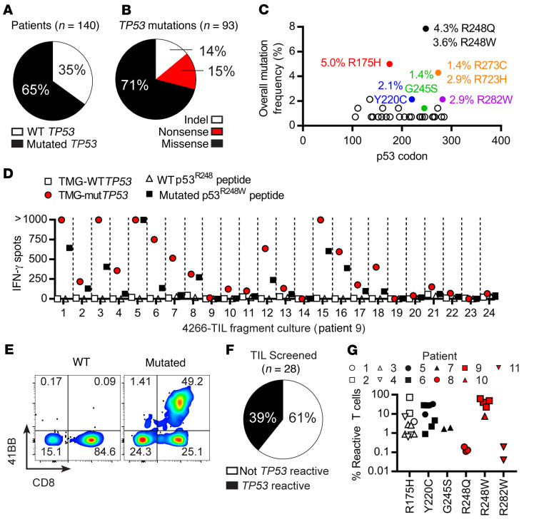
Patients with metastatic epithelial cancers were enrolled in clinical trials (NCT01174121, NCT02133196, and NCT01585428) at the National Cancer Institute (NCI). Metastases were resected for growth of TILs from fragment cultures in high-dose interleukin 2 (IL-2), and peripheral blood lymphocytes (PBLs) were collected for germline sequencing controls and to generate autologous antigen-presenting cells (APCs) for screening assays. A total of 140 patients diagnosed with cancers of the bile duct, breast, colon, cervix, endometrium, gastroesophagus, head and neck, lung, ovary, pancreas, and rectum were evaluated. Of these patients, 91 (65%) had a tumor that expressed a mutation in TP53 (Figure 1A and Supplemental Table 1; supplemental material available online with this article; https://doi.org/10.1172/JCI123791DS1). A total of 93 nonsynonymous TP53 mutations were detected, including 14 insertions or deletions (indels), 13 nonsense mutations, and 66 missense mutations (Figure 1B). Tumors from 33 patients, representing 24% of all patients evaluated, possessed mutations within codons 175, 220, 245, 248, 273, and 282 (Figure 1C and Supplemental Tables 1 and 2), positions that corresponded to previously identified TP53 hot spots in a wide variety of cancer types (Supplemental Tables 3 and 4). There was no explicit bias toward a particular disease type in patient accrual at the NCI Surgery Branch during this time even though there was a preponderance of colorectal patients enrolled in the protocol. Studies of fresh tumor with available RNA-seq also demonstrated high levels of TP53 gene expression, regardless of TP53 mutation status (Supplemental Table 1). The high levels of TP53 gene expression for the targeted mutations, which ranged between the 89th and 100th percentile of all expressed transcripts, raised the possibility that one or more of the peptides arising from these common mutations might be immunogenic in these patients.
Figure 1. TP53 hot spot mutations are immunogenic and elicit intratumoral T cell responses to p53 neoantigens.
(A) Patients with a tumor expressing a TP53 mutation. (B) Classification of TP53 mutations from resected tumors. (C) The overall frequencies of each missense mutation in TP53 from all patients sequenced by p53 codon where the specific amino acid change frequency is given for selected mutations. (D) Screening results from patient 9 as measured by IFN-γ ELISPOT. (E) Expression of 41BB from patient 9’s TIL culture 1 in response to TP53 TMGs. (F) Overall frequency of TIL responses to mutated TP53, which include TMG, peptide, or both (Supplemental Table 5). (G) Frequencies of verified positive TIL responses from selected fragment cultures (some patients had >1) to p53 R175H, Y220C, G245S, R248Q, R248W, and R282W neoantigens. The upregulation of 41BB minus the background in response to mutated p53 peptide (CD4) or TMG (CD8) is reported. Types of T cell responses can be found in Table 1.
The Catalogue of Somatic Mutations in Cancer (COSMIC) database (https://cancer.sanger.ac.uk/cosmic) showed that the 12 most common TP53 hot spot mutations across all tumor types were R175H, Y220C, G245S, G245D, R248L, R248Q, R248W, R249S, R273C, R273H, R273L, and R282W. A novel high-throughput technique was developed using minigenes encoding 25 amino acid peptides containing each TP53 mutation and flanked by 12 amino acids of WT sequence. The peptides were concatenated in tandem to generate a tandem minigene (TMG) as has been performed in testing patient-specific neoepitopes (Supplemental Figure 1) (2, 3, 5). A similar TMG encoding the corresponding WT sequences was also generated to facilitate analysis of T cell specificity to the mutated variants. The TMGs were synthesized de novo as DNA, cloned into an expression vector plasmid DNA, and in vitro transcribed into mRNA. The same 25 amino acid sequences (WT or mutated) were also synthesized as individual peptides at greater than 95% purity using high-performance liquid chromatography. Immature dendritic cells (APCs) were generated from the autologous patients’ peripheral blood and were either electroporated with TMGs (irrelevant, WT, or mutated TP53; 12–16 minigenes each) or pulsed with individual WT or mutated peptides corresponding only to the autologous TP53 mutation. Cocultures of TIL fragment cultures and prepared APCs were incubated overnight at 37°C, and T cell responses were measured by OX40 and 41BB upregulation on the cell surface (flow cytometry) and secretion of interferon-γ (IFN-γ) using an enzyme-linked immunospot (ELISPOT) assay. The upregulation of 41BB was more consistent and robust than OX40 for assessing CD4 T cell reactivities and was thus reported (Supplemental Figure 2). Negative controls for screening were dimethyl sulfoxide (DMSO; peptide vehicle) and T cells only (media), and a mixture of phorbol myristate acetate (PMA) and ionomycin as positive control. A positive response in the initial screening was measured if the IFN-γ secretion and/or 41BB expression was more than twice background (Supplemental Table 5). Selected TIL fragment cultures with the most cells and robust responses were then rescreened in additional independent experiments where they had to display at least 10-fold higher avidity to the mutated gene relative to the relevant WT counterpart to be considered positive.
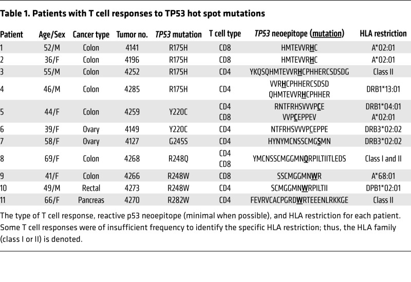
Patient 9 exemplified the speed at which mutated TP53 neoantigen–reactive T cells could be identified for possible cell-based treatment. Lung metastases were resected from this patient, 24 fragment cultures were grown, the screen was performed, and p53R248W T cell responses were identified 34 days after surgery (Figure 1, D and E). Secretion of IFN-γ was off scale (>1000 spots) for TIL cultures 1, 3, 5, and 15 when cocultured with the mutated TP53 TMG (Figure 1D, red circles), and this was specific for the p53R248W peptide (Figure 1D, closed squares). Upregulation of 41BB in response to the p53R248W neoantigen demonstrated this was a CD8+ T cell response (Figure 1E). Autologous PBLs and TILs were available for 28 of 33 patients (Supplemental Table 2). In aggregate, 39% (n = 11) of patients displayed a T cell response to one of the TP53 hot spot mutations under investigation (Figure 1F), and there was a wide range of TP53-reactive T cell frequencies found either through IFN-γ secretion, 41BB upregulation, or both in response to TMG, peptide, or both (Figure 1G and Supplemental Table 5). T cell responses were readily detected against R175H, Y220C, G245S, R248Q, R248W, and R282W with a range of frequencies (0.1%–80% of CD3+ 41BB+ cells) in the 11 patients with mutated TP53-reactive T cells (Figure 1G and Supplemental Figures 3–8).
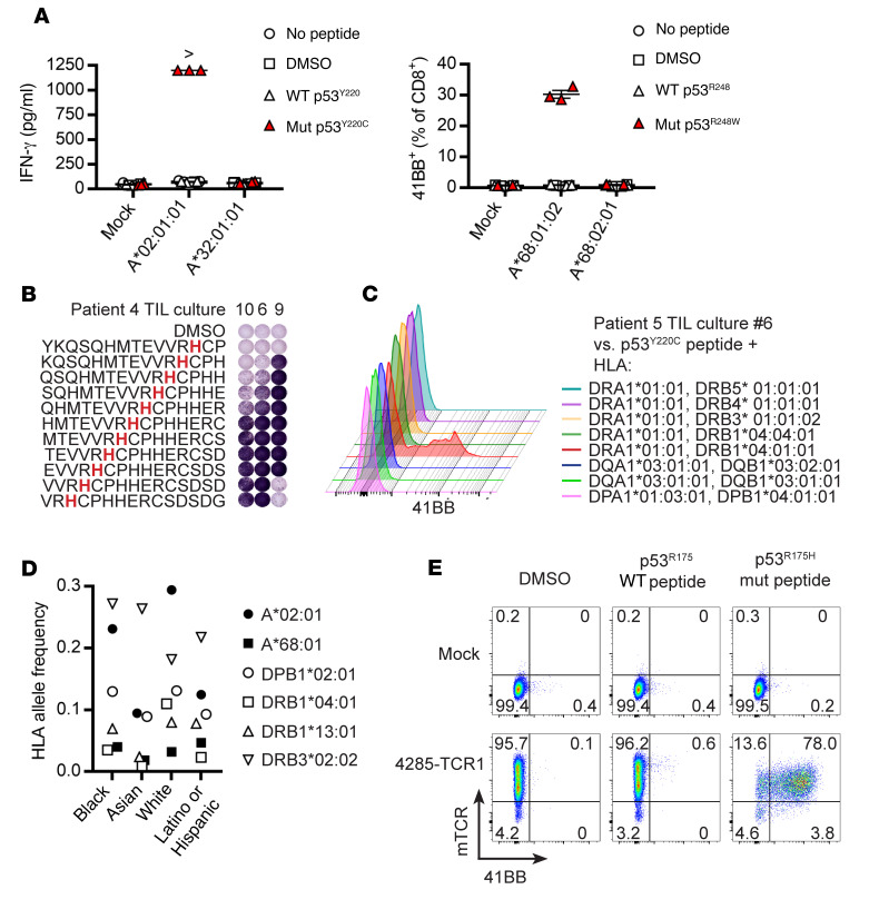
We next evaluated the HLA restriction elements for these TP53 neoantigens by transfecting DNA plasmids encoding HLA alleles into the COS7 cell line (15). TILs from patients 1 and 5 recognized HLA-A*0201 neoepitopes containing p53R175H and p53Y220C, respectively, and TILs from patient 9 recognized an HLA-A*68:01–restricted neoepitope containing p53R248W (Figure 2A and Supplemental Figure 9). Of note, the only shared mutated TP53 neoepitope we identified was the HMTEVVRHC peptide, which was recognized in the context of HLA-A*02:01 by TILs from patients 1 and 2 (Table 1). To identify HLA class II–restricted minimal neoepitopes within the 25 amino acid p53 neoantigen, we tested peptides containing 15 amino acids that overlapped in 14 amino acids, similar to a previous study evaluating patient-specific neoepitopes where we identified HLA-DRB3*02:02–restricted p53Y220C (HYNYMCNSSCMGSMN) and p53G245S (NTFRHSVVVPCEPPE) neoantigens from patients 6 and 7, respectively (15). For example, TIL cultures 4285-F9 and 4285-F6/4285-F10 from patient 4 responded to peptides containing the EVVRHCPH and VRHCPHHER core sequences, respectively (Figure 2B). Both of these p53R175H peptides were restricted by HLA-DRB1*13:01, and the CD4+ T cell response to p53R248W from patient 10 was restricted by HLA-DPB1*02:01 (Supplemental Figures 10–12). CD4+ and CD8+ T cells elicited reactivity to the HLA-DRB1*04:01–restricted peptide RNTFRHSVVVPCE (Figure 2C) and to the HLA-A*02:01–restricted peptide VVPCEPPEV (Figure 2A) p53Y220C neoepitopes, respectively, from patient 5. Some of the HLA alleles that presented the immunogenic p53 neoantigens in our patient cohort were frequent in Black, Asian, White, and/or Latino and Hispanic American populations (Figure 2D), as measured by allele frequency, which is roughly half the population frequency (allele frequency of 0.1 corresponds to ~20% of the population), indicating that a diverse cohort of patients could benefit from mutated TP53-specific T cell immunotherapy.
Figure 2. Immunogenic TP53 hot spot mutations could be restricted by common HLA alleles and targeted by TCRs isolated from p53 neoantigen–reactive TILs.
(A) HLA class I restriction of p53Y220C (patient 5, TIL culture 4259-F1) and p53R248W (patient 9, TIL culture 4266-F1) neoepitopes. Data are mean ± SEM, n = 3 technical replicates. > indicates 1,250 pg/ml IFN-γ. (B) Peptide mapping of CD4+ T cell responses from patient 4 to p53R175H neoepitopes. (C) HLA class II restriction for p53Y220C neoantigen from patient 5. (D) HLA allele frequencies from selected populations (http://www.allelefrequencies.net/default.asp). (E) Specificity of HLA-DRB*13:01/p53R175H–reactive TCR to 25 amino acid p53R175H peptides.
Table 1. Patients with T cell responses to TP53 hot spot mutations.
A library of p53 neoantigen–specific TCRs was also generated following coculture of TILs with p53 neoantigen, sorting for 41BB+ cells, and single-cell reverse transcriptase PCR with TCR gene-specific primers (17). Peripheral blood T cells were genetically modified with TCRs using the nonviral Sleeping Beauty transposon/transposase system (18), and TCR-transposed T cells were cocultured with target p53 neoepitopes. We previously identified p53Y220C- and p53G245S-reactive TCRs with a common restriction element (HLA-DRB3*02:02) from patients 6 and 7 (15). In this study, we isolated TCRs specific for p53R175H/HLA-A*02:01 (patient 1), p53R175H/HLA-DRB1*13:01 (patient 4), p53Y220C/HLA-DRB1*04:01 (patient 5), p53R248W/HLA-A*68:01 (patient 9), and p53R248W/HLA-DPB1*02:01 (patient 10) (Figure 2E and Supplemental Figures 9 and 11–16). A total of 9 TCRs recognizing 7 different p53 neoepitopes have been identified, which were likely unique as they were not found in the NCBI (https://www.ncbi.nlm.nih.gov/) and VDJDB (https://vdjdb.cdr3.net/) databases (Supplemental Table 6). Thus, off-the-shelf TCR gene therapy could be used for patients with tumors expressing the combination of one of the above HLA and p53 neoantigen combinations.
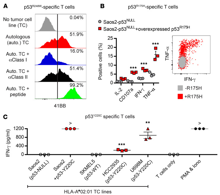
We evaluated the ability of mutated TP53-specific T cells to recognize naturally processed p53 neoepitopes. An autologous tumor cell line generated from mouse xenografts in immunocompromised mice with endogenous expression of both mutated p53R248W and HLA-A*68:01 (Supplemental Figure 17) was recognized by HLA-A*68:01/p53R248W–reactive T cells from patient 9, which was inhibited by class I–blocking antibody (Figure 3A). Saos2 is a TP53 knockout (p53NULL) tumor cell line commonly used to study TP53 biology that naturally expresses HLA-A*02:01. Cocultures of HLA-A*02:01/p53R175H–specific CD8+ T cells from patient 1 with Saos2 overexpressing p53R175H demonstrated significant specific intracellular expression of IFN-γ, tumor necrosis factor-α (TNF-α), and CD107a, which is indicative of T cell degranulation and tumor cytolysis, compared with cocultures with parental Saos2 cells (Figure 3B). CD4+ T cells also degranulated in response to p53R175H, p53Y220C, and p53R248W neoepitopes, indicating that these cells may have the capability to directly lyse tumor cells (Supplemental Figures 18–20). A panel of HLA-A*02:01–expressing tumor cells from unrelated donors was cocultured with HLA-A*02:01/p53Y220C–reactive T cells from patient 5, which secreted significant IFN-γ in response to HCC2935 and U698M tumor cell lines with endogenous expression of p53Y220C and Saos2 overexpressing p53Y220C, but lacked IFN-γ secretion in cocultures with Saos2 (p53NULL) and SKMEL5 (WT TP53) tumor cell lines (Figure 3C). These data indicate that TP53 mutational hot spot mutations can be processed and presented on the surfaces of tumor cells in the context of relevant HLA molecules and can be recognized by T cells.
Figure 3. Tumor cells naturally process and present p53 neoantigens to T cells.
(A) Expression of 41BB on CD8+ T cells expressing HLA-A*68:01/p53R248W neoantigen–specific TCR following coculture with autologous tumor cell line (TC), which was incubated with anti-HLA class I or II blocking antibodies or p53R248W peptide. (B) Intracellular cytokine staining of cocultures of CD8+ T cells expressing HLA-A*02:01/p53R175H neoantigen–specific TCR and Saos2 cells alone (HLA-A*02:01+ and p53NULL; gray circles on left graph or gray dots on right plot) or overexpressing p53R175H full-length protein (red squares on left graph or red dots on right plot). (C) Secretion of IFN-γ in cocultures of HLA-A*02:01/p53Y220C neoantigen–specific T cells (CD8+-enriched TIL culture 4259-F1 from patient 5) with HLA-A*02:01–positive tumors from unrelated donors and Saos2 tumor cells overexpressing full-length p53Y220C protein. > indicates 1,250 pg/ml IFN-γ. Data in panels B and C are mean ± SEM, n = 3 technical replicates, and 2-tailed Student’s t tests were performed for statistical analyses (**P < 0.01 and ***P < 0.001).
Both CD4+ and CD8+ T cells responded to mutated TP53 neoantigens, and 2 of the previously reported ovarian cancer patients were included with the 9 new responses to accurately describe the totality of our experience detecting T cell responses to mutated TP53 in 11 patients (Table 1). Our previous experience in detecting TP53 mutation–specific T cells in ovarian cancer (15, 16) was expanded in this study to colorectal and pancreas cancers and 4 more TP53 hot spot mutations were determined to be immunogenic in the context of 5 new HLA restrictions. This led to an additional 7 mutated TP53-specific TCRs for the potential benefit of future patients with matching HLA and TP53 mutations. Moreover, once it was determined that the tumor expressed a TP53 hot spot mutation, TIL screening could occur because the TMG and peptides were pre-prepared, thus eliminating the time and cost associated with personalized neoantigen screening. It is currently unknown why T cell responses were frequently observed to p53 neoepitopes, but high TP53 expression (Supplemental Table 1) or increased stability of the mutant p53 protein could influence their immunogenicity (19). Individual codon substitutions also appeared to differ in terms of the frequency with which they gave rise to an immunogenic epitope (Supplemental Table 7), although the sample size was small, and future studies can evaluate this in more detail with a larger patient cohort. The frequency of these responses was likely to be influenced by the HLA alleles expressed by patients’ cells. Two, three, or six patients had both, only CD8+, or only CD4+ T cell responses to mutated TP53, respectively (Table 1), suggesting that there are complex dynamics driving the immunogenicity of TP53 hot spot mutations, which may include expression of HLA on the tumor or APC, TCR avidity, and T cell trafficking to the tumor microenvironment.
The observation that 11 of 28 patients (39%) recognized an autologous p53 neoepitope suggests that TP53 mutations may be of value in developing cell transfer immunotherapy. CD4+ or CD8+ TILs selected for IFN-γ secretion or 41BB upregulation in response to KRAS and passenger mutations have been grown to large numbers and infused into cancer patients, which resulted in long-term objective tumor regressions of breast cancer, colon cancer, and cholangiocarcinoma (1, 2, 4), and suggests that targeting TP53 mutations could result in similar clinical benefit. Our focused TP53 screening approach could also be a model for targeting other high-value mutated tumor neoantigens present in large numbers of unrelated patients, e.g., KRAS, PIK3CA, and EGFR. Neoantigen loss through T cell–selective pressure in vivo could be a mechanism of resistance, especially when targeting tumor suppressors. However, the TP53 hot spot mutations may not follow this general rule, as the WT TP53 allele is typically lost through loss of heterozygosity (Supplemental Table 1), and the TP53 mutation can inactivate WT p53 tumor suppressor function and sometimes also have gain-of-function activity, which may be critical for tumor survival (20). We plan to directly test the ability of T cells specific for TP53 mutations (TILs and TCRs) to eliminate metastatic cancer in clinical trials at the NCI Surgery Branch.
Methods
Antibodies used in this study can be found in Supplemental Table 8. Additional methods are in the Supplemental Material.
Study approval.
Written, informed consent was granted for all patients prior to enrollment in National Institutes of Health protocols (NCT01174121, NCT02133196, and NCT01585428), which were approved by the Institutional Review Board. Studies in mice were approved by the National Institutes of Health Institutional Animal Care and Use Committee.
Author contributions
PM, SAR, and DCD conceptualized and developed the project. PM, AP, PFR, MRP, BCP, LJ, JJG, ZY, AS, ET, VH, WL, and DCD designed and performed experiments and analyzed data. RPTS supervised patient samples and NPR supervised mouse studies. PM, PFR, SAR, and DCD wrote the manuscript, and all authors edited and approved the manuscript.
Supplementary Material
Acknowledgments
We thank the NCI Surgery Branch Cell Production Facility for its efforts growing the tumor infiltrating lymphocytes, and Arnold Mixon and Shawn Farid for their assistance with FACS. This work was supported by intramural funding of the Center for Cancer Research, NCI.
Version 1. 02/04/2019
Electronic publication
Version 2. 03/01/2019
Print issue publication
Version 3. 07/01/2021
Correction to supplemental table 6
Footnotes
Conflict of interest: PM, AP, PFR, MRP, WL, SAR, and DCD have a patent application (no. 62/565,383) for TCRs described in this manuscript. PM, SAR, and DCD have a patent application (no. 62/565,464) for methods described in this manuscript.
License: Copyright 2019, American Society for Clinical Investigation.
Reference information: J Clin Invest. 2019;129(3):1109–1114.https://doi.org/10.1172/JCI123791.
See the related Commentary at Cancer neoantigens targeted by adoptive T cell transfer: private no more.
Contributor Information
Parisa Malekzadeh, Email: parisa.malekzadeh@nih.gov.
Anna Pasetto, Email: anna.pasetto@ki.se.
Paul F. Robbins, Email: Paul_Robbins@nih.gov.
Maria R. Parkhurst, Email: Maria_Parkhurst@nih.gov.
Biman C. Paria, Email: biman_paria@nih.gov.
Li Jia, Email: li.jia2@nih.gov.
Jared J. Gartner, Email: jared.gartner@nih.gov.
Victoria Hill, Email: victoria.hill@nih.gov.
Zhiya Yu, Email: zhiyayu@mail.nih.gov.
Nicholas P. Restifo, Email: restifo@nih.gov.
Abraham Sachs, Email: abraham.sachs@nih.gov.
Eric Tran, Email: Eric.Tran@providence.org.
Winifred Lo, Email: winifred.m.lo@gmail.com.
Robert P.T. Somerville, Email: robert.somerville@nih.gov.
Steven A. Rosenberg, Email: sar@nih.gov.
Drew C. Deniger, Email: drew.deniger@nih.gov.
References
- 1.Tran E, et al. T-cell transfer therapy targeting mutant KRAS in cancer. N Engl J Med. 2016;375(23):2255–2262. doi: 10.1056/NEJMoa1609279. [DOI] [PMC free article] [PubMed] [Google Scholar]
- 2.Zacharakis N, et al. Immune recognition of somatic mutations leading to complete durable regression in metastatic breast cancer. Nat Med. 2018;24(6):724–730. doi: 10.1038/s41591-018-0040-8. [DOI] [PMC free article] [PubMed] [Google Scholar]
- 3.Tran E, et al. Immunogenicity of somatic mutations in human gastrointestinal cancers. Science. 2015;350(6266):1387–1390. doi: 10.1126/science.aad1253. [DOI] [PMC free article] [PubMed] [Google Scholar]
- 4.Tran E, et al. Cancer immunotherapy based on mutation-specific CD4+ T cells in a patient with epithelial cancer. Science. 2014;344(6184):641–645. doi: 10.1126/science.1251102. [DOI] [PMC free article] [PubMed] [Google Scholar]
- 5.Stevanović S, et al. Landscape of immunogenic tumor antigens in successful immunotherapy of virally induced epithelial cancer. Science. 2017;356(6334):200–205. doi: 10.1126/science.aak9510. [DOI] [PMC free article] [PubMed] [Google Scholar]
- 6.Bailey MH, et al. Comprehensive characterization of cancer driver genes and mutations. Cell. 2018;173(2):371–385.e18. doi: 10.1016/j.cell.2018.02.060. [DOI] [PMC free article] [PubMed] [Google Scholar]
- 7.Freed-Pastor WA, Prives C. Mutant p53: one name, many proteins. Genes Dev. 2012;26(12):1268–1286. doi: 10.1101/gad.190678.112. [DOI] [PMC free article] [PubMed] [Google Scholar]
- 8.Theoret MR, et al. Relationship of p53 overexpression on cancers and recognition by anti-p53 T cell receptor-transduced T cells. Hum Gene Ther. 2008;19(11):1219–1232. doi: 10.1089/hum.2008.083. [DOI] [PMC free article] [PubMed] [Google Scholar]
- 9.Yanuck M, et al. A mutant p53 tumor suppressor protein is a target for peptide-induced CD8+ cytotoxic T-cells. Cancer Res. 1993;53(14):3257–3261. [PubMed] [Google Scholar]
- 10.Noguchi Y, Chen YT, Old LJ. A mouse mutant p53 product recognized by CD4+ and CD8+ T cells. Proc Natl Acad Sci USA. 1994;91(8):3171–3175. doi: 10.1073/pnas.91.8.3171. [DOI] [PMC free article] [PubMed] [Google Scholar]
- 11.Vermeij R, Leffers N, van der Burg SH, Melief CJ, Daemen T, Nijman HW. Immunological and clinical effects of vaccines targeting p53-overexpressing malignancies. J Biomed Biotechnol. 2011;2011:702146. doi: 10.1155/2011/702146. [DOI] [PMC free article] [PubMed] [Google Scholar]
- 12.Carbone DP, et al. Immunization with mutant p53- and K-ras-derived peptides in cancer patients: immune response and clinical outcome. J Clin Oncol. 2005;23(22):5099–5107. doi: 10.1200/JCO.2005.03.158. [DOI] [PubMed] [Google Scholar]
- 13.Ito D, et al. Immunological characterization of missense mutations occurring within cytotoxic T cell-defined p53 epitopes in HLA-A*0201+ squamous cell carcinomas of the head and neck. Int J Cancer. 2007;120(12):2618–2624. doi: 10.1002/ijc.22584. [DOI] [PubMed] [Google Scholar]
- 14.Couch ME, et al. Alteration of cellular and humoral immunity by mutant p53 protein and processed mutant peptide in head and neck cancer. Clin Cancer Res. 2007;13(23):7199–7206. doi: 10.1158/1078-0432.CCR-07-0682. [DOI] [PubMed] [Google Scholar]
- 15.Deniger DC, et al. T-cell responses to TP53 “hotspot” mutations and unique neoantigens expressed by human ovarian cancers. Clin Cancer Res. 2018;24(22):5562–5573. doi: 10.1158/1078-0432.CCR-18-0573. [DOI] [PMC free article] [PubMed] [Google Scholar]
- 16.Yossef R, et al. Enhanced detection of neoantigen-reactive T cells targeting unique and shared oncogenes for personalized cancer immunotherapy. JCI Insight. 2018;3(19):e122467. doi: 10.1172/jci.insight.122467. [DOI] [PMC free article] [PubMed] [Google Scholar]
- 17.Pasetto A, et al. Tumor- and neoantigen-reactive T-cell receptors can be identified based on their frequency in fresh tumor. Cancer Immunol Res. 2016;4(9):734–743. doi: 10.1158/2326-6066.CIR-16-0001. [DOI] [PMC free article] [PubMed] [Google Scholar]
- 18.Deniger DC, et al. Stable, nonviral expression of mutated tumor neoantigen-specific T-cell receptors using the sleeping beauty transposon/transposase system. Mol Ther. 2016;24(6):1078–1089. doi: 10.1038/mt.2016.51. [DOI] [PMC free article] [PubMed] [Google Scholar]
- 19.Kastenhuber ER, Lowe SW. Putting p53 in context. Cell. 2017;170(6):1062–1078. doi: 10.1016/j.cell.2017.08.028. [DOI] [PMC free article] [PubMed] [Google Scholar]
- 20.Baugh EH, Ke H, Levine AJ, Bonneau RA, Chan CS. Why are there hotspot mutations in the TP53 gene in human cancers? Cell Death Differ. 2018;25(1):154–160. doi: 10.1038/cdd.2017.180. [DOI] [PMC free article] [PubMed] [Google Scholar]
Associated Data
This section collects any data citations, data availability statements, or supplementary materials included in this article.


