Abstract
Concha bullosa is one of the most common anatomical variant found in the patients of chronic rhinosinusitis. To study the role of concha bullosa in patients of chronic rhinosinusitis a prospective cross sectional study was done comprising of 60 patients who were having symptoms of sinusitis for more than 12 weeks. These were evaluated with the help of nasal endoscope and CT scan. DNS and concha bullosa are the most common anatomical variant seen in the patients of chronic rhinosinusitis with percentage being 88.3% and 76.6% respectively. Patients with deviated nasal septum and large concha bullosa (bulbous and extensive) were associated with higher incidence of ostiomeatal complex obstruction. There seems a strong relationship between unilateral concha bullosa and contralateral deviated nasal septum in development of chronic rhinosinusitis. (1) To find out the percentage and role of concha bullosa in patients of chronic rhinosinusitis. (2) To screen the patients of chronic rhinosinusitis for concha bullosa. (3) To do the nasal endoscopy and CT scan of the screened patients. Study design: cross sectional study. Sample size: 60. Inclusion criteria: all patients of chronic rhinosinusitis. Exclusion criteria: patients with previous sinus surgeries, malignancy and acute cases of chronic rhinosinusitis.
Keywords: Concha bullosa, DNS, Ostiomeatal complex, Chronic rhinosinusitis, CT scan
Introduction
Chronic rhino sinusitis is a common disease and a major cause of morbidity among patients. Its diagnosis relies on clinical judgement based on often vague physical complaints and with the help of anterior and posterior rhinoscopic examination. Currently, computed tomography is the method of choice for assessment of paranasal sinuses, nasal fossae and their anatomical variants. Computed tomography offers detailed study of anatomical variations.
Few organs of human body are subject to remarkable inter and intra subject variations as paranasal sinuses. The range of anatomic variants that can interfere with the mucociliary drainage of ostiomeatal complex includes concha bullosa, deviated nasal septum, uncinate process variations, ethmoid bulla, paradoxical middle turbinate, agger nasi and haller cells. Concha bullosa is the pneumatization of the middle turbinate and is one of the most common variation of the sinonasal anatomy. Concha based on location is of three types lamellar concha bullosa, bulbous concha bullosa and extensive concha bullosa [4]. Concha bullosa intruding into the middle meatus may fill the space between septum and lateral wall impinging on the infundibulum and maxillary sinus ostium creating areas of mucosal contact and alters normal airflow thus producing negative influence on paranasal sinus ventilation and on the mucocilliary clearance in ostiomeatal complex thus making the relevant sinus prone to infection [1]. Bulbous type of concha bullosa especially the large ones were considered to predispose to sinusits. Concha bullosa is best diagnosed radiographically as they are identified on CT scan, appearing as an air space of the middle turbinate surrounded by an oval bony rim.
Materials and Methods
A prospective cross sectional was carried out in the Department of Otorhinolaryngology, People’s Medical College, Bhopal (M.P.) from 1/1/2013 to 1/1/2014 and a total of 60 patients who were clinically and radiologically diagnosed as having chronic rhino sinusitis, were evaluated with the help of CT scan paranasal sinuses and by nasal endoscopy. Patients with acute sinusitis or malignant disease or those who had previously undergone nasal or sinus surgery, either open or endoscopic, were excluded from the study.
Observations and Results
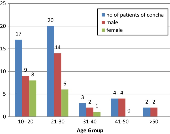
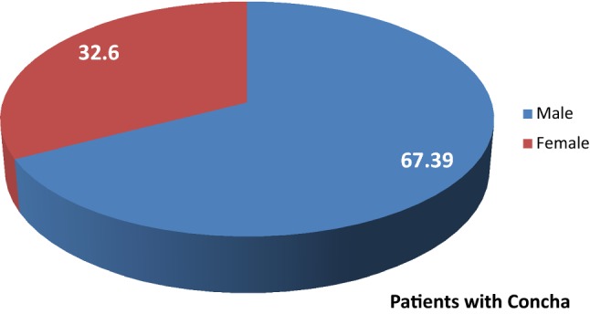
A total of 60 patients of chronic rhinosinusitis were examined for the presence of various anatomical variants. Out of 60 patients, 40 were males and 20 were females with male female ratio being 2:1. The maximum number of patients were seen in age group 21–30 followed by age group 10–20 years, and least in age group > 50 years.
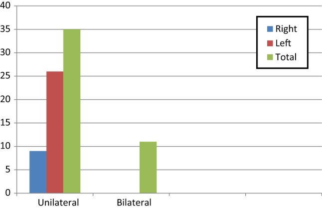
Regarding the symptoms, the most common symptom noticed was nasal obstruction. (91.6%) followed by headache (63.3% and facial pain (65%). The most common anatomical variants found was DNS (53) followed by concha bullosa (46) and prominent bulla (38). The concha bullosa was more common among males and in age group of 21–30 years (Figs. 1, 2). Out of 46 patients of concha bullosa, 35 had unilateral and 11 had bilateral concha bullosa (Fig. 3). The most common type of concha bullosa was of bulbous type (63%) followed by extensive (24%) and lamellar type (13%).
Fig. 1.

Distribution of gender according to age group in patients of concha bullosa
Fig. 2.

Sex ratio in patients of concha bullosa
Fig. 3.

Side distribution of concha bullosa in patients of chronic rhinosinusitis
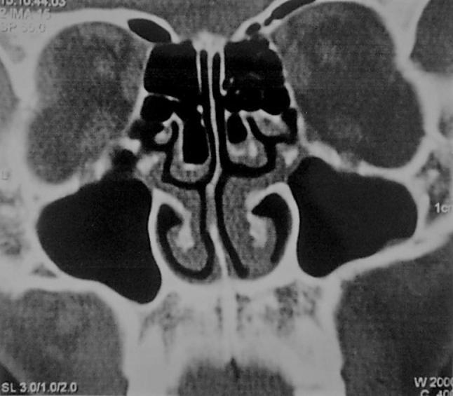
41 patients had both DNS and concha bullosa. The concha bullosa was seen significantly higher on side contralateral to DNS (26 patients) than ipsilateral to DNS (9 patients) in the study group with p value = 0.001 (Fig. 4). There was presence of air column between medial aspect of concha bullosa and the adjacent DNS in 38 cases while there was loss of air column in 3 cases p value 0.0001, which is significant.
Fig. 4.

CT PNS coronal view showing bilateral bulbous concha bullosa
Regarding the sinus involvement the most frequently involved sinus was maxillary sinus (85%) followed by anterior ethmoid (73.3%), sphenoid sinus was the least commonly involved (3.3%).
Discussion
Computerized tomographic imaging of sinonasal region has become the gold standard in the evaluation of patients with chronic sinusitis. A cross sectional study was carried out in the Department of Otorhinolaryngology, People’s Medical College, Bhopal (M.P.) comprising of 60 patients of chronic rhino sinusitis. These cases were examined clinically and provisional diagnosis was made on the basis of nasal endoscopy and CT scan. CT scan was evaluated to study the effect of concha bullosa on lateral nasal wall and ostiomeatal complex.
Stammberger and Wolf have documented the prominent role of ostiomeatal complex as a gateway to sinus disease. As a consequence of various anatomic and functional relationships immediately adjacent to OMC, proliferation of disease into anterior sinuses is the natural corollary. However, few investigators have examined the role of the concha bullosa and deviated nasal septum and its ultimate effect on the sinus disease. Concha bullosa infringe on the patency of already narrow intricate ostiomeatal complex thus predisposing to sinusitis by interfering with the mucociliary clearance of ostiomeatal area.
Since Zukerlandl E observed an air cavity within the middle turbinate and termed its pneumatization as “concha bullosa”, many studies have been undertaken to study the incidence and its etiological role on sinusitis. On endoscopy, it appears as an enlarged head of middle turbinate. Concha bullosa are best diagnosed radiographically by CT scan PNS coronal section which enables visualization of even very small pneumatization.
The percentage of concha bullosa in our study was 76.4%. The prevalence of concha bullosa varies from 5 to 53%. Lothrop HA. Noted middle turbinate pneumatization in 9% of the 1000 lateral nasal specimens examined [2]. As per Turner AL it was in 20% [3] Bolger WE et al found the incidence to be 53.6% [4] and Maru YK et al found it out to be 41.3% [5] while only 15% as found out by Bharathi MB et al [6]. Bogler WE et al divided middle turbinate pneumatisation into three types, bulbous type: pneumatization of bulbous segment of middle turbinate, lamellar type: pneumatization of vertical lamella of middle turbinate and extensive type: pneumatization of vertical and bulbous portion. In our study percentage of lamellar, bulbous, extensive concha bullosa is 13, 63 and 24% respectively. Calhoun Kh et al stated that concha bullosa was found more frequently in the symptomatic group of patients with sinusitis [7], Zinreich SJ et al showed that concha bullosa is often associated with ostiomeatal complex disease [8]. Yousem DM et al remarked “it appears that the size not just the presence of concha bullosa and another anatomic variant is the critical factor”. A large size concha bullosa impinges directly on OMC and can predispose maxillary sinus ostium obstruction and subsequently chronic rhinosinusitis [9]. In our study we found significant relationship between deviated nasal septum and contralateral concha bullosa.
In our study the percentage of bulbous type of concha bullosa was the largest that is 63 percent. It was found that there was always a maintenance of nasal air channel between the medial aspect of concha (unilateral/dominant) and the adjacent surface of nasal septum (97.8% cases). This implies deviation of the septum away from concha is not the result of concha pushing the septum, rather there appears to be some as yet unknown developmental relationship between a concha and the nasal septum.
In our study we found significant relationship between deviated nasal septum and contralateral concha bullosa. We found there was always maintenance of nasal air channel between the medial aspect of concha (unilateral/dominant) and the adjacent surface of nasal septum was maintained in 97.8% cases. This implies that the deviation of septum away from concha is not the result of concha pushing the septum, rather there appears to be some as yet unknown developmental relationship between a concha and nasal septum.
Conclusion
Chronic rhino sinusitis is fairly a common disease condition affecting most commonly the age group between 21 and 30 years Combination of CT scan PNS and fiber optic diagnostic nasal endoscopy is excellent for precise evaluation of nasal cavity. Concha bullosa and deviated nasal septum are the two most common anatomical variants seen in patients with chronic rhniosinusitis. Patients with nasal septum deviation and large concha bullosa (bulbous and extensive) types were associated with higher incidence of ostiomeatal complex obstruction. There is a strong relationship between the presence of unilateral concha and contra-lateral nasal septal deviation.
In the above study it is concluded that concha bullosa plays major role in development of chronic rhinosinusitis. Thus after confirming the presence of concha bullosa surgeon should keep in mind the possibility of development of chronic rhinosinusitis or vice versa.
Conflict of interest
All authors declare that they have no conflict of interest.
Ethical approval
All procedures performed in studies involving human participants were in accordance with the ethical standards of the institutional and/or national research committee and with the 1964 Helsinki declaration and its later amendments or comparable ethical standards.
Informed consent
Informed consent was obtained from all individual participants included in the study. Being a retrospective study, for this type of study formal consent is not required.
References
- 1.Stammberger H, Wolf G. Headaches and sinus diseases: the endoscopic approach. Ann Otol Rhinol Laryngol. 1988;97(Suppl 134):3–23. doi: 10.1177/00034894880970S501. [DOI] [PubMed] [Google Scholar]
- 2.Lothrop HA. The anatomy of the inferior ethmoidal turbinate bone with particular reference to cell formation: surgical Importance of such ethmoid cells. Ann Surg. 1903;38:233–255. doi: 10.1097/00000658-190308000-00005. [DOI] [PMC free article] [PubMed] [Google Scholar]
- 3.Turner AL (1927) Disease of the nose, throat and ear for practitioners and students, 2nd edn. John Wright and Sons, Bristol, p 17 (quoted by—Bolger WE, Butzin CA, Parson DS (1991) Paranasal sinus bony anatomic variations and mucosal abnormalities; CT analysis for endoscopic sinus surgery. Laryngoscope 101:56–64) [DOI] [PubMed]
- 4.Bolger WE, Butzin CA, Parson DS. Paranasal sinus bony anatomic variations and mucosal abnormalities; CT analysis for endoscopic sinus surgery. Laryngoscope. 1991;101:56–64. doi: 10.1288/00005537-199101000-00010. [DOI] [PubMed] [Google Scholar]
- 5.Maru YK, Gupta Y. Concha bullosa: frequency and appearance on sinonasal CT. Indian J Otolaryngol Head Neck Surg. 2000;52:40–44. doi: 10.1007/BF02996431. [DOI] [PMC free article] [PubMed] [Google Scholar]
- 6.Bharathi MB, Mamtha H, Prasanna LC. Variations of ostiomeatal complex and its applied anatomy: a CT scan study. Indian J Sci Technol. 2010;3(8):904–907. [Google Scholar]
- 7.Calhoun KH, Waggenspack GA, Simpson CB, Hokanson JA, Bailey BJ. CT evaluation of Paranasal sinuses in symptomatic and asymptomatic populations. Otolaryngology Head & Neck Surgery. 1991;104:480–483. doi: 10.1177/019459989110400409. [DOI] [PubMed] [Google Scholar]
- 8.Zinreich SJ. Paranasal sinus imaging. Otolaryngol Head Neck Surg 1990 Nov. 1990;103(5):863–868. doi: 10.1177/01945998901030S505. [DOI] [PubMed] [Google Scholar]
- 9.Yousem DM.Imaging of sinonasal inflammatory disease. Radiology 1993Aug;19(2):303–3014(quoted by Vincent TES,Gendeh B S :the association of concha bullosa and deviated nasal septum with chronic rhinosinusitis in functional endoscopic sinus surgery patients:Med J Malaysia Vol 65No 2 June 2010;105-111) [PubMed]


