Abstract
Background
Whether use of high‐intensity statins is more important than achieving low‐density lipoprotein cholesterol (LDL‐C) target remains controversial in patients with coronary artery disease. We sought to investigate the association between statin intensity and long‐term clinical outcomes in patients achieving treatment target for LDL‐C after percutaneous coronary intervention.
Methods and Results
Between February 2003 and December 2014, 1746 patients who underwent percutaneous coronary intervention and achieved treatment target for LDL‐C (<70 mg/dL or >50% reduction from baseline level) were studied. We classified patients into 2 groups according to an intensity of statin prescribed after index percutaneous coronary intervention: high‐intensity statin group (atorvastatin 40 or 80 mg, and rosuvastatin 20 mg, 372 patients) and non‐high‐intensity statin group (the other statin treatment, 1374 patients). The primary outcome was a composite of cardiac death, myocardial infarction, or stroke. Difference in time‐averaged LDL‐C during follow‐up was significant, but small, between the high‐intensity statin group and non‐high‐intensity statin group (59±13 versus 61±12 mg/dL; P=0.04). At 5 years, patients receiving high‐intensity statins had a significantly lower incidence of the primary outcome than those treated with non‐high‐intensity statins (4.1% versus 9.9%; hazard ratio, 0.42; 95% confidence interval, 0.23–0.79; P<0.01). Results were consistent after propensity‐score matching (4.2% versus 11.2%; hazard ratio, 0.36; 95% confidence interval, 0.19–0.69; P<0.01) and across various subgroups.
Conclusions
Among patients achieving treatment target for LDL‐C after percutaneous coronary intervention, high‐intensity statins were associated with a lower risk of major adverse cardiovascular events than non‐high‐intensity statins despite a small difference in achieved LDL‐C level.
Keywords: cardiovascular events, low‐density lipoprotein cholesterol, percutaneous coronary intervention, secondary prevention, statin
Subject Categories: Percutaneous Coronary Intervention, Secondary Prevention, Lipids and Cholesterol
Clinical Perspective
What Is New?
In patients undergoing coronary revascularization who achieved a low‐density lipoprotein‐cholesterol <70 mg/dL, high‐intensity statin therapy was significantly associated with a lower risk of primary outcome compared with non‐high‐intensity statin therapy.
What Are the Clinical Implications?
In patients undergoing percutaneous coronary intervention, high‐intensity statin therapy is associated with additional lowering of risk of major adverse cardiovascular events beyond what may be achieved by low‐density lipoprotein cholesterol target alone.
High‐intensity statins have demonstrated consistent benefits for secondary prevention of adverse cardiovascular events compared with moderate‐intensity statins in several randomized trials.1, 2 Therefore, the 2013 American College of Cardiology/American Heart Association (ACC/AHA) guideline on treatment of blood cholesterol recommends high‐intensity statins, including atorvastatin 40 or 80 mg and rosuvastatin 20 or 40 mg, for patients with atherosclerotic cardiovascular disease.3 However, whether beneficial effects of high‐intensity statins can be attributable to statin intensity per se or to merely lower low‐density lipoprotein cholesterol (LDL‐C) level achieved by high‐intensity statins compared with moderate‐intensity statins is uncertain. It has been reported that lowering of LDL‐C with statin therapy reduces major cardiovascular events regardless of types or intensities of statins.4 Therefore, European Society of Cardiology/European Atherosclerosis Society (ESC/EAS) guidelines for management of dyslipidemia still propose the target goal for LDL‐C of <1.8 mmol/L (70 mg/dL) or a reduction of at least 50% from baseline for subjects at very high risk without specific recommendation for type or intensity of statin.5 The differences between the 2 guidelines have caused many debates and much confusion in daily practice.6 To date, it remains controversial whether outcomes differ according to statin intensity in patients with similar LDL‐C level. Therefore, we sought to investigate the association between statin intensity and long‐term clinical outcomes in patients undergoing percutaneous coronary intervention (PCI) and achieving treatment target for LDL‐C.
Methods
Study Population
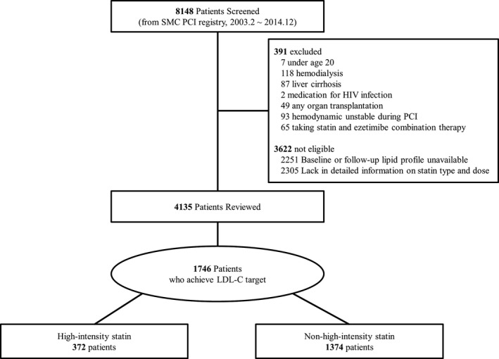
The study population was selected from the Samsung Medical Center (Seoul, Korea) PCI registry. Between February 2003 and December 2014, 8148 patients underwent PCI with drug‐eluting stents. Patients were ineligible for the study if there were no data on blood cholesterol level or medication information at discharge. Exclusion criteria were: (1) <20 years of age; (2) on maintenance hemodialysis; (3) had a history of liver cirrhosis; (4) received treatment for human immunodeficiency virus infection; (5) had undergone any organ transplantation; (6) had cardiopulmonary resuscitation or mechanical circulatory support during PCI; or (7) taking statin and ezetimibe combination therapy. After exclusion of 4013 patients, whether treatment target for LDL‐C was achieved or not during follow‐up after index PCI was reviewed in 4135 patients. We calculated time‐averaged LDL‐C using follow‐up LDL‐C levels measured from 4 weeks to 3 years after index PCI and regarded target for LDL‐C being achieved when time‐averaged LDL‐C was <1.8 mmol/L (70 mg/dL) or reduced at least 50% from baseline during follow‐up after index PCI based on the ESC/EAS guideline.5 Time‐averaged LDL‐C was calculated by the formula:
where Li=consecutively measured follow‐up LDL‐C level, Ti=time period of measurement between Li‐1 and Li, and T1=time period between index PCI and L1.
Finally, 1746 patients who achieved target for time‐averaged LDL‐C during follow‐up were included in this study. We classified patients into 2 groups according to the intensity of statin prescribed at hospital discharge from admission for the index PCI: high‐intensity statin group (n=372) and non‐high‐intensity statin group (n=1374). Definition of statin intensity was based on the guideline from ACC/AHA.3 Atorvastatin 40 or 80 mg and rosuvastatin 20 mg were defined as high‐intensity statins, and the other statins were classified as non‐high‐intensity statins. (Details about statins used in the non‐high‐intensity statin group are shown in Table S1.) Rosuvastatin 40 mg has not been approved in Korea. The exclusion and division of the study is shown in a flow diagram in Figure 1. The Institutional Review Board of Samsung Medical Center approved this study and waived the requirement for written informed consent. The data, analytical methods, and study materials will not be made available to other researchers for purposes of reproducing the results or replicating the procedure.
Figure 1.

Scheme of group distribution. HIV indicates human immunodeficiency virus; LDL‐C, low‐density lipoprotein cholesterol; PCI, percutaneous coronary intervention; SMC, Samsung Medical Center.
Data Collection and Follow‐up
Baseline characteristics and clinical outcome data were prospectively collected in our PCI registry by trained research coordinators using a standardized case report form and protocol. Patients were routinely followed up at 1, 6, and 12 months after the index procedure and annually thereafter. Further information was collected by telephone contact or medical records, if necessary. Data on statin intensity were collected from the electronic prescribing system of Samsung Medical Center. To assess whether the intensity of statin was maintained during follow‐up, information on medications that had been prescribed during the 3 years after index PCI were collected. We also collected high‐sensitivity C‐reactive protein (hs‐CRP) level measured during the 3 years after index PCI. Time‐averaged hs‐CRP was calculated by the same formula used in calculating time‐averaged LDL‐C. Follow‐up was considered complete if mortality was confirmed from the National Population Registry of the Korea National Statistical Office using a unique personal identification number or if the patient was contacted at the planned follow‐up interval.
Study Outcomes and Definition
The primary outcome was the occurrence of major adverse cardiovascular events (MACEs) during follow‐up, defined as a composite of cardiac death, myocardial infarction (MI), or stroke. Secondary outcomes included all‐cause death, target lesion revascularization, target vessel revascularization, and individual components of the primary outcome. All deaths were considered to be cardiac death unless a definite noncardiac cause could be established.7 MI was defined as elevated cardiac enzymes (troponin or myocardial band fraction of creatinine kinase) greater than the upper‐normal limit with ischemic symptoms or electrocardiography findings indicative of ischemia or MI at readmission requiring subsequent hospitalization (defined as emergency admission with principal diagnosis of MI).8, 9 Stroke was defined as a neurological deficit attributed to an acute focal injury of the central nervous system by a vascular cause, including cerebral infarction, intracerebral hemorrhage, and subarachnoid hemorrhage.10 Target lesion revascularization was defined as repeat PCI of the lesion within 5 mm of stent deployment or bypass graft surgery of the target vessel. Target vessel revascularization was repeat revascularization of the target vessel by PCI or bypass graft surgery.7, 8 All end points in this study were censored at 5 years after index PCI.
Statistical Analysis
Categorical variables are presented as numbers of events and percentages and were compared using the chi‐square test or Fisher's exact test (for sparse data). Continuous variables are presented as mean±SD and were compared using the t test or Wilcoxon rank‐sum test. We assessed survival curves using the Kaplan–Meier method, along with a log‐rank test. We estimated the hazard ratios (HRs) of high versus non‐high‐intensity statin group in both univariate and multiple Cox regression models. In multiple Cox regression, variables that appeared to be related in the univariate analysis with a P<0.2 were considered in a step‐wise method to select predictors of outcomes. HRs of high versus non‐high‐intensity statin group were reported with their 95% confidence intervals (CIs).
In addition, to adjust the 2 groups for any inherent imbalance in their demographic and clinical characteristics, we advocated the propensity score method and balanced the 2 groups so that they were comparable with no undue influences from confounding factors. Propensity scores, which are the probability of being treated with a high‐intensity statin for each patient, were estimated using predicted probabilities from a multiple logistic regression analysis. Typically a full nonparsimonious model was fit using all variables in Table 1 (except aspirin and P2Y12 inhibitors) and baseline lipid profile in Table 2 to predict the probability to be treated with a high‐intensity statin. The variable ratio, parallel, and pair‐wise nearest neighbor matching method created a matched data set to avoid a significant data loss from unmatched patients. A standardized mean difference of less than 10% for each matching covariate suggested an appropriate balance between the groups, as did a variance ratio near 1.0 between the 2 groups. To confirm the balance, McNemar's or Bowker's test of symmetry were used to compare categorical variables, whereas continuous variables were compared using a paired t test. Using the matched data set, the risk of outcomes was assessed using a conditional Cox regression model to obtain another set of HRs. For all analyses, we used SAS (version 9.2; SAS Institute Inc., Cary, NC) and R software (version 3.3; “MatchIt” and “survival” packages; R Foundation for Statistical Computing, Vienna, Austria) for Windows for statistical analyses.
Table 1.
Baseline and Procedural Characteristics of the Patients
| Total Population | Propensity‐Matched Population | |||||||
|---|---|---|---|---|---|---|---|---|
| High‐Intensity Statin (n=372) | Non‐High‐Intensity Statin (n=1374) | P Value | Standardized Difference | High‐Intensity Statin (n=367) | Non‐High‐Intensity Statin (n=798) | P Value | Standardized Difference | |
| Age, y | 62.0±11.8 | 62.6±10.7 | 0.63 | −5.4 | 62.0±11.8 | 62.1±11.1 | 0.90 | −0.7 |
| Sex (male) | 302 (81.2) | 1079 (78.5) | 0.26 | 6.3 | 298 (81.2) | 639 (80.1) | 0.65 | 2.9 |
| BMI ≥25 kg/m2 | 170 (45.7) | 573 (41.8) | 0.17 | 9.0 | 172 (46.9) | 366 (45.9) | 0.76 | 1.9 |
| Medical history | ||||||||
| Diabetes mellitus | 212 (57.0) | 631 (45.9) | <0.01 | 22.8 | 209 (57.0) | 442 (55.4) | 0.61 | 3.2 |
| Hypertension | 205 (55.1) | 760 (55.3) | 0.94 | −0.3 | 202 (55.0) | 438 (54.9) | 0.97 | 0.3 |
| Current smoker | 133 (35.8) | 349 (25.4) | <0.01 | 21.7 | 132 (36.0) | 269 (33.7) | 0.46 | 4.6 |
| Chronic kidney disease | 18 (4.8) | 92 (6.7) | 0.19 | −8.4 | 18 (4.9) | 37 (4.7) | 0.87 | 1.1 |
| Previous history of MI | 38 (10.2) | 172 (12.5) | 0.23 | −7.5 | 38 (10.4) | 83 (10.4) | 1.00 | 0.0 |
| Previous history of PCI | 23 (6.2) | 116 (8.4) | 0.15 | −9.5 | 23 (6.3) | 62 (7.8) | 0.36 | −6.2 |
| Previous history of CABG surgery | 3 (0.8) | 34 (2.5) | 0.06 | −18.8 | 3 (0.8) | 5 (0.6) | 0.73 | 2.0 |
| Previous CVA | 24 (6.5) | 55 (4.0) | 0.04 | 10.2 | 24 (6.5) | 51 (6.4) | 0.91 | 0.7 |
| Peripheral artery disease | 3 (0.8) | 22 (1.6) | 0.33 | −9.0 | 3 (0.8) | 5 (0.6) | 0.66 | 2.5 |
| Family history of CAD | 41 (11.0) | 128 (9.3) | 0.32 | 5.3 | 39 (10.6) | 89 (11.1) | 0.80 | −1.6 |
| Previous statin use | 67 (18.0) | 272 (19.8) | 0.44 | −4.6 | 67 (18.3) | 125 (15.7) | 0.27 | 7.0 |
| Clinical presentation | <0.01 | 0.75 | ||||||
| Stable ischemic heart disease | 119 (32.0) | 706 (51.4) | 119 (32.4) | 266 (33.4) | ||||
| Acute coronary syndrome | 253 (68.0) | 668 (48.6) | 41.7 | 248 (67.6) | 532 (66.6) | 2.0 | ||
| Disease extent | 0.16 | 0.95 | ||||||
| 1‐vessel disease | 159 (42.7) | 568 (41.3) | 156 (42.5) | 341 (42.8) | ||||
| 2‐vessel disease | 140 (37.6) | 473 (34.4) | 6.8 | 138 (37.6) | 293 (36.7) | 1.8 | ||
| 3‐vessel disease | 73 (19.6) | 333 (24.2) | −11.5 | 73 (19.9) | 163 (20.5) | −1.5 | ||
| Concomitant therapies at hospital discharge | ||||||||
| Aspirin | 369 (99.2) | 1367 (99.4) | 0.45 | −3.7 | 364 (99.2) | 795 (99.7) | 0.25 | −5.7 |
| P2Y12 inhibitorsa | 372 (100.0) | 1368 (99.6) | 0.35 | 9.4 | 367 (100.0) | 794 (99.6) | 0.20 | 9.9 |
| DAPT | 369 (99.2) | 1361 (99.1) | 1.00 | 1.5 | 364 (99.2) | 791 (99.1) | 1.00 | 0.7 |
| β‐blockers | 215 (57.8) | 765 (55.7) | 0.47 | 4.4 | 210 (57.2) | 445 (55.8) | 0.64 | 2.9 |
| ACE inhibitors or ARBs | 241 (64.8) | 869 (63.3) | 0.58 | 3.1 | 236 (64.3) | 496 (62.2) | 0.49 | 4.4 |
| DAPT duration, mo | 18.1±17.2 | 18.9±18.2 | 0.47 | −4.3 | 18.0±17.1 | 17.4±20.2 | 0.63 | 3.2 |
| Procedural characteristics | ||||||||
| First‐generation DESb | 32 (8.6) | 449 (32.7) | <0.01 | −85.0 | 32 (8.7) | 75 (9.4) | 0.73 | −2.3 |
| No. of stents | 1.4±0.7 | 1.5±0.8 | 0.53 | −9.5 | 1.4±0.7 | 1.4±0.7 | 0.72 | 2.3 |
| Mean stent diameter, mm | 3.1±0.4 | 3.1±0.4 | 0.27 | −7.7 | 3.1±0.4 | 3.1±0.4 | 0.63 | −3.2 |
| Intervention on LM or LAD | 233 (62.6) | 847 (61.6) | 0.73 | 1.9 | 230 (62.7) | 499 (62.6) | 0.98 | 0.2 |
| SYNTAX score at baseline | 14.3±8.3 | 14.6±9.6 | 0.63 | −2.8 | 13.9±11.4 | 14.4±8.3 | 0.39 | −5.1 |
| Population with available LV ejection fraction data | (n=256) | (n=957) | (n=253) | (n=576) | ||||
| LV ejection fraction at baseline, % | 59±11 | 60±11 | 0.32 | 7.0 | 59±11 | 60±28 | 0.55 | 3.8 |
Values are mean±SD or n (%). ACE indicates angiotensin‐converting enzyme; ARB, angiotensin II receptor blocker; BMI, body mass index; CABG, coronary artery bypass graft; CAD, coronary artery disease; CVA, cerebrovascular accident; DAPT, dual antiplatelet therapy; DES, drug‐eluting stents; LAD, left anterior descending artery; LM, left main coronary artery; LV, left ventricle; MI, myocardial infarction; PCI, percutaneous coronary intervention; SYNTAX, Synergy Between PCI With Taxus and Cardiac Surgery.
P2Y12 inhibitors included clopidogrel, ticagrelor, prasugrel, and ticlopidine.
First‐generation DES included sirolimus‐eluting stents and paclitaxel‐eluting stents.
Table 2.
Baseline Lipid Profile and Changes in LDL‐C and hs‐CRP
| Total Population | Propensity‐Matched Population | |||||
|---|---|---|---|---|---|---|
| High‐Intensity Statin (n=372) | Non‐High‐Intensity Statin (n=1374) | P Value | High‐Intensity Statin (n=367) | Non‐High‐Intensity Statin (n=798) | P Value | |
| Baseline lipid profile, mg/dL | ||||||
| LDL‐C | 126±41 | 112±41 | <0.01 | 125±41 | 123±41 | 0.54 |
| HDL‐C | 46±12 | 45±12 | 0.14 | 46±12 | 46±12 | 0.89 |
| Triglycerides | 155±114 | 151±102 | 0.57 | 155±114 | 154±97 | 0.90 |
| Time‐averaged LDL‐C, mg/dLa | 59±13 | 61±12 | 0.04 | 59±13 | 62±13 | 0.03 |
| Reduction from baseline LDL‐C, mg/dL | 66±36 | 51±35 | <0.01 | 66±36 | 61±35 | 0.10 |
| Percent reduction from baseline LDL‐C, % | 48.0 | 39.4 | <0.01 | 47.9 | 42.9 | <0.01 |
| Population with available hs‐CRP data | (n=292) | (n=924) | (n=287) | (n=556) | ||
| Baseline hs‐CRP, mg/L | 9.6±35.3 | 6.9±21.5 | 0.22 | 9.7±35.6 | 6.2±16.9 | 0.11 |
| Time‐averaged hs‐CRP, mg/La | 3.0±9.0 | 4.8±15.7 | 0.01 | 3.0±9.0 | 4.1±13.7 | 0.15 |
| Reduction from baseline hs‐CRP, mg/L | 6.6±34.8 | 2.1±24.6 | 0.04 | 6.7±35.1 | 2.1±19.0 | 0.04 |
Values are mean±SD. HDL‐C indicates high‐density lipoprotein cholesterol; hs‐CRP, high‐sensitivity C‐reactive protein; LDL‐C, low‐density lipoprotein cholesterol; PCI, percutaneous coronary intervention.
Time‐averaged LDL‐C and time‐averaged hs‐CRP were calculated from follow‐up LDL‐C and hs‐CRP values collected during the periods from 4 weeks to 3 years after index PCI.
Results
Baseline Characteristics
Overall population
Baseline and procedure characteristics between the high‐intensity statin group (n=372) and the non‐high‐intensity statin group (n=1374) are presented in Table 1. The high‐intensity statin group had a higher prevalence of diabetes mellitus, current smoker, previous cerebrovascular accident event, and acute coronary syndrome on admission. Aspirin, P2Y12 inhibitors, beta blockers, and angiotensin‐converting enzyme inhibitors or angiotensin II receptor blockers were similarly prescribed at hospital discharge in both groups. At index PCI, first‐generation coronary stents were more frequently used in the non‐high‐intensity statin group. But total number of stents, mean stent diameter, and whether stents were implanted on the left main coronary artery or left anterior descending artery and were not different between the 2 groups.
Propensity‐matched population
After performing propensity‐score matching for the entire population, 367 patients in the high‐intensity statin group and 798 in the non‐high‐intensity statin group were matched using a variable 1:N matching (Table 1). There were no significant differences in baseline characteristics, medications at hospital discharge, and procedural characteristics for the propensity‐matched subjects.
Adherence to Statin Treatment
Information about statin intensity during follow‐up is shown in Table 3 and Table S2. In the high‐intensity statin group, adherence to high‐intensity statin was 98.5% at 2 years and 87.3% at 3 years after the index PCI. In the non‐high‐intensity statin group, adherence to non‐high‐intensity statin was 99.2% at 2 years and 94.8% at 3 years after the index PCI. At the 3 years of follow‐up, adherence in the high‐intensity statin group was significantly lower than in the non‐high‐intensity statin group (87.3% versus 94.8%; P<0.01).
Table 3.
Maintenance of Statin Intensity During Follow‐up
| Year of Follow‐upa | Total Population | Propensity‐Matched Population | ||
|---|---|---|---|---|
| High‐Intensity Statin (n=372) | Non‐High‐Intensity Statin (n=1374) | High‐Intensity Statin (n=367) | Non‐High‐Intensity Statin (n=798) | |
| 1 y | 302/303 (99.7%) | 1127/1133 (99.5%) | 297/298 (99.7%) | 645/649 (99.4%) |
| 2 y | 191/194 (98.5%) | 869/876 (99.2%) | 187/190 (98.4%) | 480/486 (98.8%) |
| 3 y | 124/142 (87.3%) | 620/654 (94.8%) | 122/139 (87.8%) | 313/333 (94.0%) |
PCI indicates percutaneous coronary intervention.
We investigated the maintenance of statin intensity in the patients who were not censored and had information about the follow‐up medication at 1, 2, and 3 years after index PCI, respectively. The denominators of each cell were calculated by subtracting the number of patients without statin information from the patients under follow‐up at each year. The numbers of patients under follow‐up and without statin information are summarized in Table S2.
Changes in LDL‐C and hs‐CRP
Overall population
Baseline lipid profile, time‐averaged LDL‐C, and hs‐CRP between the high‐intensity statin group and non‐high‐intensity statin group are shown in Table 2. The high‐intensity statin group showed higher baseline LDL‐C than the non‐high‐intensity statin group (126±41 versus 112±41 mg/dL; P<0.01). Baseline level of high‐density lipoprotein cholesterol and triglyceride were similar between the 2 groups. Difference in the time‐averaged LDL‐C during follow‐up was significant, but small (59±13 versus 61±12 mg/dL; P=0.04). A high‐intensity statin lowered LDL‐C further from baseline by approximately 15 mg/dL than a non‐high‐intensity statin, and the difference between the 2 groups in percent reduction from baseline LDL‐C was 8.6% (48.0% versus 39.4%; P<0.01). Reduction from baseline hs‐CRP was significantly greater in the high‐intensity statin group than in the non‐high‐intensity statin group (6.6±34.8 versus 2.1±24.6 mg/L; P=0.04).
Propensity‐matched population
Baseline level of LDL‐C was similar in the 2 groups (125±41 versus 123±41 mg/dL; P=0.54). In the high‐intensity statin group, time‐averaged LDL‐C was significantly lower than that of the non‐high‐intensity statin group (59±13 versus 62±13 mg/dL; P=0.03). A high‐intensity statin lowered LDL‐C further from baseline by approximately 5 mg/dL than a non‐high‐intensity statin, and the difference between the 2 groups in percent reduction from baseline LDL‐C was 5.0% (47.9% versus 42.9%; P<0.01). Reduction from baseline hs‐CRP was significantly greater in the high‐intensity statin group than in the non‐high‐intensity statin group (6.7±35.1 versus 2.1±19.0 mg/L; P=0.04).
Clinical Outcomes
Overall population
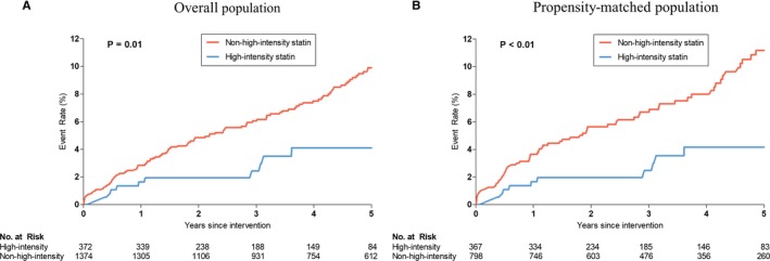
Median follow‐up duration was 4.2 years (interquartile range, 2.2–5.0). Observed clinical outcomes are shown in Table 4. MACEs occurred in 119 patients, including 53 cardiac deaths, 30 MIs, and 51 strokes. Incidence of MACEs was significantly lower in the high‐intensity statin group than that of the non‐high‐intensity statin group (4.1% versus 9.9%; adjusted HR, 0.42; 95% CI, 0.23–0.79; P<0.01; Figure 2A). Cardiac death also occurred less frequently in the high‐intensity statin group than the non‐high‐intensity statin group (0.8% versus 4.8%; adjusted HR, 0.29; 95% CI, 0.09–0.94; P=0.04). However, in the analysis that we classified only those deaths proven to have a definite cardiac etiology as “cardiac death,” incidence of cardiac death was not significantly different between the 2 groups (0.6% versus 2.8%; adjusted HR, 0.31; 95% CI, 0.07–1.34; P=0.12). Although all‐cause death and stroke tended to occur less frequently in the high‐intensity statin group than in the non‐high‐intensity statin group, statistically significance was not achieved.
Table 4.
Clinical Outcomes
| High‐Intensity Statin | Non‐High‐Intensity Statin | Unadjusted HR (95% CI) | P Value | Adjusted HR (95% CI)a | P Value | |
|---|---|---|---|---|---|---|
| Total population (n=1746) | (n=372) | (n=1374) | ||||
| Primary end point | ||||||
| Cardiac death, MI, stroke | 11 (4.1) | 108 (9.9) | 0.44 (0.24–0.82) | 0.01 | 0.42 (0.23–0.79) | <0.01 |
| Secondary end points | ||||||
| All‐cause death | 12 (5.0) | 100 (9.6) | 0.56 (0.31–1.02) | 0.06 | 0.56 (0.30–1.01) | 0.06 |
| Cardiac death | 3 (0.8) | 50 (4.8) | 0.27 (0.09–0.87) | 0.03 | 0.29 (0.09–0.94) | 0.04 |
| MI | 4 (1.4) | 26 (2.4) | 0.64 (0.22–1.83) | 0.40 | 0.64 (0.22–1.84) | 0.41 |
| Stroke | 5 (2.2) | 46 (4.2) | 0.48 (0.19–1.20) | 0.12 | 0.45 (0.18–1.13) | 0.09 |
| Target lesion revascularization | 11 (4.2) | 65 (5.2) | 0.66 (0.35–1.26) | 0.21 | 0.68 (0.36–1.29) | 0.24 |
| Target vessel revascularization | 15 (5.4) | 110 (9.0) | 0.54 (0.31–0.92) | 0.02 | 0.64 (0.37–1.11) | 0.11 |
| Propensity‐matched population (n=1165) | (n=367) | (n=798) | ||||
| Primary end point | ||||||
| Cardiac death, MI, stroke | 11 (4.2) | 66 (11.2) | 0.39 (0.20–0.75) | <0.01 | 0.36 (0.19–0.69) | <0.01 |
| Secondary end points | ||||||
| All‐cause death | 12 (5.1) | 50 (9.0) | 0.57 (0.30–1.07) | 0.08 | 0.58 (0.31–1.07) | 0.08 |
| Cardiac death | 3 (0.9) | 27 (4.9) | 0.26 (0.08–0.86) | 0.03 | 0.27 (0.08–0.90) | 0.03 |
| MI | 4 (1.4) | 15 (2.6) | 0.56 (0.18–1.72) | 0.31 | 0.53 (0.17–1.63) | 0.27 |
| Stroke | 5 (2.3) | 33 (5.6) | 0.40 (0.19–1.03) | 0.06 | 0.41 (0.16–1.06) | 0.07 |
| Target lesion revascularization | 11 (4.3) | 37 (4.8) | 0.68 (0.34–1.34) | 0.26 | 0.67 (0.34–1.34) | 0.26 |
| Target vessel revascularization | 15 (5.4) | 52 (7.3) | 0.66 (0.37–1.19) | 0.17 | 0.66 (0.37–1.19) | 0.17 |
Figure 2.

Kaplan–Meier estimates of the incidence of the primary end point in overall and propensity‐matched population. A, Kaplan–Meier curves for major cardiovascular events (a composite of cardiac death, myocardial infarction, or stroke) in the high‐intensity statin group vs the non‐high‐intensity statin group in the overall population. B, Kaplan–Meier curves for major cardiovascular events (a composite of cardiac death, myocardial infarction, or stroke) in the high‐intensity statin group vs the non‐high‐intensity statin group in the propensity‐matched population.
Propensity‐matched population
There were 77 instances of MACEs with a median follow‐up of 4.1 years (interquartile range, 2.1–5.0) in the matched patients. Incidence of MACEs was significantly lower in the high‐intensity statin group than that of the non‐high‐intensity statin group (4.2% versus 11.2%; adjusted HR, 0.36; 95% CI, 0.19–0.69; P<0.01; Table 4; Figure 2B). Cardiac death also occurred less frequently in the high‐intensity statin group than the non‐high‐intensity statin group (0.9% versus 4.9%; adjusted HR, 0.27; 95% CI, 0.08–0.90; P=0.03). Although all‐cause death and stroke tended to occur less frequently in the high‐intensity statin group than in the non‐high‐intensity statin group, statistically significance was not achieved.
Subgroup Analysis
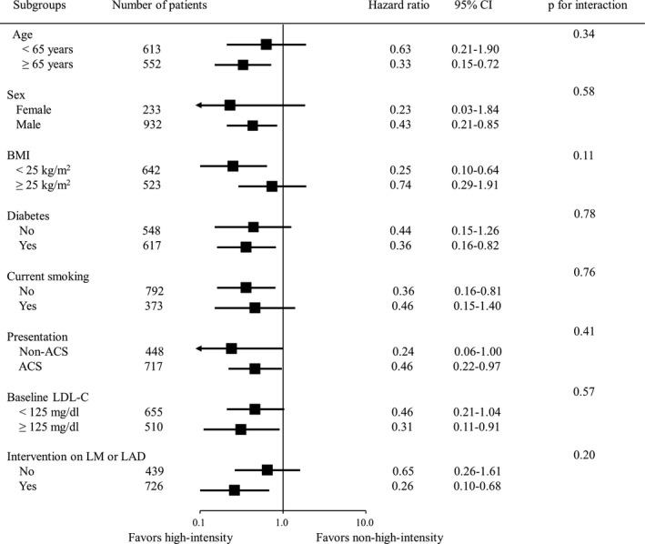
To determine whether the treatment benefits of high‐intensity statin observed in the overall population were consistent, we calculated the unadjusted HR for the MACEs in various subgroups (Figure 3). Benefits of high‐intensity statin were consistent, and there was no significant interaction between statin intensity and primary outcome in any subgroups.
Figure 3.

Comparative unadjusted hazard ratios of primary end point for subgroups. ACS indicates acute coronary syndrome; BMI, body mass index; CI, confidence interval; LDL‐C, low‐density lipoprotein cholesterol; LAD, left anterior descending artery; LM, left main coronary artery.
Discussion
In the present study, we compared the long‐term clinical outcomes according to intensity of statin in patients achieving treatment target for LDL‐C during follow‐up after PCI. The major findings of this study were: (1) Patients in the high‐intensity statin group had a significantly lower incidence of MACEs than those in the non‐high‐intensity statin; (2) this finding was consistently observed in the propensity‐matched population and the various subgroups; (3) time‐averaged LDL‐C, which was calculated from follow‐up LDL‐C values after index PCI, was significantly lower in the high‐intensity statin group than in the non‐high‐intensity statin group, but the difference was small; and (4) reduction from baseline hs‐CRP was significantly greater in the high‐intensity statin group than in the non‐high‐intensity statin group.
Limited Data on Comparison Between Statin Intensity‐Based Strategy Versus LDL‐C Target‐Based Strategy
Statin therapy should be initiated as early as possible to all patients who undergo coronary revascularization for coronary artery disease, regardless of baseline serum cholesterol level.11, 12, 13 However, 2 major guidelines from the ACC/AHA and ESC/EAS recommend different lipid‐lowering strategies for secondary prevention in patients who undergo PCI.3, 5 Whereas the ESC/EAS guideline focuses on decreasing LDL‐C to specific treatment target, ACC/AHA recommends the treatment using evidence‐based intensity statin therapy without specific cholesterol target.14 Such a distinction between the 2 guidelines has led to confusion in the clinical setting.6 During the study period, a high‐intensity statin was prescribed in 14.2% (774 of 5452) of all patients with available information on statin type and dose in our PCI registry. Although the rate of prescription of a high‐intensity statin increased to 41.9% (137 of 327) after the ACC/AHA published the guideline on the treatment of blood cholesterol in November 2013, non‐high‐intensity statins have still been used frequently for secondary prevention in patients undergoing PCI. These observations imply a lack of consensus in the treatment of LDL‐C for secondary prevention. In a meta‐analysis by the Cholesterol Treatment Trialists’ Collabolation, each 1‐mmol/L (approximately 40 mg/dL) reduction in LDL‐C reduced the risk of major vascular events by around 22%, regardless of statin type and dose.4 Moreover, recent trials demonstrated that nonstatin lipid‐lowering drugs, such as ezetimibe and cholesteryl ester transfer protein inhibitor, when added to statin, can further decrease LDL‐C and improve outcomes.15, 16 These findings support LDL‐C target‐based strategy. However, several landmark studies on statin therapy for secondary prevention compared high‐intensity statin versus moderate‐intensity statin, not target LDL‐C, and demonstrated that high‐intensity statin was superior to moderate‐intensity statin for reducing the incidence of MACEs.1, 2 So far, there are limited data on comparison between statin intensity‐based strategy versus LDL‐C target‐based strategy, especially in patients with similar LDL‐C levels. Therefore, we compared long‐term clinical outcomes between the high‐intensity statin group and the non‐high‐intensity statin group among patients who achieved LDL‐C target after PCI.
Plausible Explanations for the Benefit of High‐Intensity Statin Therapy
In the present study, high‐intensity statin therapy was more effective in preventing MACE than non‐high‐intensity statin therapy in patients achieving treatment target for LDL‐C after PCI. There are several plausible explanations for our results. First, level of follow‐up time‐averaged LDL‐C was lower in the high‐intensity statin group than in the non‐high‐intensity statin group. As mentioned above, greater reduction in LDL‐C resulted in greater reduction in risk of MACE in meta‐analyses and randomized controlled trials. Second, percent LDL‐C reduction was greater in the high‐intensity statin group than in the non‐high‐intensity statin group. In a pooled individual patient‐level analysis of 3 large statin trials, Bangalore et al have reported that, among patients with attained LDL‐C ≤70 mg/dL, those with percent LDL‐C reduction of <50% had a significantly higher risk of cardiovascular event when compared with the group with percent LDL‐C reduction of ≥50%.17 Greater percent LDL‐C reduction in the high‐intensity statin group, compared with the non‐high‐intensity statin group, might have partly explain difference in the risk of MACEs. Third, however, observed benefit in the high‐intensity statin group was greater than expected benefit from the difference in time‐averaged LDL‐C between the 2 groups, when compared with previous data from meta‐analyses.4 This finding suggests that observed benefit in the high‐intensity statin group could not be explained by only the lipid‐lowering effect of statins and might be, at least partly, attributable to a non‐lipid‐mediated mechanism or pleotropic effects of statins. Statins have been reported to have diverse protective effects on the cardiovascular system: improvement of endothelial dysfunction18; modulation of inflammatory response and thrombogenesis19; and stabilization of plaque.20 In particular, several trials demonstrated that pleotropic effects of statins are more potent in high‐intensity statins than in non‐high‐intensity statin.21, 22, 23 In the present study, we investigated follow‐up hs‐CRP to compare anti‐inflammatory effects between the 2 groups. Follow‐up hs‐CRP was significantly lower and reduction from baseline hs‐CRP was significantly greater in the high‐intensity statin group than in the non‐high‐intensity statin group, suggesting benefit from non‐lipid‐lowering effects of high‐intensity statins. Additionally, to find an explanation for the extreme differences between the 2 groups, we compared the clinical outcomes between the high‐intensity statin group (n=618) and the non‐high‐intensity statin group (n=3517) in all reviewed patients (n=4135), including the patients who failed to achieve treatment target for LDL‐C. Unlike the results of the patients who achieved LDL‐C goal, incidence of the primary outcome was not significantly different between the 2 groups (7.3% versus 10.0%; adjusted HR, 0.80; 95% CI, 0.55–1.16; P=0.24). Although it is difficult for us to explain the exact causes, the relatively small sample size, especially for the high‐intensity statin group, and nonrandomized nature of our study may have resulted in the extreme differences between the 2 groups.
Limitations
Our study had several limitations. First, the study was a nonrandomized, observational study. Statin intensity was determined at the discretion of the attending physician and might have been influenced by several factors such as underlying demographics, clinical presentation at admission, baseline lipid values, and physician's preference. Although we performed propensity‐score–matched analysis and adjustments to overcome the potential bias that can influence the study outcome, unmeasured factors might have affected study outcomes. Second, 3622 patients were excluded because of lack of information on statin prescription and LDL‐C during follow‐up, although 8148 patients were screened at first. For this reason, a selection bias could have influenced the study results. Third, patients’ compliance to statin was not accurately evaluated. To evaluate the compliance indirectly, we used information of medication prescribed during follow‐up after PCI. As shown in Table 3, statin intensity at discharge after PCI was well maintained during follow‐up. Moreover, because we included only patients who achieved treatment target for LDL‐C, we believe most patients might have a good compliance to statins. The main cause of sudden drop of adherence to high‐intensity statin at the 3 years of follow‐up might be that some physicians reduced the intensity of statin from high to moderate because LDL‐C level had been well maintained under the treatment target during follow‐up. Fourth, hs‐CRP data were only available for 69.6% of the total population (1216 of 1746). Fifth, the median follow‐up duration of the high‐intensity statin group was shorter than that of the non‐high‐intensity statin group (3.1 [1.4–4.9] versus 4.5 [2.5–5.0] years). To assess whether difference in follow‐up duration influenced the study outcome, we truncated follow‐up period to 3 years and re‐evaluated the clinical outcome. Incidence of primary outcome was significantly lower in the high‐intensity statin group than that of the non‐high‐intensity statin group (2.4% versus 6.1%; adjusted HR, 0.41; 95% CI, 0.20–0.86; P=0.02). An adjusted HR of the primary outcome obtained from 3‐year follow‐up data was similar to that using 5‐year follow‐up data. Based on this similarity, we could estimate that the difference in follow‐up duration between the 2 groups did not significantly affect the clinical outcome. Last, MI and stroke might be relatively under‐reported given that this was not a randomized, controlled trial with rigorous follow‐up. However, ratios of MI and stroke to cardiac death or all‐cause death were comparable to, or higher than, those of the randomized, controlled trials24, 25 conducted in Korea. Moreover, it was highly unlikely that MI and stroke were selectively under‐reported in the high‐intensity statin group than in the non‐high‐intensity statin group.
Conclusions
In patients who the achieved LDL‐C target recommended by the ESC/EAS guideline for secondary prevention after PCI, patients treated with high‐intensity statin had a significantly lower incidence of MACE than those treated with non‐high‐intensity statin. Our data suggest that high‐intensity statins should be considered even in patients achieving LDL‐C target with non‐high‐intensity statins.
Sources of Funding
This research was supported by a grant of the Korea Health Technology R&D Project through the Korea Health Industry Development Institute (KHIDI), funded by the Ministry of Health & Welfare, Republic of Korea (grant number: HI10C2020).
Disclosures
Dr Hahn has received speaker's fees from AstraZeneca, Daiichi Sankyo, MSD Korea, and Pfizer. The remaining authors have no disclosures to report.
Supporting information
Table S1. Statins Used in the Non‐High‐Intensity Statin Group
Table S2. Numbers of Patients Under Follow‐up and Without Statin Information
(J Am Heart Assoc. 2018;7:e009517 DOI: 10.1161/JAHA.118.009517)
References
- 1. Cannon CP, Braunwald E, McCabe CH, Rader DJ, Rouleau JL, Belder R, Joyal SV, Hill KA, Pfeffer MA, Skene AM; Pravastatin or Atorvastatin Evaluation and Infection Therapy‐Thrombolysis in Myocardial Infarction 22 Investigators . Intensive versus moderate lipid lowering with statins after acute coronary syndromes. N Engl J Med. 2004;350:1495–1504. [DOI] [PubMed] [Google Scholar]
- 2. LaRosa JC, Grundy SM, Waters DD, Shear C, Barter P, Fruchart JC, Gotto AM, Greten H, Kastelein JJ, Shepherd J, Wenger NK; Treating to New Targets (TNT) Investigators . Intensive lipid lowering with atorvastatin in patients with stable coronary disease. N Engl J Med. 2005;352:1425–1435. [DOI] [PubMed] [Google Scholar]
- 3. Stone NJ, Robinson JG, Lichtenstein AH, Bairey Merz CN, Blum CB, Eckel RH, Goldberg AC, Gordon D, Levy D, Lloyd‐Jones DM, McBride P, Schwartz JS, Shero ST, Smith SC Jr, Watson K, Wilson PW, Eddleman KM, Jarrett NM, LaBresh K, Nevo L, Wnek J, Anderson JL, Halperin JL, Albert NM, Bozkurt B, Brindis RG, Curtis LH, DeMets D, Hochman JS, Kovacs RJ, Ohman EM, Pressler SJ, Sellke FW, Shen WK, Smith SC Jr, Tomaselli GF; American College of Cardiology/American Heart Association Task Force on Practice Guidelines . 2013 ACC/AHA guideline on the treatment of blood cholesterol to reduce atherosclerotic cardiovascular risk in adults: a report of the American College of Cardiology/American Heart Association Task Force on Practice Guidelines. Circulation. 2014;129(Suppl 2):S1–S45. [DOI] [PubMed] [Google Scholar]
- 4. Cholesterol Treatment Trialists C , Baigent C, Blackwell L, Emberson J, Holland LE, Reith C, Bhala N, Peto R, Barnes EH, Keech A, Simes J, Collins R. Efficacy and safety of more intensive lowering of LDL cholesterol: a meta‐analysis of data from 170,000 participants in 26 randomised trials. Lancet. 2010;376:1670–1681. [DOI] [PMC free article] [PubMed] [Google Scholar]
- 5. Piepoli MF, Hoes AW, Agewall S, Albus C, Brotons C, Catapano AL, Cooney MT, Corra U, Cosyns B, Deaton C, Graham I, Hall MS, Hobbs FD, Lochen ML, Lollgen H, Marques‐Vidal P, Perk J, Prescott E, Redon J, Richter DJ, Sattar N, Smulders Y, Tiberi M, van der Worp HB, van Dis I, Verschuren WM; ESC Scientific Document Group . 2016 European Guidelines on cardiovascular disease prevention in clinical practice: the Sixth Joint Task Force of the European Society of Cardiology and Other Societies on Cardiovascular Disease Prevention in Clinical Practice (constituted by representatives of 10 societies and by invited experts). Developed with the special contribution of the European Association for Cardiovascular Prevention & Rehabilitation (EACPR). Eur Heart J. 2016;37:2315–2381. [DOI] [PMC free article] [PubMed] [Google Scholar]
- 6. Ray KK, Kastelein JJ, Boekholdt SM, Nicholls SJ, Khaw KT, Ballantyne CM, Catapano AL, Reiner Z, Luscher TF. The ACC/AHA 2013 guideline on the treatment of blood cholesterol to reduce atherosclerotic cardiovascular disease risk in adults: the good the bad and the uncertain: a comparison with ESC/EAS guidelines for the management of dyslipidaemias 2011. Eur Heart J. 2014;35:960–968. [DOI] [PubMed] [Google Scholar]
- 7. Song YB, Hahn JY, Choi SH, Choi JH, Lee SH, Jeong MH, Kim HS, Seong IW, Yang JY, Rha SW, Jang Y, Yoon JH, Tahk SJ, Seung KB, Park SJ, Gwon HC. Sirolimus‐ versus paclitaxel‐eluting stents for the treatment of coronary bifurcations results: from the COBIS (Coronary Bifurcation Stenting) Registry. J Am Coll Cardiol. 2010;55:1743–1750. [DOI] [PubMed] [Google Scholar]
- 8. Cutlip DE, Windecker S, Mehran R, Boam A, Cohen DJ, van Es GA, Steg PG, Morel MA, Mauri L, Vranckx P, McFadden E, Lansky A, Hamon M, Krucoff MW, Serruys PW; Academic Research Consortium . Clinical end points in coronary stent trials: a case for standardized definitions. Circulation. 2007;115:2344–2351. [DOI] [PubMed] [Google Scholar]
- 9. Hannan EL, Wu C, Walford G, Culliford AT, Gold JP, Smith CR, Higgins RS, Carlson RE, Jones RH. Drug‐eluting stents vs. coronary‐artery bypass grafting in multivessel coronary disease. N Engl J Med. 2008;358:331–341. [DOI] [PubMed] [Google Scholar]
- 10. Sacco RL, Kasner SE, Broderick JP, Caplan LR, Connors JJ, Culebras A, Elkind MS, George MG, Hamdan AD, Higashida RT, Hoh BL, Janis LS, Kase CS, Kleindorfer DO, Lee JM, Moseley ME, Peterson ED, Turan TN, Valderrama AL, Vinters HV; American Heart Association Stroke Council, Council on Cardiovascular Surgery and Anesthesia; Council on Cardiovascular Radiology and Intervention; Council on Cardiovascular and Stroke Nursing; Council on Epidemiology and Prevention; Council on Peripheral Vascular Disease; Council on Nutrition, Physical Activity and Metabolism. An updated definition of stroke for the 21st century: a statement for healthcare professionals from the American Heart Association/American Stroke Association. Stroke. 2013;44:2064–2089. [DOI] [PMC free article] [PubMed] [Google Scholar]
- 11. O'Gara PT, Kushner FG, Ascheim DD, Casey DE Jr, Chung MK, de Lemos JA, Ettinger SM, Fang JC, Fesmire FM, Franklin BA, Granger CB, Krumholz HM, Linderbaum JA, Morrow DA, Newby LK, Ornato JP, Ou N, Radford MJ, Tamis‐Holland JE, Tommaso CL, Tracy CM, Woo YJ, Zhao DX, Anderson JL, Jacobs AK, Halperin JL, Albert NM, Brindis RG, Creager MA, DeMets D, Guyton RA, Hochman JS, Kovacs RJ, Kushner FG, Ohman EM, Stevenson WG, Yancy CW; American College of Cardiology Foundation/American Heart Association Task Force on Practice Guidelines . 2013 ACCF/AHA guideline for the management of ST‐elevation myocardial infarction: a report of the American College of Cardiology Foundation/American Heart Association Task Force on Practice Guidelines. Circulation. 2013;127:e362–e425. [DOI] [PubMed] [Google Scholar]
- 12. Amsterdam EA, Wenger NK, Brindis RG, Casey DE Jr, Ganiats TG, Holmes DR Jr, Jaffe AS, Jneid H, Kelly RF, Kontos MC, Levine GN, Liebson PR, Mukherjee D, Peterson ED, Sabatine MS, Smalling RW, Zieman SJ; ACC/AHA Task Force Members. 2014 AHA/ACC guideline for the management of patients with non‐ST‐elevation acute coronary syndromes: a report of the American College of Cardiology/American Heart Association Task Force on Practice Guidelines. Circulation. 2014;130:e344–e426. [DOI] [PubMed] [Google Scholar]
- 13. Fihn SD, Gardin JM, Abrams J, Berra K, Blankenship JC, Dallas AP, Douglas PS, Foody JM, Gerber TC, Hinderliter AL, King SB III, Kligfield PD, Krumholz HM, Kwong RY, Lim MJ, Linderbaum JA, Mack MJ, Munger MA, Prager RL, Sabik JF, Shaw LJ, Sikkema JD, Smith CR Jr, Smith SC Jr, Spertus JA, Williams SV, Anderson JL; American College of Cardiology Foundation/American Heart Association Task Force . 2012 ACCF/AHA/ACP/AATS/PCNA/SCAI/STS guideline for the diagnosis and management of patients with stable ischemic heart disease: a report of the American College of Cardiology Foundation/American Heart Association Task Force on Practice Guidelines, and the American College of Physicians, American Association for Thoracic Surgery, Preventive Cardiovascular Nurses Association, Society for Cardiovascular Angiography and Interventions, and Society of Thoracic Surgeons. Circulation. 2012;126:e354–e471. [DOI] [PubMed] [Google Scholar]
- 14. Smith SC Jr, Grundy SM. 2013 ACC/AHA guideline recommends fixed‐dose strategies instead of targeted goals to lower blood cholesterol. J Am Coll Cardiol. 2014;64:601–612. [DOI] [PubMed] [Google Scholar]
- 15. Cannon CP, Blazing MA, Giugliano RP, McCagg A, White JA, Theroux P, Darius H, Lewis BS, Ophuis TO, Jukema JW, De Ferrari GM, Ruzyllo W, De Lucca P, Im K, Bohula EA, Reist C, Wiviott SD, Tershakovec AM, Musliner TA, Braunwald E, Califf RM; IMPROVE‐IT Investigators . Ezetimibe added to statin therapy after acute coronary syndromes. N Engl J Med. 2015;372:2387–2397. [DOI] [PubMed] [Google Scholar]
- 16. Group HTRC , Bowman L, Hopewell JC, Chen F, Wallendszus K, Stevens W, Collins R, Wiviott SD, Cannon CP, Braunwald E, Sammons E, Landray MJ. Effects of anacetrapib in patients with atherosclerotic vascular disease. N Engl J Med. 2017;377:1217–1227. [DOI] [PubMed] [Google Scholar]
- 17. Bangalore S, Fayyad R, Kastelein JJ, Laskey R, Amarenco P, DeMicco DA, Waters DD. 2013 cholesterol guidelines revisited: percent LDL cholesterol reduction or attained LDL cholesterol level or both for prognosis? Am J Med. 2016;129:384–391. [DOI] [PubMed] [Google Scholar]
- 18. Sposito AC, Chapman MJ. Statin therapy in acute coronary syndromes: mechanistic insight into clinical benefit. Arterioscler Thromb Vasc Biol. 2002;22:1524–1534. [DOI] [PubMed] [Google Scholar]
- 19. Ray KK, Cannon CP. The potential relevance of the multiple lipid‐independent (pleiotropic) effects of statins in the management of acute coronary syndromes. J Am Coll Cardiol. 2005;46:1425–1433. [DOI] [PubMed] [Google Scholar]
- 20. Rosenson RS, Tangney CC. Antiatherothrombotic properties of statins: implications for cardiovascular event reduction. JAMA. 1998;279:1643–1650. [DOI] [PubMed] [Google Scholar]
- 21. Nissen SE, Tuzcu EM, Schoenhagen P, Brown BG, Ganz P, Vogel RA, Crowe T, Howard G, Cooper CJ, Brodie B, Grines CL, DeMaria AN; REVERSAL Investigators . Effect of intensive compared with moderate lipid‐lowering therapy on progression of coronary atherosclerosis: a randomized controlled trial. JAMA. 2004;291:1071–1080. [DOI] [PubMed] [Google Scholar]
- 22. Wiklund O, Mattsson‐Hulten L, Hurt‐Camejo E, Oscarsson J. Effects of simvastatin and atorvastatin on inflammation markers in plasma. J Intern Med. 2002;251:338–347. [DOI] [PubMed] [Google Scholar]
- 23. Oikonomou E, Siasos G, Zaromitidou M, Hatzis G, Mourouzis K, Chrysohoou C, Zisimos K, Mazaris S, Tourikis P, Athanasiou D, Stefanadis C, Papavassiliou AG, Tousoulis D. Atorvastatin treatment improves endothelial function through endothelial progenitor cells mobilization in ischemic heart failure patients. Atherosclerosis. 2015;238:159–164. [DOI] [PubMed] [Google Scholar]
- 24. Park KW, Kang SH, Kang HJ, Koo BK, Park BE, Cha KS, Rhew JY, Jeon HK, Shin ES, Oh JH, Jeong MH, Kim S, Hwang KK, Yoon JH, Lee SY, Park TH, Moon KW, Kwon HM, Hur SH, Ryu JK, Lee BR, Park YW, Chae IH, Kim HS; HOST–ASSURE Investigators . A randomized comparison of platinum chromium‐based everolimus‐eluting stents versus cobalt chromium‐based Zotarolimus‐Eluting stents in all‐comers receiving percutaneous coronary intervention: HOST‐ASSURE (harmonizing optimal strategy for treatment of coronary artery stenosis‐safety & effectiveness of drug‐eluting stents & anti‐platelet regimen), a randomized, controlled, noninferiority trial. J Am Coll Cardiol. 2014;63:2805–2816. [DOI] [PubMed] [Google Scholar]
- 25. Lee SW, Park SW, Kim YH, Yun SC, Park DW, Lee CW, Kang SJ, Rhee KS, Chae JK, Ko JK, Park JH, Lee JH, Choi SW, Jeong JO, Seong IW, Cho YH, Lee NH, Kim JH, Chun KJ, Kim HS, Park SJ. A randomized comparison of sirolimus‐ versus paclitaxel‐eluting stent implantation in patients with diabetes mellitus: 4‐year clinical outcomes of DES‐DIABETES (drug‐eluting stent in patients with DIABETES mellitus) trial. JACC Cardiovasc Interv. 2011;4:310–316. [DOI] [PubMed] [Google Scholar]
Associated Data
This section collects any data citations, data availability statements, or supplementary materials included in this article.
Supplementary Materials
Table S1. Statins Used in the Non‐High‐Intensity Statin Group
Table S2. Numbers of Patients Under Follow‐up and Without Statin Information


