Key Clinical Message
Previous reports have suggested that mucosal barrier failure allows lanthanum to stray into the lamina propria, and macrophages play an important role for the clearance. However, there is no report to analyze the polarity of phagocytizing macrophages. We suggested that M2 macrophage potentially played a major role in the clearance of lanthanum.
Keywords: dialysis, lanthanum, M2 macrophage, phagocytosis
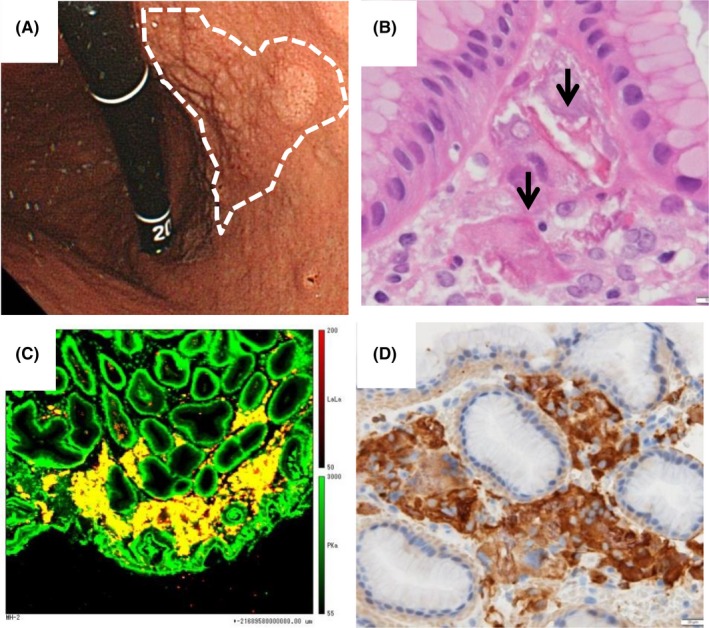
Lanthanum carbonate (LC) is an oral phosphate binder to prevent hyperphosphatemia for patients undergoing dialysis. Recently, lanthanum‐deposited cases in the stomach have been reported (Table 1).1 On endoscopy, such deposits are recognized as minute whitish granules within mucosa (Figure 1A). Lanthanum deposition in patients taking LC is diagnosed based on detection of macrophages containing foreign body in the lamina propria (Figure 1B). Quantitative analysis of elemental composition by electron probe microanalysis confirms both lanthanum and phosphorus (Figure 1C). Macrophages generally can be divided into pro‐inflammatory type M1 and anti‐inflammatory type M2.2 However, the macrophage polarization involved in lanthanum clearance in the gastrointestinal tract has not been elucidated. We found that biopsy specimens from the stomach of 62‐year‐old man with diabetes mellitus, 2‐year history of dialysis, 9‐month history of LC administration, and no specific abnormal physical examination revealed massive eosinophilic infiltration positive for CD68 and CD206 (Figure 1D). CD68 is a common and general marker for both M1 and M2 macrophages, while CD206 is a marker to indicate M2 polarization. Therefore, we assume that phagocytizing M2 macrophages play an important role and M2 polarity induction can accelerate the clearance of lanthanum.2
Table 1.
Previously reported cases of lanthanum carbonate deposition
| Authors | Year | Number of patients | Age | Co‐existing symptoms | Duration of LC administration (mo) | Dose of LC (mg/d) | Deposition site | Immunohistochemistry |
|---|---|---|---|---|---|---|---|---|
| Yasunaga | 2015 | 1 | 64 | Epigastric discomfort | 50 | 1500 | Stomach | CD68, S100, Cam 5.2 |
| Makino | 2015 | 1 | 63 | Heartburn and hiccups | 41 | 500‐750 | Stomach | CD68 |
| Haratake | 2015 | 6 | 61‐69 | Abdominal discomfort and pain | 21‐49 | 500‐1500 | Stomach and duodenum | CD68, S100, Cam 5.2, Ki‐67 |
| Rothenberg | 2015 | 1 | 64 | Low‐grade fever, nausea, diarrhea, and anorexia | NA | NA | Stomach and duodenum | CD68, CAM5.2, CD1a, S100 |
| Tonooka | 2015 | 1 | 81 | Abdominal discomfort | 7 | 750 | Stomach | NA |
| Goto | 2016 | 19 | NA | NA | NA | NA | Stomach and colon | NA |
| Yabuki | 2016 | 3 | 68‐77 | None | 3‐36 | 750‐1500 | Stomach and duodenum | CD68 |
| Shitomi | 2017 | 23 | 34‐82 | Nausea, melena, abdominal pain, and discomfort | 3‐67 | 500‐750 | Stomach | CD68 |
| Hoda | 2017 | 5 | 29‐66 | Dysphagia, reflux, nausea, epigastric burn, melena, and early satiety | 12‐72 | 3000‐6000 | Stomach and duodenum | NA |
| Murakami | 2017 | 7 | 50‐79 | Difficulty swallowing and appetite loss | 5‐45 | NA | Stomach | CD68 |
| Iwamuro | 2017 | 2 | 42, 73 | None | 25, 69 | NA | Stomach and duodenum | CD68 |
| Hattori | 2017 | 16 | 47‐79 | None | 4‐65 | 500‐1500 | Stomach and duodenum | CD68 |
| Nishikawa | 2018 | 1 | 64 | Nausea | 72 | 750‐1500 | Stomach | NA |
| Iwamuro | 2018 | 10 | 42‐76 | NA | 12‐86 | NA | Stomach and duodenum | NA |
| Eso | 2018 | 1 | 26 | Epigastric discomfort and appetite loss | 84 | 750 | Stomach and duodenum | CD68, PU.1 |
Figure 1.

Lanthanum deposition and their analysis (A‐D). Lanthanum deposits are recognized as minute whitish granules within mucosa by endoscopy (A). They are also recognized as eosinophilic infiltration in the lamina propria with hematoxylin and eosin staining (B). Quantitative analysis of elemental composition by electron probe microanalysis reveals macrophages include both lanthanum and phosphorus (yellow area; C). Macrophages are positive for CD206, indicating M2 polarization (D)
CONFLICT OF INTEREST
None declared.
AUTHOR CONTRIBUTION
All the authors: made substantial contribution to the preparation of this manuscript and approved the final version for submission. TN and AT: drafted the manuscript. AT: corresponding author. MK and MN: provided clinical support. ST: reviewed the manuscript.
Nakamura T, Tsuchiya A, Kobayashi M, Naito M, Terai S. M2‐polarized macrophages relate the clearance of gastric lanthanum deposition. Clin Case Rep. 2019;7:570–572. 10.1002/ccr3.1989
REFERENCES
- 1. Murakami N, Yoshioka M, Iwamuro M, et al. Clinical characteristics of seven patients with lanthanum phosphate deposition in the stomach. Int Med. 2017;56(16):2089‐2095. [DOI] [PMC free article] [PubMed] [Google Scholar]
- 2. Gordon S. Alternative activation of macrophages. Nat Rev Immunol. 2003;3(1):23‐35. [DOI] [PubMed] [Google Scholar]


