
FIGURE 5.
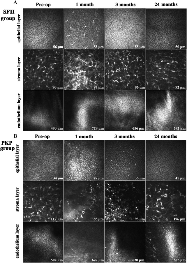
Preoperative and postoperative images of corneal confocal microscopy using the HRT III microscope (magnification, 400 × 400 μm). A, In the SFII group, some dendritic cells were found in the subepithelial region and stromal layer at 1 month postoperatively. They disappeared 3 months after surgery. Endothelial cells showed no particular change in shape or density during the study period. B, In the PKP group, many dendritic cells and inflammatory cells were dispersed in the basal epithelium and stroma within 3 months after surgery.