



Summary
Facioscapulohumeral muscular dystrophy (FSHD) also known as Landouzy-Dejerine disease, is an autosomal-dominant disorder of the skeletal muscles with the name according to the various muscle groups it affects: the face, shoulders and upper arms. It is the third most common genetic degenerative disorder of the skeletal muscles without specific patterns in all the affected individuals. At present there is no cure for the disease but numerous management strategies are available to improve the quality of life and prevent further degeneration of various muscle groups. This review aims to provide an insight on the management strategies for FSHD patients including both lifestyle and medical intervention.
Keywords: Facioscapulohumeral muscular dystrophy, Landouzy-Dejerine disease, clinical manifestations, management, surgical intervention
1. Introduction
Facioscapulohumeral muscular dystrophy (FSHD) also known as Landouzy-Dejerine disease, is the third most common genetic abnormality of the skeletal muscles characterized by progressive muscular dystrophy commonly appearing in individuals before the age of 20 (1,2). Individuals with mild symptoms may manifest FSHD in their later stages of life, but in rare cases, FSHD may also manifest early during infancy or childhood. It is an autosomal dominant genetic disorder which obtains its name from the muscle groups it affects: skeletal muscles of the face (facio), muscle groups surrounding the shoulder blades (scapulo), and the upper arms (humeral). There has not been an agreement on the prevalence on FSHD, but several authors reported a prevalence of 4 to 10 in 100,000 people (3-5).
FSHD commonly presents early in childhood with 95 percent of the affected individuals manifesting the disease by the age of 20 (6). Muscle weakness is commonly non-specific but asymmetrical in occurrence. Individuals with FSHD manifesting with diaphragmatic weakness are at risk of respiratory insufficiency and 20 percent of individuals with FSHD become physically dependent requiring the use of wheelchairs or mobility scooters (7). Most of the cases of FSHD are associated with disorders of D4Z4 repeat in the 4q35 subtelomeric region of chromosome 4. A 2010 report on a unifying theory for the pathogenesis of FSHD explains a second mechanism resulting in toxic gain of function of the DUX4 gene. This recent finding proposed the first pathophysiologic definition of the disease and the possible therapeutic approaches for management of FSHD (8).
Numerous management strategies are available to improve the quality of life of FSHD patients. A prompt diagnosis and lifestyle adjustments are detrimental in preventing unwanted mortality. This review aims to provide an insight into the numerous management strategies in FSHD patients according to each symptomatic presentation.
2. Clinical manifestations of FSHD
Different variations of FSHD exist resulting in the absence of an exact set of symptoms (1,9). FSHD commonly presents as progressive muscle weakness which involves various muscle groups of the face (difficulty whistling, drooping eyelid, decreased facial expressions, speech impairment), shoulder girdle (difficulty raising arms laterally, sloping shoulder), and upper arms (asymmetrical weakness of the biceps and triceps brachii) (1,10,11) . Muscle weakness prominent on one side of the body and scapular winging are the two main characteristics for FSHD. Scapular winging results from weakness of the lower trapezius muscle causing an upward movement of the scapula during flexion of the arm (posterior protrusion), which becomes obvious when the affected individual tries to raise their arms laterally (10). The shoulders tend to have a forward slant with straight clavicles and prominent atrophy of the pectoral muscles.
Typical symptoms of FSHD also involve muscle groups of the face, abdomen and diaphragm. Early presentation of FSHD is often characterized by a pronounced weakness of the facial muscles, which are then followed by symptoms of the shoulders (scapular winging) (12). Early signs also include difficulty whistling due to being unable to purse their lips or sleeping with their eyes partially open due to being unable to completely close their eyelids (13,14). In most cases, extraocular muscles, the eyelid and bulbar muscles are spared.
Individuals with FSHD often have unaffected deltoid muscles in the early stages. The biceps and triceps muscles are asymmetrically involved resulting in a prominent atrophy of the upper arms without affecting the forearm muscles. This leads to the appearance of a large forearm in comparison to the upper arm (Popeye arms) (11). Abdominal muscle weakness may lead to abdominal protuberance and in some cases, lumbar lordosis (15). Weakness of the lower abdominal muscles results in an upward displacement of the umbilicus upon flexion of the neck during supination (Beevor's sign) (5,16).
Muscular weakness of the hip and pelvis results in difficulty climbing stairs or prolonged walking. This condition is further worsened by an aggravated curvature in individuals presenting with lumbar lordosis (12). FSHD individuals affected with progressive movement impairment then become physically dependent on mobility aids.
In rare cases, weakness of the diaphragm leads to respiratory insufficiency, increasing the overall mortality of this disease (2). Retinal telangiectasia and macular edema mimicking Coat's disease may also arise due to failure of vascularization of the vascular peripheries (17). Telangiectatic changes and microaneurysms can be detected by fluorescein angiography in up to 60 percent of the affected individuals (18,19). Bilateral high-tone sensorineural hearing loss can be detected in up to 60 percent of affected individuals. Subclinical sensorineural hearing loss occurs in a higher proportion of individuals (20).
In a study by Zatz et al. (19), FSHD penetrance was found to differ according to age and gender. Penetrance by the age of 30 was estimated to be at 83 percent for both genders, but significantly higher in males (95%) compared to females (69%). They also reported that de novo mutations were more common in females compared to the male participants. No particular evidence of X-chromosome related recessive inheritance is seen, which may be related to excess of symptomatic female individuals in their familial studies including asymptomatic mothers.
3. Subtypes of FSHD
3.1. FSHD Type 1
At least 95 percent of the affected individuals are associated with the pathogenic contraction of D4Z4 repeat (3.2kb unit, normally at 11-100 repeats) at the subtelomeric region 4q35 on chromosome 4. D4Z4 comprises both heterochromatin and euchromatin structures as well as the putative DUX4 gene. A decreased D4Z4 repeat below a certain threshold results in a corresponding relaxation of the chromatin, causing an inappropriate expression of DUX4 in muscle cells which leads to disease progression. In a study in 2014, FSHD was proposed as an "inefficient repeat-mediated epigenetic repression of the D4Z4 macrosatellite repeat array on chromosome 4, resulting in the variegated expression of the DUX4 retrogene, encoding a double-homeobox transcription factor in skeletal muscle" (15). In a regular individual, the D4Z4 repeats are above a certain threshold value and keep the expression of DUX4 repressed. Individuals affected with FSHD possess drastically lower amounts of D4Z4 in addition to a haplotype polymorphism on chromosome 4, causing expression of DUX4.
3.2. FSHD Type 2
The majority of FSHD occurrences are Type 1 cases, accounting for approximately 95 percent. The remaining 5 percent of the cases are Type 2, possessing a phenotype indistinguishable from FSHD but without any evidence of pathological changes of D4Z4 (8,21). Individuals with FSHD type 2 have an open chromatin structure at the D4Z4 loci characterized by CpG methylation loss at D4Z4 repeats in chromosome 4 and 10. Around 80 percent of the cases of FSHD type 2 chromatin relaxation are caused by mutation of SMCHD1 (Structural maintenance of chromosome flexible hinge domain containing gene 1) on chromosome 18. SMCHD1 mutation results in the shortening of DNA of the D4Z4 region, which is permissive for the misexpression of the DUX4 gene by repressing transcription through CpG DNA methylation (22). FSHD type 2 phenotype occurs in individuals who inherit both the SMCHD1 mutation as well as a permissive DUX4 allele, presenting with a digenic inheritance pattern (23). A structural representation of both FSHD type I and type II are represented in Figure 1.
Figure 1.
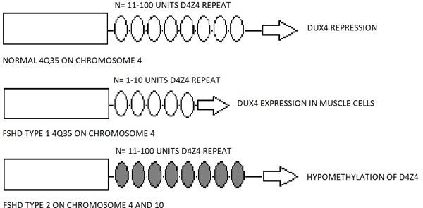
Schematic representation of a pathologic Chromosome 4 in FSHD Type 1 compared to a normal allele.The normal allele normally has 11-100 repeats of D4Z4 (represented by ovals) which is contracted to less than 10 units in FSHD Type 1. In FSHD Type II there is no evidence of pathological change of D4Z4 but there is hypomethylation loss in chromosome 4 and 10.
4. Treatment modalities
Currently there are no specific therapeutic cures for FSHD. But in 2010, the standard of care and management of FSHD were discussed and a census was agreed on at the 171st ENMC International Workshop (24). The main treatment strategies are divided upon the different aspects of the muscle systems it affects (25).
Individuals affected with FSHD are recommended to seek consultation with a physical therapist. The initial consultation may include the overall assessment of the balance, gait, posture, and the probable need of walking aids. According to each individual, a suitable exercise regimen including both aerobic and anaerobic exercises may be suggested to prevent movement impairment. Affected individuals will require a progressive follow-up to monitor the progress of muscular dystrophy and movement impairments during everyday activities. Stretching exercises and low intensity anaerobic exercises are beneficial in the management of symptoms and improve the overall cardiovascular function and muscle strength without having detrimental effects (26).
Affected individuals with severe mobility impairments will require continuous input from a physical therapist and may require mobility aids from an orthotist. Patients presenting with a prominent foot drop will benefit from the use of ankle-foot orthoses. Simple prostheses however may further provide a hindrance to patients who present with an accompanying quadriceps atrophy leading to weakness of the extensor mechanisms (27). A simple ankle-foot orthoses will prevent the full extension of the affected knee and further worsen the walking gait. A floor-reaction ankle-foot orthoses or knee-ankle-foot orthoses are advised for patients presenting with foot drop and inadequate extensor mechanisms simultaneously.
Pain and fatigue are common findings in individuals with FSHD and are often neglected. In a study by van der Kooi et al. (28) they reported that pain occurs in 77 percent of their study population and is reported to occur daily. The most common source of pain is recorded to be localized around the joints where muscle weakness is observed: shoulders, upper back, knees and lumbosacral regions due to hyperlordosis. Pain may also reflect changes in psychological conditions. Chronic pain should be managed with appropriate analgesics and antidepressants if necessary, to improve the quality of life of FSHD individuals.
Respiratory insufficiency may occur in less than 1 percent of individuals with FSHD if muscular dystrophy affects the respiratory muscles (7). Patients presenting with severe symptoms of FSHD should be screened for the possibility of breathing disorders to reduce the overall mortality. Individuals requiring surgical correction are advised to undergo a comprehensive pulmonary function test (PFT) in case of anesthetic reactions. A routine pulmonary workup is also recommended for individuals with severe mobility deficiency, presenting with pelvic weakness accompanied with underlying pulmonary comorbidities, and individuals presenting with obvious spinal deformities causing an obvious abnormality in the thoracic cavity.
Cardiac abnormalities are present in approximately 5 percent of individuals with FSHD without other risk factors for cardiac diseases (29,30). Individuals commonly present with complaints of chest discomfort accompanied with a cardiac arrythmia on an electrocardiogram (ECG) caused by right bundle branch block, which may also be asymptomatic. At present, an ECG is not a routine exam in individuals with FSHD but may be recommended for those at risk.
In a multicenter study by Trevisan et al. (31), individuals affected with FSHD underwent auditory examination. Four patients with FSHD were tested to have auditory problems but none had awareness of any hearing loss. Innate FSHD occurring in infants may lead to auditory and verbal delays in development without being detected and be mistaken for cognitive abnormalities. Cognitive and hearing testing should be routinely examined in children diagnosed with FSHD to prevent auditory and speech impairment due to hearing loss.
Retinal changes in individuals with FSHD commonly are not life-threatening conditions. Retinal vasculopathy is commonly found in individuals affected with FSHD, but the localized pathological changes of the ocular vessels may be effectively treated with laser therapy (24,32). Common findings also include keratitis in individuals who are unable to completely shut their eyelids during sleep. A consultation with an Ophthalmologist is advised for all individuals with FSHD.
Orthopedic intervention is advised for individuals presenting with an obvious scapular winging in a rapidly progressive FSHD. Surgical fixation of the scapula to the thoracic wall (33-35) improves the overall range of motion of the arm in the affected side of the body, but may be temporary. In a series by Giannini et al. (10), 13 FSHD patients presenting with winged scapula underwent bilateral surgical fixation. All 13 patients experienced an obvious increase in active range of motion and a fix for their winged scapula. The scapulothoracic fixation provides a support for deltoid contraction which allows the affected arm to abduct and flex. This however does not provide a full range of motion because the treated joint will not be able to contribute to the arc of motion (10).
Hyperlordosis is a common presentation of FSHD, which subsequently causes pelvic extensor and paraspinal muscle insufficiency (36,37). The spinal deformity is a direct cause of incorrect posture and movement disorders, and patients do not benefit from simple bracing. There is no consensus on surgical intervention for hyperlordosis in FSHD patients due to the controversial results. But in a case resport by Tan et al. (38), they reported a successful partial correction of hyperlordosis with an improvement in sitting posture after surgical correction. Surgical intervention in FSHD patients presenting with hyperlordosis requires further investigation to achieve a definite indication.
Two conflicting studies by Ciafaloni et al. (39) and Rudnik-Schoneborn (40) report on the risk factors for caesarian surgeries and preterm births, and FSHD. In cases of pregnant women affected with FSHD, 25 percent of the individuals report a progressive decrease in motor function which corresponds to the occurrence in other neuromuscular disorders. A comprehensive consult by an obstetrician is advised for pregnant women with FSHD to prevent pregnancy and delivery complications. Pregnant women with FSHD are also advised to closely monitor cardiopulmonary functions to prevent unwanted morbidities.
5. Conclusion
FSHD is one of the most common skeletal diseases, which is easily misdiagnosed during its early stages causing undesired effects in the overall quality of life. Numerous management strategies are available and crucial to prevent undesired progression of the disease. Early diagnosis and a prompt intervention are necessary in improving the life expectancy of FSHD patients.
References
- 1. Tawil R, Van Der Maarel SM. Facioscapulohumeral muscular dystrophy. Muscle Nerve. 2006; 34:1-15. [DOI] [PubMed] [Google Scholar]
- 2. Kilmer DD, Abresch RT, McCrory MA, Carter GT, Fowler WM Jr, Johnson ER, McDonald CM. Profiles of neuromuscular diseases. Facioscapulohumeral muscular dystrophy. Am J Phys Med Rehabil. 1995; 74:S131-S139. [DOI] [PubMed] [Google Scholar]
- 3. Sposito R, Pasquali L, Galluzzi F, Rocchi A, Solito B, Soragna D, Tupler R, Siciliano G. Facioscapulohumeral muscular dystrophy type 1A in northwestern Tuscany: A molecular genetics-based epidemiological and genotype-phenotype study. Genet Test. 2005; 9:30-36. [DOI] [PubMed] [Google Scholar]
- 4. Deenen JC, Arnts H, van der Maarel SM, Padberg GW, Verschuuren JJ, Bakker E, Weinreich SS, Verbeek AL, van Engelen BG. Population-based incidence and prevalence of facioscapulohumeral dystrophy. Neurology. 2014; 83:1056-1059. [DOI] [PMC free article] [PubMed] [Google Scholar]
- 5. Deenen JC, Horlings CG, Verschuuren JJ, Verbeek AL, van Engelen BG. The epidemiology of neuromuscular disorders: A comprehensive overview of the literature. J Neuromuscul Dis. 2015; 2:73-85. [PubMed] [Google Scholar]
- 6. Kissel JT. Facioscapulohumeral muscular dystrophy. Semin Neurol. 1999; 19:35-43. [DOI] [PubMed] [Google Scholar]
- 7. Wohlgemuth M, van der Kooi EL, van Kesteren RG, van der Maarel SM, Padberg GW. Ventilatory support in facioscapulohumeral muscular dystrophy. Neurology. 2004; 63:176-178. [DOI] [PubMed] [Google Scholar]
- 8. Lemmers RJ, van der Vliet PJ, Klooster R, Sacconi S, Camaño P, Dauwerse JG, Snider L, Straasheijm KR, van Ommen GJ, Padberg GW, Miller DG, Tapscott SJ, Tawil R, Frants RR, van der Maarel SM. A unifying genetic model for facioscapulohumeral muscular dystrophy. Science. 2010; 329:1650-1653. [DOI] [PMC free article] [PubMed] [Google Scholar]
- 9. van der Kooi AJ, Visser MC, Rosenberg N, van der Berg- Vos R, Wokke JH, Bakker E, de Visser M. Extension of the clinical range of facioscapulohumeral muscular dystrophy: Report of six cases. J Neurol Neurosurg Psychiatry. 2000; 69:114-116. [DOI] [PMC free article] [PubMed] [Google Scholar]
- 10. Giannini S, Faldini C, Pagkrati S, Grandi G, Digennaro V, Luciani D, Merlini L. Fixation of winged scapula in facioscapulohumeral muscular dystrophy. Clin Med Res. 2007; 5:155-162. [DOI] [PMC free article] [PubMed] [Google Scholar]
- 11. Statland JM, Tawil R. Facioscapulohumeral muscular dystrophy. Continuum (Minneap Minn). 2016; 22:1916-1931. [DOI] [PMC free article] [PubMed] [Google Scholar]
- 12. Kottlors M, Kress W, Meng G, Glocker FX. Facioscapulohumeral muscular dystrophy presenting with isolated axial myopathy and bent spine syndrome. Muscle Nerve. 2010; 42:273-275. [DOI] [PubMed] [Google Scholar]
- 13. Jordan B, Eger K, Koesling S, Zierz S. Camptocormia phenotype of FSHD: A clinical and MRI study on six patients. J Neurol. 2011; 258:866-873. [DOI] [PubMed] [Google Scholar]
- 14. Krasnianski M, Eger K, Neudecker S, Jakubiczka S, Zierz S. Atypical phenotypes in patients with facioscapularhumeral muscular dystrophy 4q35 deletion. Arch Neurol. 2003; 60:1421-1425. [DOI] [PubMed] [Google Scholar]
- 15. Ta w i l R, v a n d e r M a a r e l SM, Ta p s c o t t S J. Facioscapulohumeral dystrophy: The path to consensus on pathophysiology. Skelet Muscle. 2014; 4:12. [DOI] [PMC free article] [PubMed] [Google Scholar]
- 16. Eger K, Jordan B, Habermann S, Zierz S. Beevor's sign in facioscapulohumeral muscular dystrophy: An old sign with new implications. J Neurol. 2010; 257:436-438. [DOI] [PubMed] [Google Scholar]
- 17. Osborne RJ, Welle S, Venance SL, Thornton CA, Tawil R. Expression profile of FSHD supports a link between retinal vasculopathy and muscular dystrophy. Neurology. 2007; 68:569-577. [DOI] [PubMed] [Google Scholar]
- 18. Lindner M, Holz FG, Charbel Issa P. Spontaneous resolution of retinal vascular abnormalities and macular oedema in facioscapulohumeral muscular dystrophy. Clin Exp Ophthalmol. 2016; 44:627-628. [DOI] [PubMed] [Google Scholar]
- 19. Zatz M, Marie SK, Cerqueira A, Vainzof M, Pavanello RC, Passos-Bueno MR. The facioscapulohumeral muscular dystrophy (FSHD1) gene affects males more severely and more frequently than females. Am J Med Genet. 1998; 77:155-161. [PubMed] [Google Scholar]
- 20. Padberg GW, Brouwer OF, de Keizer RJ, Dijkman G, Wijmenga C, Grote JJ, Frants RR. On the significance of retinal vascular disease and hearing loss in facioscapulohumeral muscular dystrophy. Muscle Nerve Suppl. 1995; S73-S80. [PubMed] [Google Scholar]
- 21. de Greef JC, Lemmers RJ, van Engelen BG, Sacconi S, Venance SL, Frants RR, Tawil R, van der Maarel SM. Common epigenetic changes of D4Z4 in contraction-dependent and contraction independent FSHD. Hum Muta. 2009; 30:1449-1459. [DOI] [PubMed] [Google Scholar]
- 22. Blewitt ME, Gendrel AV, Pang Z, Sparrow DB, Whitelaw N, Craig JM, Apedaile A, Hilton DJ, Dunwoodie SL, Brockdorff N, Kay GF, Whitelaw E. SmcHD1, containing a structural-maintenance-of-chromosomes hinge domain, has a critical role in X inactivation. Nat Genet. 2008; 40:663-669. [DOI] [PubMed] [Google Scholar]
- 23. Lemmers RJ, Tawil R, Petek LM, et al. Digenic inheritance of an SMCHD1 mutation and an FSHD-permissive D4Z4 allele causes facioscapulohumeral muscular dystrophy type 2. Nat Genet. 2012; 44:1370-1374. [DOI] [PMC free article] [PubMed] [Google Scholar]
- 24. Tawil R, van der Maarel S, Padberg GW, van Engelen BG. 171st ENMC International workshop: Standards of care and management of facioscapulohumeral muscular dystrophy. Neuromuscul Disord. 2010; 20:471-475. [DOI] [PubMed] [Google Scholar]
- 25. Tawil R, Kissel JT, Heatwole C, et al. Evidence-based guideline summary: Evaluation, diagnosis, and management of facioscapulohumeral muscular dystrophy: Report of the Guideline Development, Dissemination, and Implementation Subcommittee of the American Academy of Neurology and the Practice Issues Review Panel of the American Association of Neuromuscular & Electrodiagnostic Medicine. Neurology. 2015; 85:357-364. [DOI] [PMC free article] [PubMed] [Google Scholar]
- 26. Olsen DB, Orngreen MC, Vissing J. Aerobic training improves exercise performance in facioscapulohumeral muscular dystrophy. Neurology. 2005; 64:1064-1066. [DOI] [PubMed] [Google Scholar]
- 27. Tawil R. Facioscapulohumeral muscular dystrophy. Neurotherapeutics. 2008; 5:601-606. [DOI] [PMC free article] [PubMed] [Google Scholar]
- 28. van der Kooi EL, Kalkman JS, Lindeman E, Hendriks JC, van Engelen BG, Bleijenberg G, Padberg GW. Effects of training and albuterol on pain and fatigue in facioscapulohumeral muscular dystrophy. J Neurol. 2007; 254:931-940. [DOI] [PMC free article] [PubMed] [Google Scholar]
- 29. Laforêt P, de Toma C, Eymard B, Becane HM, Jeanpierre M, Fardeau M, Duboc D. Cardiac involvement in genetically confirmed facioscapulohumeral muscular dystrophy. Nerology. 1998; 51:1454-1456. [DOI] [PubMed] [Google Scholar]
- 30. Trevisan CP, Pastorello E, Armani M, Angelini C, Nante G, Tomelleri G, Tonin P, Mongini T, Palmucci L, Galluzzi G, Tupler RG, Barchitta A. Faciosapulohumeral muscular dystrophy and occurrence of heart arrythmia. Eur Neurol. 2006; 56:1-5. [DOI] [PubMed] [Google Scholar]
- 31. Trevisan CP, Pastorello E, Ermani M, Angelini C, Tomelleri G, Tonin P, Mongini T, Palmucci L, Galluzzi G, Tupler RG, Marioni G, Rimini A. Facioscapulohumeral muscular dystrophy: A multicenter study on hearing function. Audiol Neurootol. 2008; 13:1-6. [DOI] [PubMed] [Google Scholar]
- 32. Fitzsimons RB, Gurwin EB, Bird AC. Retinal vascular abnormalities in facioscapulohumeral muscular dystrophy. A general association with genetic and therapeutic implications. Brain. 1987; 110(Pt 3):631-648. [DOI] [PubMed] [Google Scholar]
- 33. Mummery CJ, Copeland SA, Rose MR. Scapular fixation in muscular dystrophy. Chochrane Database Syst Rev. 2003; 2003; CD003278. [DOI] [PubMed] [Google Scholar]
- 34. Rhee YG, Ha JH. Long-term results of scapulothoracic arthrodesis of facioscapulohumeral muscular dystrophy. J Shoulder Elbow Surg. 2006; 15:445-450. [DOI] [PubMed] [Google Scholar]
- 35. Demirhan M, Uysal O, Atalar AC, Kilicoglu O, Serdaroglu P. Scapulothoracic arthrodes in facioscapulohumeral dystrophy with multifilament cables. Clin Orthop Relat Res. 2009; 467:2090-2097. [DOI] [PMC free article] [PubMed] [Google Scholar]
- 36. Lee CS, Kang SJ, Wang CJ, Lee SW, Ahn YJ, Kim YT, Lee DH, Lee MY. Early-onsetfacioscapulohumeral muscular dystrophy - significance of pelvic extensorsin sagittal spinal imbalance. J Pediatr Orthop B. 2009; 18:325-329. [DOI] [PubMed] [Google Scholar]
- 37. Rijken NH, van Engelen BG, de Rooy JW, Geurts AC, Weerdesteyn V. Trunk muscle involvement is most critical for the loss of balance control in patients with facioscapulohumeral muscular dystrophy. Clin Biomech (Bristol, Avon). 2014; 29:855-860. [DOI] [PubMed] [Google Scholar]
- 38. Tan H, Feng F, Lin Y, Chen C, Li Z, Shen J. Surgical correction of hyeperlordosis in facioscapulohumeral muscular dystrophy: A case report. BMC Surg. 2017; 17:83. [DOI] [PMC free article] [PubMed] [Google Scholar]
- 39. Ciafaloni E, Pressman EK, Lori AM, Smirnow AM, Guntrum DJ, Dilek N, Tawil R. Pregnancy and birth outcomes in women with facioscapulohumeralmuscular dystrophy. Neurology. 2006; 67:1887-1889. [DOI] [PubMed] [Google Scholar]
- 40. Rudnik-Schoneborn S, Golner B, Rohrig D, Zerres K. Obstetric aspects in women with facioscapulohumeral muscular dystrophy, limb-girdle muscular dystrophy, and congenital myopathies. Arch Neurol. 1997; 54:888-894. [DOI] [PubMed] [Google Scholar]