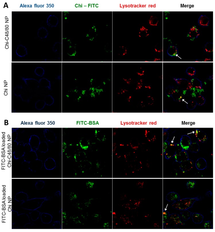
Figure 8.
Evaluation of uptake by macrophages. (A) Uptake of nanoparticles was assessed by incubating 4 h, 100 µg/mL RAW 264.7 cells with Chi-C48/80 NPs or Chi NPs prepared with fluorescein isothiocyanate (FITC)-labeled chitosan (green). (B) Uptake of the antigen loaded on nanoparticles was evaluated by incubating the cells with FITC-BSA loaded Chi-C48/80 NPs or Chi NPs. Cells were labeled with Alexa Fluor© 350 WGA (blue) to identify the membrane, and Lysotracker© Red identifies the acidic endosomes and lysosomes. Arrows in the merge image show co-localization.

