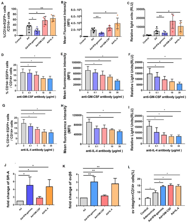
Figure 5.
Incubation of GM-CSF and IL-4 promoted the efficacy of adenovirus infection by upregulating SR-A and integrin receptor. 1250 vp/cell of Ad5-EGFP or Ad5-SEAP virus was added into PBMCs with or without recombinant IFN-γ (40 ng/mL), GM-CSF (20 ng/mL) or IL-4 (20 ng/mL) respectively, and the expression of EGFP or SEAP was detected after 48 h. (A) The change of percentage of EGFP-positive cells in the different cytokines-treated CD14+ cells (n = 6). (B) The change of expression level of EGFP protein (MFI value) in the different cytokines-treated CD14+ cells (n = 6). (C) The change of RLU (Relative light units) in the different cytokines-treated PBMCs (n = 6). 1250 vp/cell of Ad5-EGFP or Ad5-SEAP virus was added into PBMCs (n = 3) with isotype control antibody or anti-human GM-CSF monoclonal antibody (0–30 μg/mL), and the percentage of EGFP-positive cells (D), the expression level of EGFP protein (MFI value) (E) and the expression level of SEAP (RLU value) (F) were detected after 24 h. 1250 vp/cell of Ad5-EGFP or Ad5-SEAP virus was added into PBMCs (n = 3) with isotype control antibody or anti-human IL-4 monoclonal antibody (0–30 μg/mL), and the percentage of EGFP-positive cells (G), the expression level of EGFP protein (MFI value) (H) and the expression level of SEAP (RLU value) (I) were detected after 24 h. Moreover, the expression level of adenovirus-related receptors, including SR-A (J) and αV integrin (K) was detected by quantitative RT-PCR. The integrin receptor protein on the cell surface was also analyzed by flow cytometer (L). The bars represent the standard error. *: p < 0.05, **: p < 0.01.

