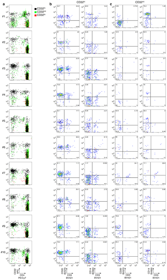
Extended Data Fig. 2. Post-sort flow cytometry of CD3+CD4+ subsets that were CD32hi, CD32int or CD32lo.
Cells were sorted as in Extended Data Fig. 1. a, Overlay plots of CD32 and CD4 expression by cells in CD32hi, CD32int and CD32lo sorted populations. Note the heterogeneous pattern of cells from the CD32hi and CD32int populations. b, c, CD20, CD14 and CD3 staining in the CD32+ cells from the CD32hi (b) and the CD32int (c) subsets. Note the large proportions of all CD32+ cells bearing surface markers consistent with B cells (CD20+CD3−) or monocytes (CD14+CD3−) after sorting.

